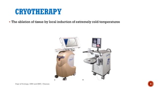
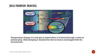
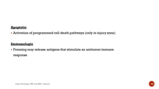
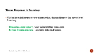
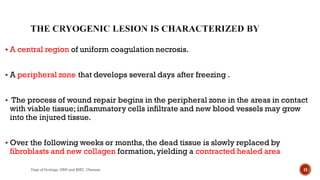
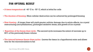
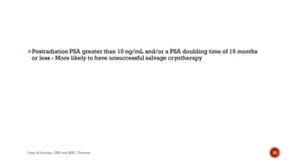
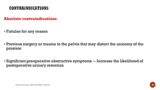
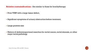
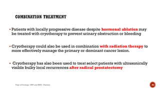
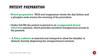
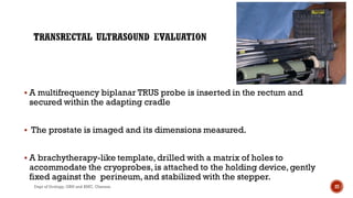
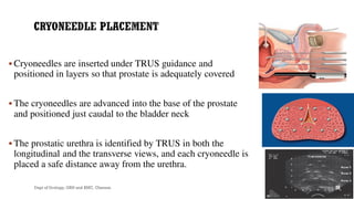
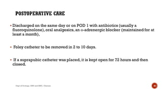
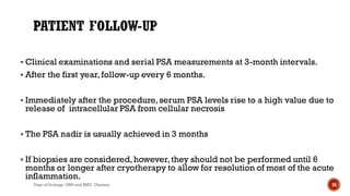
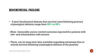
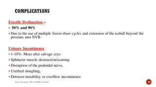
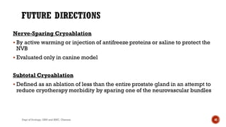
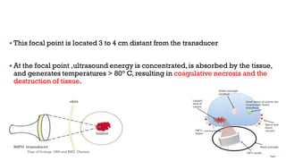
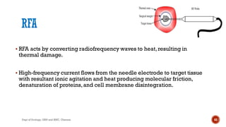
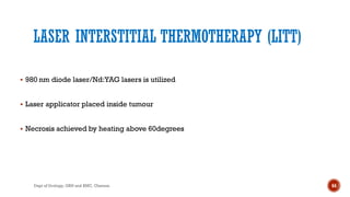
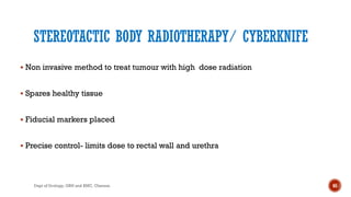
Dept of Urology
Govt Royapettah Hospital and Kilpauk Medical College
Chennai
1
Professors:
 Prof. Dr.G.Sivasankar, M.S., M.Ch.,
 Prof. Dr.A. Senthilvel, M.S., M.Ch.,
Asst Professors:
 Dr.J. Sivabalan, M.S., M.Ch.,
 Dr.R. Bhargavi, M.S., M.Ch.,
 Dr.S. Raju, M.S., M.Ch.,
 Dr.K. Muthurathinam,M.S., M.Ch.,
 Dr.D.Tamilselvan, M.S., M.Ch.,
 Dr.K. Senthilkumar,M.S., M.Ch.
Dept of Urology, GRH and KMC, Chennai. 2
 Prostate cancer is relatively slow growing, with doubling time for local tumors
estimated at 2–4 years.
 In order to cure and control localized prostate cancer,the concept of focal therapy
has emerged.
 Focal therapy is the middle ground between AS and radical therapy,offering much
less morbidity with cancer control.
 Focal destruction of cancer,with preservation of the surrounding organ
3
Dept of Urology, GRH and KMC, Chennai.
 Defined as the lesion(s) with the highest cancer suspicion score based on initial
MP-MRI,irrespective of size.
 Histopathological features of the index lesion predict the clinical behavior of the
entire gland despite multiple synchronous tumors
 Since most metastatic cancers originate from a single clonal cancer cell, it would
be reasonable and effective to identify and target this potentially lethal lesion with
focal therapy
4
Dept of Urology, GRH and KMC, Chennai.
 At present,there is neither a consensus as to a strict definition of inclusion and
exclusion criteria nor pretreatment planning guidance for focal therapy.
 Cure- the disease should be low risk and low volume in a targetable area of the
prostate.
 Disease control- treat the dominant lesion or index lesion
 Multimodal treatment approach in the high-risk patient
5
Dept of Urology, GRH and KMC, Chennai.
 Either alone or in combination
 To detect extent and laterality of tumour and target focal therapy
 12 core TRUS-Standard approach
 Transperineal (TP) biopsy with three-dimensional (3D) mapping
 MRI-targeted fusion biopsy
6
Dept of Urology, GRH and KMC, Chennai.
METHODS OF FOCAL ABLATION
 CRYOTHERAPY
 HIFU
 MICROWAVE ABLATION
 FOCAL IRREVERSIBLE ELECTROPORATION
 INTESTITIAL LASER COAGULATION
 RADIOFREQUENCY ABLATION
 RADIOSURGERY
 PHOTODYNAMIC THERAPY
7
Dept of Urology, GRH and KMC, Chennai.
CRYOTHERAPY
 The ablation of tissue by local induction of extremely cold temperatures
8
Dept of Urology, GRH and KMC, Chennai.
Temperature change of a real gas or liquid when it is forced through a valve or
porous plug while keeping it insulated so that no heat is exchanged with the
environment
9
Dept of Urology, GRH and KMC, Chennai.
The main mechanism –
Induction of targeted areas of coagulative necrosis by freezing
The key factors involved in freezing injury include
 Direct mechanical shock,
 Osmotic shock, and
 Cellular hypoxia.
10
Dept of Urology, GRH and KMC, Chennai.
MECHANISMS
 Protein denaturation via dehydration,
 Transfer of water from the intracellular
space to the extracellular space,
 Rupture of cell membranes from ice
crystal expansion,
 Toxic concentration of cellular
constituents,
 Thermal shock from rapid
supercooling,
 Slow thawing,
 Vascular stasis, and
 Increased apoptosis
11
Dept of Urology, GRH and KMC, Chennai.
Mechanical
 Rapid formation of intracellular and extracellular ice crystals exerting mechanical shear forces
on cell membranes and organelles
Ischemic
 Microvascular damage: stasis of blood leading to thrombosis
 Freezing major blood supply (neurovascular bundle)
12
Dept of Urology, GRH and KMC, Chennai.
Apoptotic
 Activation of programmed cell death pathways (only in injury zone)
Immunologic
 Freezing may release antigens that stimulate an antitumor immune
response
13
Dept of Urology, GRH and KMC, Chennai.
Tissue Response to Freezing:
 Varies from inflammatory to destructive,depending on the severity of
freezing
 Minor freezing injury - Only inflammatory responses
 Severe freezing injury - Destroys cells and tissues
14
Dept of Urology, GRH and KMC, Chennai.
 A central region of uniform coagulation necrosis.
 A peripheral zone that develops several days after freezing .
 The process of wound repair begins in the peripheral zone in the areas in contact
with viable tissue;inflammatory cells infiltrate and new blood vessels may grow
into the injured tissue.
 Over the following weeks or months,the dead tissue is slowly replaced by
fibroblasts and new collagen formation,yielding a contracted healed area
15
Dept of Urology, GRH and KMC, Chennai.
 A tissue temperature of −40° C to −50° C, which is lethal for cells
 The duration of freezing- More cellular destruction can be achieved by prolonged freezing
 Slow thawing - A longer thaw will yield greater cellular damage due to solute effects, ice crystal
restructuring (recrystallization),prolonged oxidative stress,and growth of ice crystals
 Repetition of the freeze-thaw cycle- The second cycle increases the extent of necrosis up to
80% of the previously frozen volume
 The interval between freeze-thaw cycles - Leaves the tissue in a hypothermic state and allows
time for the microcirculation to fail
16
Dept of Urology, GRH and KMC, Chennai.
 Outcomes depends on the stage and grade
 Suitable for patients with clinical stage T1c-T3
 Mainly as an alternative treatment to radiation therapy
 For T3 disease,this is more appropriate for smaller-volume disease in
which the tumor is likely to be encompassed in the freezing process.
17
Dept of Urology, GRH and KMC, Chennai.
Poor candidates for radical prostatectomy or radiation therapy (e.g.,men
with Crohn disease,ulcerative colitis,prior pelvic irradiation or pelvic
surgery,cardiac disease,morbid obesity, and/or body habitus unfavorable for
radical prostatectomy or radiation therapy)
Patients who have no evidence of metastatic disease with more than a 10-year
life expectancy
Patients who either unsatisfied or unwilling to other treatments -
prostatectomy or radiation therapy
18
Dept of Urology, GRH and KMC, Chennai.
 Post radiotherapy PSA recurrence(20% and 60% )
 Radical prostatectomy, cryotherapy,brachytherapy,and radiation
therapy are the options available for salvage treatment of
radiorecurrent prostate cancer
 Salvage prostatectomy is associated with significant morbidity
 Recent modifications in the technique of salvage cryotherapy have led
to the ability to eradicate radiorecurrent prostate cancer safely and
with decreased morbidity
19
Dept of Urology, GRH and KMC, Chennai.
 Rising PSA value after radiation
 A positive postradiation biopsy, and
 A negative metastatic survey
20
Dept of Urology, GRH and KMC, Chennai.
Postradiation PSA greater than 10 ng/mL and/or a PSA doubling time of 16 months
or less - More likely to have unsuccessful salvage cryotherapy
21
Dept of Urology, GRH and KMC, Chennai.
Absolute contraindications
 Fistulae for any reason
 Previous surgery or trauma to the pelvis that may distort the anatomy of the
prostate
 Significant preoperative obstructive symptoms -- Increase the likelihood of
postoperative urinary retention
22
Dept of Urology, GRH and KMC, Chennai.
Relative contraindications - Are similar to those for brachytherapy
 Prior TURP with a large tissue defect,
 Significant symptoms of urinary obstruction before treatment,
 Large prostate size
 History of abdominoperineal resection for rectal cancer,rectal stenosis, or other
major rectal pathology
23
Dept of Urology, GRH and KMC, Chennai.
 Patients with locally progressive disease despite hormonal ablation may
be treated with cryotherapy to prevent urinary obstruction or bleeding
 Cryotherapy could also be used in combination with radiation therapy to
more effectively manage the primary or dominant cancer lesion.
 Cryotherapy has also been used to treat select patients with ultrasonically
visible bulky local recurrences after radical prostatectomy
24
Dept of Urology, GRH and KMC, Chennai.
 Patient Preparation
 Transrectal Ultrasound Evaluation
 Cryoneedle Placement
 Thermocouple and Urethral Warmer Placement
 Monitoring of Freezing Process
25
Dept of Urology, GRH and KMC, Chennai.
 Bowel preparation -With oral magnesium citrate the day before and
a phospho-soda enema the morning of the procedure.
 Under GA/RA the patient is placed in an exaggerated dorsal
lithotomy position,which provides excellent transperineal access to
the prostate.
 A Foley catheter is inserted and clamped to allow the bladder to
distend,thereby displacing the intraperitoneal contents.
26
Dept of Urology, GRH and KMC, Chennai.
 A multifrequency biplanar TRUS probe is inserted in the rectum and
secured within the adapting cradle
 The prostate is imaged and its dimensions measured.
 A brachytherapy-like template,drilled with a matrix of holes to
accommodate the cryoprobes,is attached to the holding device,gently
fixed against the perineum,and stabilized with the stepper.
27
Dept of Urology, GRH and KMC, Chennai.
 Cryoneedles are inserted under TRUS guidance and
positioned in layers so that prostate is adequately covered
 The cryoneedles are advanced into the base of the prostate
and positioned just caudal to the bladder neck
 The prostatic urethra is identified by TRUS in both the
longitudinal and the transverse views, and each cryoneedle is
placed a safe distance away from the urethra.
28
Dept of Urology, GRH and KMC, Chennai.
 Thermocouples may be placed in the mid gland, at the level of the
external sphincter, at each NVB, and in Denonvilliers fascia to minimize the
risk of incontinence and rectourethral fistula
 The temperature in the sphincter be maintained above 15° C.
29
Dept of Urology, GRH and KMC, Chennai.
 MultiThermal Sensor (MTS) needles,which enable four separate
temperature readings from each sensor,has improved significantly the
safety of cryosurgical ablation and decreased dramatically the risk of
postablation fistulae
 A meticulous 360-degree examination of the urethra is performed
using a flexible cystoscope.
 The bladder is also inspected to ensure that no misplaced probes have
pierced the bladder neck
30
Dept of Urology, GRH and KMC, Chennai.
 A 0.038-inch super-stiff guidewire is inserted and a heavily
lubricated urethral warming catheter is introduced into the bladder
over the guidewire and under sonographic guidance.
 During the procedure,the bladder is kept nearly full to prevent injury
from the rigid tip of the warmer device.
31
Dept of Urology, GRH and KMC, Chennai.
UrethralWarming Device
 To protect the urethra and the external urinary sphincter
 This is a closed double-lumen catheter through which saline heated to
43° C is continuously circulated by a water pump at a rate of 350 to 500
mL/min
 preserves a thin layer of the mucosa
 significantly reducing urethral sloughing, stricture formation,
and urinary incontinence
32
Dept of Urology, GRH and KMC, Chennai.
 Discharged on the same day or on POD 1 with antibiotics (usually a
fluoroquinolone),oral analgesics,an α-adrenergic blocker (maintained for at
least a month),
 Foley catheter to be removed in 2 to 10 days.
 If a suprapubic catheter was placed, it is kept open for 72 hours and then
closed.
33
Dept of Urology, GRH and KMC, Chennai.
 Clinical examinations and serial PSA measurements at 3-month intervals.
 After the first year,follow-up every 6 months.
 Immediately after the procedure,serum PSA levels rise to a high value due to
release of intracellular PSA from cellular necrosis
 The PSA nadir is usually achieved in 3 months
 If biopsies are considered,however,they should not be performed until 6
months or longer after cryotherapy to allow for resolution of most of the acute
inflammation.
34
Dept of Urology, GRH and KMC, Chennai.
Local Control
 The positive biopsy rate after cryotherapy ranges from 7.7% to 25%.
 In a series with longer-term follow-up, a 10-year negative biopsy rate is 77%.
 Higher pretreatment PSA levels and clinical T stage were independently
associated with an increased risk of positive postcryosurgical biopsy
 Recurrence was more common in cancers located at the apex (9.5%) and the
seminal vesicles(43.8%),in contrast to those located in the mid gland (4.1%)
and base (0%).
35
Dept of Urology, GRH and KMC, Chennai.
 5-year biochemical disease-free survival rates following primary
cryosurgical ablation range from 60% to 90%
 More favourable cancer control outcomes reported for patients with
low- and intermediate-risk cancers
 There are no long-term data available regarding metastasis-free or
overall survival following cryosurgical ablation of the prostate
36
Dept of Urology, GRH and KMC, Chennai.
Erectile Dysfunction :-
 50% and 90%
 Due to the use of multiple freeze-thaw cycles and extension of the iceball beyond the
prostate, into NVB
Urinary Incontinence
 1-10%- More after salvage cryo
 Sphincter muscle destruction/scarring
 Disruption of the pudendal nerve,
 Urethral sloughing,
 Detrusor instability or overflow incontinence
37
Dept of Urology, GRH and KMC, Chennai.
 Urethral Sloughing
- Less than 5% to 15%
- Treatment is antibiotic and adequate drainage
-Transurethral resection of necrotic tissue may be required if the
condition persists
 Urethral stricture
- It is rare & are treated with either balloon dilation or transurethral
incision
38
Dept of Urology, GRH and KMC, Chennai.
 Pelvic and/or Rectal Pain
-0 to 50%
- Due to osteitis pubis
- More in previously irradiated patients
- Treated with antiinflammatory drugs
 Rectourethral Fistula
- 0% to 3%
- Salvage > primary
- Watery diarrhea, pneumaturia,or fecaluria
- VCUG or CT will confirm the diagnosis & location of fistula
39
Dept of Urology, GRH and KMC, Chennai.
 Treatment
- Foleys drainage may be useful in primary cryo
- Early fecal and urinary diversion in salvage cryo
- If the fistula tract matures,fulguration may facilitate
spontaneous closure.
- Fistula repair should be delayed for 4 to 6 months.
A transperineal, posterior, or anterior approach for closure is
recommended
40
Dept of Urology, GRH and KMC, Chennai.
OTHER COMPLICATIONS
 Penile and scrotal swelling
 Hydronephrosis is a relatively uncommon may be due to extensive freezing
of the bladder neck area or placement of a cryoprobe deep into the seminal
vesicle leading to freezing of the ureteral orifices or distal ureters
 Small bowel obstruction may occur if the iceball is allowed to extend into
the cul-de-sac of the peritoneal cavity
41
Dept of Urology, GRH and KMC, Chennai.
Nerve-Sparing Cryoablation
 By active warming or injection of antifreeze proteins or saline to protect the
NVB
 Evaluated only in canine model
Subtotal Cryoablation
 Defined as an ablation of less than the entire prostate gland in an attempt to
reduce cryotherapy morbidity by sparing one of the neurovascular bundles
42
Dept of Urology, GRH and KMC, Chennai.
HIGH-INTENSITY FOCUSED ULTRASOUND
43
Dept of Urology, GRH and KMC, Chennai.
 High-energy ultrasound waves to destroy the tissue at the focal point of a
transducer without injuring the intervening tissue.
 HIFU is different from cryotherapy in that the focal zone is extremely discrete with
little or no tissue effects in areas immediately adjacent to the treatment zone.
44
Dept of Urology, GRH and KMC, Chennai.
 Strong ultrasound waves in the inaudible sound range 10,000 times stronger than
diagnostic ultrasound are generated by a transducer with a parabolic
configuration
 Focuses these sound waves into a discrete focal point measuring approximately 3
mm ×3 mm × 11 mm.
45
Dept of Urology, GRH and KMC, Chennai.
 This focal point is located 3 to 4 cm distant from the transducer
 At the focal point ,ultrasound energy is concentrated,is absorbed by the tissue,
and generates temperatures > 80° C, resulting in coagulative necrosis and the
destruction of tissue.
46
Dept of Urology, GRH and KMC, Chennai.
Thermal effect
 Absorption of ultrasound energy into the tissue, which is converted
into heat.
 The temperature within sonicated tissue will rise to a level sufficient
to induce irreversible damages.
 Sharp increases in temperature up to 70°C to 100°C can be achieved
in a few seconds with each pulse.
47
Dept of Urology, GRH and KMC, Chennai.
Cavitation:
 As a result of the interaction of ultrasound and micro-bubbles in the
sonicated tissue
 This interaction may lead to oscillation of these micro-bubbles,violent
collapses,and dispersion of energy that enhances tissue ablation
 Both of these mechanisms lead to the destruction of the cells by a
coagulative necrosis
48
Dept of Urology, GRH and KMC, Chennai.
MICROSTREAMING- rapid movement of liquid outside an oscillating
bubble.
Release of free radicals
Induction of apoptosis
49
Dept of Urology, GRH and KMC, Chennai.
 Sonablate (Focus Surgery Inc,Indianapolis,Ind) and Ablatherm
(EDAP-TMS SA,Vaulx enVelin,France).
 The technology of both systems is basically the same,but there are a
few technical differences between these two devices
50
Dept of Urology, GRH and KMC, Chennai.
 GA or RA
 A HF transducer probe placed in a balloon filled with cooled liquid is inserted
into the rectum- to both serve as an acoustical interface and to cool the rectal
wall.
51
Dept of Urology, GRH and KMC, Chennai.
 Low-energy transducers (3 to 4 MHz) for imaging and high-energy
transducers for treatment.
 The prostate is imaged in both the sagittal and coronal planes,and the
target treatment zone is outlined.
 There is a treatment cycle in which the treatment zone is heated and
then a cooling period during which the computer-controlled device
moves to the next treatment zone distant from the first.
52
Dept of Urology, GRH and KMC, Chennai.
 Localized prostate cancer with clinical stage T1–T2 Nx–N0 M0
 Not suitable for a radical prostatectomy (eg, age >70 years of age, life
expectancy ≤10 years,major comorbidities precluding surgery)
 Unwilling for surgery.
 Locally proven recurrence of prostate cancer after radiation or
brachytherapy failures.
53
Dept of Urology, GRH and KMC, Chennai.
 The gland volume should not be > 40 mL.
 Rectal pathology
 Major prostatic calcifications
 can lead to an ultrasonic wave transmission impairment
54
Dept of Urology, GRH and KMC, Chennai.
 Urinary retention-swollen gland or the passage of necrotic debris (sloughing) induced by
coagulated adenoma
 Bladder outlet obstruction-reduced with Ablatherm devices
 Stricture rate of 22% that required intermittent dilatations
 UTI
 Impotence ranged from 20% to 49.8%
 Incontinence,reported between 0.6% and 15.4%,
55
Dept of Urology, GRH and KMC, Chennai.
High-Intensity Ultrasound after Radiation Failure
 Positive biopsy rates after radiotherapy for prostate cancer range between 25%
and 32%
 The majority of these patients have traditionally been treated expectantly or with
hormonal deprivation
 The complication rates of salvage HIFU are higher than those for HIFU as a
primary procedure
56
Dept of Urology, GRH and KMC, Chennai.
HIFU Re-treatment:
 The management of recurrence after HIFU differs from that after
radiation failure in that with HIFU there is no maximum dose and
sessions can be repeated.
57
Dept of Urology, GRH and KMC, Chennai.
NANO KNIFE – FOCAL IRREVERSIBLE ELECTROPORATION
 A kind of "electronic scalpel" that opens the cell membrane-
 The electricity creates very tiny openings (called pores) in the tumor’s cells,
leading to the death of the cells.
 An ultrasound or a CT scan- focus the current precisely on the tumor, sparing blood
vessels and other tissues.
 The cell matrix of the healthy tissue in the area of treatment remains preserved and
is able to regenerate itself
58
Dept of Urology, GRH and KMC, Chennai.
PHOTODYNAMIC THERAPY
 Phase II trials for preliminary treatment of localized disease.
 3 elements:a photo-sensitizer,light source,and oxygen.
 hematoporphyrin or 5 Aminolevulinic acid - as the photosensitizer
 Therapeutic action- ROS formed by light activation of photosensitizer
 Cell death by cytoplasmic and mitochondrial activation of apoptosis
59
Dept of Urology, GRH and KMC, Chennai.
60
Dept of Urology, GRH and KMC, Chennai.
VASCULAR TARGETED PHOTODYNAMIC THERAPY (WST-09 VTP)
 Because the photosensitizer (Padeliporfin ) is confined to the
vasculature,the mechanism for cell death is primarily attributed to
vascular occlusion and other processes that originate in a vascular
oxidative stress.
 Post irradiation recurrences
61
Dept of Urology, GRH and KMC, Chennai.
62
Dept of Urology, GRH and KMC, Chennai.
RFA
 RFA acts by converting radiofrequency waves to heat,resulting in
thermal damage.
 High-frequency current flows from the needle electrode to target tissue
with resultant ionic agitation and heat producing molecular friction,
denaturation of proteins,and cell membrane disintegration.
63
Dept of Urology, GRH and KMC, Chennai.
LASER INTERSTITIAL THERMOTHERAPY (LITT)
 980 nm diode laser/Nd:YAG lasers is utilized
 Laser applicator placed inside tumour
 Necrosis achieved by heating above 60degrees
64
Dept of Urology, GRH and KMC, Chennai.
STEREOTACTIC BODY RADIOTHERAPY/ CYBERKNIFE
 Non invasive method to treat tumour with high dose radiation
 Spares healthy tissue
 Fiducial markers placed
 Precise control- limits dose to rectal wall and urethra
65
Dept of Urology, GRH and KMC, Chennai.
66
Dept of Urology, GRH and KMC, Chennai.
67
Dept of Urology, GRH and KMC, Chennai.
 The heat sink effect relates to one area that overheats in the HIFU pulses’ pathway
and thus prevents adequate ultrasound propagation to the targeted area; such a
phenomenon occurs if the time between HIFU pulses is inadequate for tissue
cooling or if an area is high in water content,such as a cyst.
68
Dept of Urology, GRH and KMC, Chennai.
Computerized Planning
 Provide improved intraoperative treatment planning with a potential for more
precise and complete tissue destruction
 Used to determine optimal placement of cryoprobes in the prostate to maximize
ablation of the prostate while minimizing collateral damage
69
Dept of Urology, GRH and KMC, Chennai.

Prostate carcinoma- focal therapy

  • 1.
    Dept of Urology GovtRoyapettah Hospital and Kilpauk Medical College Chennai 1
  • 2.
    Professors:  Prof. Dr.G.Sivasankar,M.S., M.Ch.,  Prof. Dr.A. Senthilvel, M.S., M.Ch., Asst Professors:  Dr.J. Sivabalan, M.S., M.Ch.,  Dr.R. Bhargavi, M.S., M.Ch.,  Dr.S. Raju, M.S., M.Ch.,  Dr.K. Muthurathinam,M.S., M.Ch.,  Dr.D.Tamilselvan, M.S., M.Ch.,  Dr.K. Senthilkumar,M.S., M.Ch. Dept of Urology, GRH and KMC, Chennai. 2
  • 3.
     Prostate canceris relatively slow growing, with doubling time for local tumors estimated at 2–4 years.  In order to cure and control localized prostate cancer,the concept of focal therapy has emerged.  Focal therapy is the middle ground between AS and radical therapy,offering much less morbidity with cancer control.  Focal destruction of cancer,with preservation of the surrounding organ 3 Dept of Urology, GRH and KMC, Chennai.
  • 4.
     Defined asthe lesion(s) with the highest cancer suspicion score based on initial MP-MRI,irrespective of size.  Histopathological features of the index lesion predict the clinical behavior of the entire gland despite multiple synchronous tumors  Since most metastatic cancers originate from a single clonal cancer cell, it would be reasonable and effective to identify and target this potentially lethal lesion with focal therapy 4 Dept of Urology, GRH and KMC, Chennai.
  • 5.
     At present,thereis neither a consensus as to a strict definition of inclusion and exclusion criteria nor pretreatment planning guidance for focal therapy.  Cure- the disease should be low risk and low volume in a targetable area of the prostate.  Disease control- treat the dominant lesion or index lesion  Multimodal treatment approach in the high-risk patient 5 Dept of Urology, GRH and KMC, Chennai.
  • 6.
     Either aloneor in combination  To detect extent and laterality of tumour and target focal therapy  12 core TRUS-Standard approach  Transperineal (TP) biopsy with three-dimensional (3D) mapping  MRI-targeted fusion biopsy 6 Dept of Urology, GRH and KMC, Chennai.
  • 7.
    METHODS OF FOCALABLATION  CRYOTHERAPY  HIFU  MICROWAVE ABLATION  FOCAL IRREVERSIBLE ELECTROPORATION  INTESTITIAL LASER COAGULATION  RADIOFREQUENCY ABLATION  RADIOSURGERY  PHOTODYNAMIC THERAPY 7 Dept of Urology, GRH and KMC, Chennai.
  • 8.
    CRYOTHERAPY  The ablationof tissue by local induction of extremely cold temperatures 8 Dept of Urology, GRH and KMC, Chennai.
  • 9.
    Temperature change ofa real gas or liquid when it is forced through a valve or porous plug while keeping it insulated so that no heat is exchanged with the environment 9 Dept of Urology, GRH and KMC, Chennai.
  • 10.
    The main mechanism– Induction of targeted areas of coagulative necrosis by freezing The key factors involved in freezing injury include  Direct mechanical shock,  Osmotic shock, and  Cellular hypoxia. 10 Dept of Urology, GRH and KMC, Chennai.
  • 11.
    MECHANISMS  Protein denaturationvia dehydration,  Transfer of water from the intracellular space to the extracellular space,  Rupture of cell membranes from ice crystal expansion,  Toxic concentration of cellular constituents,  Thermal shock from rapid supercooling,  Slow thawing,  Vascular stasis, and  Increased apoptosis 11 Dept of Urology, GRH and KMC, Chennai.
  • 12.
    Mechanical  Rapid formationof intracellular and extracellular ice crystals exerting mechanical shear forces on cell membranes and organelles Ischemic  Microvascular damage: stasis of blood leading to thrombosis  Freezing major blood supply (neurovascular bundle) 12 Dept of Urology, GRH and KMC, Chennai.
  • 13.
    Apoptotic  Activation ofprogrammed cell death pathways (only in injury zone) Immunologic  Freezing may release antigens that stimulate an antitumor immune response 13 Dept of Urology, GRH and KMC, Chennai.
  • 14.
    Tissue Response toFreezing:  Varies from inflammatory to destructive,depending on the severity of freezing  Minor freezing injury - Only inflammatory responses  Severe freezing injury - Destroys cells and tissues 14 Dept of Urology, GRH and KMC, Chennai.
  • 15.
     A centralregion of uniform coagulation necrosis.  A peripheral zone that develops several days after freezing .  The process of wound repair begins in the peripheral zone in the areas in contact with viable tissue;inflammatory cells infiltrate and new blood vessels may grow into the injured tissue.  Over the following weeks or months,the dead tissue is slowly replaced by fibroblasts and new collagen formation,yielding a contracted healed area 15 Dept of Urology, GRH and KMC, Chennai.
  • 16.
     A tissuetemperature of −40° C to −50° C, which is lethal for cells  The duration of freezing- More cellular destruction can be achieved by prolonged freezing  Slow thawing - A longer thaw will yield greater cellular damage due to solute effects, ice crystal restructuring (recrystallization),prolonged oxidative stress,and growth of ice crystals  Repetition of the freeze-thaw cycle- The second cycle increases the extent of necrosis up to 80% of the previously frozen volume  The interval between freeze-thaw cycles - Leaves the tissue in a hypothermic state and allows time for the microcirculation to fail 16 Dept of Urology, GRH and KMC, Chennai.
  • 17.
     Outcomes dependson the stage and grade  Suitable for patients with clinical stage T1c-T3  Mainly as an alternative treatment to radiation therapy  For T3 disease,this is more appropriate for smaller-volume disease in which the tumor is likely to be encompassed in the freezing process. 17 Dept of Urology, GRH and KMC, Chennai.
  • 18.
    Poor candidates forradical prostatectomy or radiation therapy (e.g.,men with Crohn disease,ulcerative colitis,prior pelvic irradiation or pelvic surgery,cardiac disease,morbid obesity, and/or body habitus unfavorable for radical prostatectomy or radiation therapy) Patients who have no evidence of metastatic disease with more than a 10-year life expectancy Patients who either unsatisfied or unwilling to other treatments - prostatectomy or radiation therapy 18 Dept of Urology, GRH and KMC, Chennai.
  • 19.
     Post radiotherapyPSA recurrence(20% and 60% )  Radical prostatectomy, cryotherapy,brachytherapy,and radiation therapy are the options available for salvage treatment of radiorecurrent prostate cancer  Salvage prostatectomy is associated with significant morbidity  Recent modifications in the technique of salvage cryotherapy have led to the ability to eradicate radiorecurrent prostate cancer safely and with decreased morbidity 19 Dept of Urology, GRH and KMC, Chennai.
  • 20.
     Rising PSAvalue after radiation  A positive postradiation biopsy, and  A negative metastatic survey 20 Dept of Urology, GRH and KMC, Chennai.
  • 21.
    Postradiation PSA greaterthan 10 ng/mL and/or a PSA doubling time of 16 months or less - More likely to have unsuccessful salvage cryotherapy 21 Dept of Urology, GRH and KMC, Chennai.
  • 22.
    Absolute contraindications  Fistulaefor any reason  Previous surgery or trauma to the pelvis that may distort the anatomy of the prostate  Significant preoperative obstructive symptoms -- Increase the likelihood of postoperative urinary retention 22 Dept of Urology, GRH and KMC, Chennai.
  • 23.
    Relative contraindications -Are similar to those for brachytherapy  Prior TURP with a large tissue defect,  Significant symptoms of urinary obstruction before treatment,  Large prostate size  History of abdominoperineal resection for rectal cancer,rectal stenosis, or other major rectal pathology 23 Dept of Urology, GRH and KMC, Chennai.
  • 24.
     Patients withlocally progressive disease despite hormonal ablation may be treated with cryotherapy to prevent urinary obstruction or bleeding  Cryotherapy could also be used in combination with radiation therapy to more effectively manage the primary or dominant cancer lesion.  Cryotherapy has also been used to treat select patients with ultrasonically visible bulky local recurrences after radical prostatectomy 24 Dept of Urology, GRH and KMC, Chennai.
  • 25.
     Patient Preparation Transrectal Ultrasound Evaluation  Cryoneedle Placement  Thermocouple and Urethral Warmer Placement  Monitoring of Freezing Process 25 Dept of Urology, GRH and KMC, Chennai.
  • 26.
     Bowel preparation-With oral magnesium citrate the day before and a phospho-soda enema the morning of the procedure.  Under GA/RA the patient is placed in an exaggerated dorsal lithotomy position,which provides excellent transperineal access to the prostate.  A Foley catheter is inserted and clamped to allow the bladder to distend,thereby displacing the intraperitoneal contents. 26 Dept of Urology, GRH and KMC, Chennai.
  • 27.
     A multifrequencybiplanar TRUS probe is inserted in the rectum and secured within the adapting cradle  The prostate is imaged and its dimensions measured.  A brachytherapy-like template,drilled with a matrix of holes to accommodate the cryoprobes,is attached to the holding device,gently fixed against the perineum,and stabilized with the stepper. 27 Dept of Urology, GRH and KMC, Chennai.
  • 28.
     Cryoneedles areinserted under TRUS guidance and positioned in layers so that prostate is adequately covered  The cryoneedles are advanced into the base of the prostate and positioned just caudal to the bladder neck  The prostatic urethra is identified by TRUS in both the longitudinal and the transverse views, and each cryoneedle is placed a safe distance away from the urethra. 28 Dept of Urology, GRH and KMC, Chennai.
  • 29.
     Thermocouples maybe placed in the mid gland, at the level of the external sphincter, at each NVB, and in Denonvilliers fascia to minimize the risk of incontinence and rectourethral fistula  The temperature in the sphincter be maintained above 15° C. 29 Dept of Urology, GRH and KMC, Chennai.
  • 30.
     MultiThermal Sensor(MTS) needles,which enable four separate temperature readings from each sensor,has improved significantly the safety of cryosurgical ablation and decreased dramatically the risk of postablation fistulae  A meticulous 360-degree examination of the urethra is performed using a flexible cystoscope.  The bladder is also inspected to ensure that no misplaced probes have pierced the bladder neck 30 Dept of Urology, GRH and KMC, Chennai.
  • 31.
     A 0.038-inchsuper-stiff guidewire is inserted and a heavily lubricated urethral warming catheter is introduced into the bladder over the guidewire and under sonographic guidance.  During the procedure,the bladder is kept nearly full to prevent injury from the rigid tip of the warmer device. 31 Dept of Urology, GRH and KMC, Chennai.
  • 32.
    UrethralWarming Device  Toprotect the urethra and the external urinary sphincter  This is a closed double-lumen catheter through which saline heated to 43° C is continuously circulated by a water pump at a rate of 350 to 500 mL/min  preserves a thin layer of the mucosa  significantly reducing urethral sloughing, stricture formation, and urinary incontinence 32 Dept of Urology, GRH and KMC, Chennai.
  • 33.
     Discharged onthe same day or on POD 1 with antibiotics (usually a fluoroquinolone),oral analgesics,an α-adrenergic blocker (maintained for at least a month),  Foley catheter to be removed in 2 to 10 days.  If a suprapubic catheter was placed, it is kept open for 72 hours and then closed. 33 Dept of Urology, GRH and KMC, Chennai.
  • 34.
     Clinical examinationsand serial PSA measurements at 3-month intervals.  After the first year,follow-up every 6 months.  Immediately after the procedure,serum PSA levels rise to a high value due to release of intracellular PSA from cellular necrosis  The PSA nadir is usually achieved in 3 months  If biopsies are considered,however,they should not be performed until 6 months or longer after cryotherapy to allow for resolution of most of the acute inflammation. 34 Dept of Urology, GRH and KMC, Chennai.
  • 35.
    Local Control  Thepositive biopsy rate after cryotherapy ranges from 7.7% to 25%.  In a series with longer-term follow-up, a 10-year negative biopsy rate is 77%.  Higher pretreatment PSA levels and clinical T stage were independently associated with an increased risk of positive postcryosurgical biopsy  Recurrence was more common in cancers located at the apex (9.5%) and the seminal vesicles(43.8%),in contrast to those located in the mid gland (4.1%) and base (0%). 35 Dept of Urology, GRH and KMC, Chennai.
  • 36.
     5-year biochemicaldisease-free survival rates following primary cryosurgical ablation range from 60% to 90%  More favourable cancer control outcomes reported for patients with low- and intermediate-risk cancers  There are no long-term data available regarding metastasis-free or overall survival following cryosurgical ablation of the prostate 36 Dept of Urology, GRH and KMC, Chennai.
  • 37.
    Erectile Dysfunction :- 50% and 90%  Due to the use of multiple freeze-thaw cycles and extension of the iceball beyond the prostate, into NVB Urinary Incontinence  1-10%- More after salvage cryo  Sphincter muscle destruction/scarring  Disruption of the pudendal nerve,  Urethral sloughing,  Detrusor instability or overflow incontinence 37 Dept of Urology, GRH and KMC, Chennai.
  • 38.
     Urethral Sloughing -Less than 5% to 15% - Treatment is antibiotic and adequate drainage -Transurethral resection of necrotic tissue may be required if the condition persists  Urethral stricture - It is rare & are treated with either balloon dilation or transurethral incision 38 Dept of Urology, GRH and KMC, Chennai.
  • 39.
     Pelvic and/orRectal Pain -0 to 50% - Due to osteitis pubis - More in previously irradiated patients - Treated with antiinflammatory drugs  Rectourethral Fistula - 0% to 3% - Salvage > primary - Watery diarrhea, pneumaturia,or fecaluria - VCUG or CT will confirm the diagnosis & location of fistula 39 Dept of Urology, GRH and KMC, Chennai.
  • 40.
     Treatment - Foleysdrainage may be useful in primary cryo - Early fecal and urinary diversion in salvage cryo - If the fistula tract matures,fulguration may facilitate spontaneous closure. - Fistula repair should be delayed for 4 to 6 months. A transperineal, posterior, or anterior approach for closure is recommended 40 Dept of Urology, GRH and KMC, Chennai.
  • 41.
    OTHER COMPLICATIONS  Penileand scrotal swelling  Hydronephrosis is a relatively uncommon may be due to extensive freezing of the bladder neck area or placement of a cryoprobe deep into the seminal vesicle leading to freezing of the ureteral orifices or distal ureters  Small bowel obstruction may occur if the iceball is allowed to extend into the cul-de-sac of the peritoneal cavity 41 Dept of Urology, GRH and KMC, Chennai.
  • 42.
    Nerve-Sparing Cryoablation  Byactive warming or injection of antifreeze proteins or saline to protect the NVB  Evaluated only in canine model Subtotal Cryoablation  Defined as an ablation of less than the entire prostate gland in an attempt to reduce cryotherapy morbidity by sparing one of the neurovascular bundles 42 Dept of Urology, GRH and KMC, Chennai.
  • 43.
    HIGH-INTENSITY FOCUSED ULTRASOUND 43 Deptof Urology, GRH and KMC, Chennai.
  • 44.
     High-energy ultrasoundwaves to destroy the tissue at the focal point of a transducer without injuring the intervening tissue.  HIFU is different from cryotherapy in that the focal zone is extremely discrete with little or no tissue effects in areas immediately adjacent to the treatment zone. 44 Dept of Urology, GRH and KMC, Chennai.
  • 45.
     Strong ultrasoundwaves in the inaudible sound range 10,000 times stronger than diagnostic ultrasound are generated by a transducer with a parabolic configuration  Focuses these sound waves into a discrete focal point measuring approximately 3 mm ×3 mm × 11 mm. 45 Dept of Urology, GRH and KMC, Chennai.
  • 46.
     This focalpoint is located 3 to 4 cm distant from the transducer  At the focal point ,ultrasound energy is concentrated,is absorbed by the tissue, and generates temperatures > 80° C, resulting in coagulative necrosis and the destruction of tissue. 46 Dept of Urology, GRH and KMC, Chennai.
  • 47.
    Thermal effect  Absorptionof ultrasound energy into the tissue, which is converted into heat.  The temperature within sonicated tissue will rise to a level sufficient to induce irreversible damages.  Sharp increases in temperature up to 70°C to 100°C can be achieved in a few seconds with each pulse. 47 Dept of Urology, GRH and KMC, Chennai.
  • 48.
    Cavitation:  As aresult of the interaction of ultrasound and micro-bubbles in the sonicated tissue  This interaction may lead to oscillation of these micro-bubbles,violent collapses,and dispersion of energy that enhances tissue ablation  Both of these mechanisms lead to the destruction of the cells by a coagulative necrosis 48 Dept of Urology, GRH and KMC, Chennai.
  • 49.
    MICROSTREAMING- rapid movementof liquid outside an oscillating bubble. Release of free radicals Induction of apoptosis 49 Dept of Urology, GRH and KMC, Chennai.
  • 50.
     Sonablate (FocusSurgery Inc,Indianapolis,Ind) and Ablatherm (EDAP-TMS SA,Vaulx enVelin,France).  The technology of both systems is basically the same,but there are a few technical differences between these two devices 50 Dept of Urology, GRH and KMC, Chennai.
  • 51.
     GA orRA  A HF transducer probe placed in a balloon filled with cooled liquid is inserted into the rectum- to both serve as an acoustical interface and to cool the rectal wall. 51 Dept of Urology, GRH and KMC, Chennai.
  • 52.
     Low-energy transducers(3 to 4 MHz) for imaging and high-energy transducers for treatment.  The prostate is imaged in both the sagittal and coronal planes,and the target treatment zone is outlined.  There is a treatment cycle in which the treatment zone is heated and then a cooling period during which the computer-controlled device moves to the next treatment zone distant from the first. 52 Dept of Urology, GRH and KMC, Chennai.
  • 53.
     Localized prostatecancer with clinical stage T1–T2 Nx–N0 M0  Not suitable for a radical prostatectomy (eg, age >70 years of age, life expectancy ≤10 years,major comorbidities precluding surgery)  Unwilling for surgery.  Locally proven recurrence of prostate cancer after radiation or brachytherapy failures. 53 Dept of Urology, GRH and KMC, Chennai.
  • 54.
     The glandvolume should not be > 40 mL.  Rectal pathology  Major prostatic calcifications  can lead to an ultrasonic wave transmission impairment 54 Dept of Urology, GRH and KMC, Chennai.
  • 55.
     Urinary retention-swollengland or the passage of necrotic debris (sloughing) induced by coagulated adenoma  Bladder outlet obstruction-reduced with Ablatherm devices  Stricture rate of 22% that required intermittent dilatations  UTI  Impotence ranged from 20% to 49.8%  Incontinence,reported between 0.6% and 15.4%, 55 Dept of Urology, GRH and KMC, Chennai.
  • 56.
    High-Intensity Ultrasound afterRadiation Failure  Positive biopsy rates after radiotherapy for prostate cancer range between 25% and 32%  The majority of these patients have traditionally been treated expectantly or with hormonal deprivation  The complication rates of salvage HIFU are higher than those for HIFU as a primary procedure 56 Dept of Urology, GRH and KMC, Chennai.
  • 57.
    HIFU Re-treatment:  Themanagement of recurrence after HIFU differs from that after radiation failure in that with HIFU there is no maximum dose and sessions can be repeated. 57 Dept of Urology, GRH and KMC, Chennai.
  • 58.
    NANO KNIFE –FOCAL IRREVERSIBLE ELECTROPORATION  A kind of "electronic scalpel" that opens the cell membrane-  The electricity creates very tiny openings (called pores) in the tumor’s cells, leading to the death of the cells.  An ultrasound or a CT scan- focus the current precisely on the tumor, sparing blood vessels and other tissues.  The cell matrix of the healthy tissue in the area of treatment remains preserved and is able to regenerate itself 58 Dept of Urology, GRH and KMC, Chennai.
  • 59.
    PHOTODYNAMIC THERAPY  PhaseII trials for preliminary treatment of localized disease.  3 elements:a photo-sensitizer,light source,and oxygen.  hematoporphyrin or 5 Aminolevulinic acid - as the photosensitizer  Therapeutic action- ROS formed by light activation of photosensitizer  Cell death by cytoplasmic and mitochondrial activation of apoptosis 59 Dept of Urology, GRH and KMC, Chennai.
  • 60.
    60 Dept of Urology,GRH and KMC, Chennai.
  • 61.
    VASCULAR TARGETED PHOTODYNAMICTHERAPY (WST-09 VTP)  Because the photosensitizer (Padeliporfin ) is confined to the vasculature,the mechanism for cell death is primarily attributed to vascular occlusion and other processes that originate in a vascular oxidative stress.  Post irradiation recurrences 61 Dept of Urology, GRH and KMC, Chennai.
  • 62.
    62 Dept of Urology,GRH and KMC, Chennai.
  • 63.
    RFA  RFA actsby converting radiofrequency waves to heat,resulting in thermal damage.  High-frequency current flows from the needle electrode to target tissue with resultant ionic agitation and heat producing molecular friction, denaturation of proteins,and cell membrane disintegration. 63 Dept of Urology, GRH and KMC, Chennai.
  • 64.
    LASER INTERSTITIAL THERMOTHERAPY(LITT)  980 nm diode laser/Nd:YAG lasers is utilized  Laser applicator placed inside tumour  Necrosis achieved by heating above 60degrees 64 Dept of Urology, GRH and KMC, Chennai.
  • 65.
    STEREOTACTIC BODY RADIOTHERAPY/CYBERKNIFE  Non invasive method to treat tumour with high dose radiation  Spares healthy tissue  Fiducial markers placed  Precise control- limits dose to rectal wall and urethra 65 Dept of Urology, GRH and KMC, Chennai.
  • 66.
    66 Dept of Urology,GRH and KMC, Chennai.
  • 67.
    67 Dept of Urology,GRH and KMC, Chennai.
  • 68.
     The heatsink effect relates to one area that overheats in the HIFU pulses’ pathway and thus prevents adequate ultrasound propagation to the targeted area; such a phenomenon occurs if the time between HIFU pulses is inadequate for tissue cooling or if an area is high in water content,such as a cyst. 68 Dept of Urology, GRH and KMC, Chennai.
  • 69.
    Computerized Planning  Provideimproved intraoperative treatment planning with a potential for more precise and complete tissue destruction  Used to determine optimal placement of cryoprobes in the prostate to maximize ablation of the prostate while minimizing collateral damage 69 Dept of Urology, GRH and KMC, Chennai.