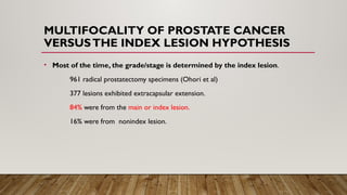
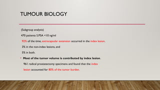
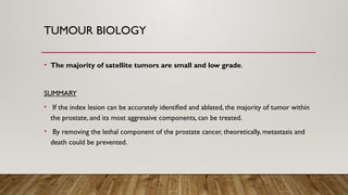
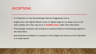
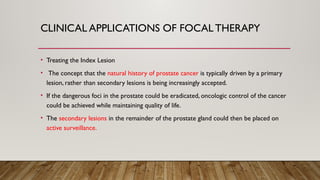
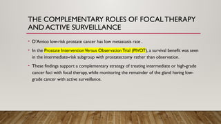
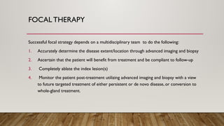
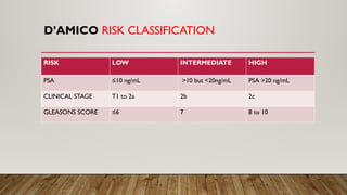
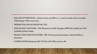
FOCAL THERAPY FOR
PROSTATE CANCER
DR.MANIKANDAN G
FOCAL THERAPY AND PROSTATE CANCER
INTRODUCTION.
• The idea of prostate focal therapy followed on the heels of the successful shift in breast
cancer treatment from Halsted mastectomy to breast preservation utilizing local
excision.
• Early attempts in prostate gland–sparing treatments involved subtotal ablations, whereby
the sparing of one, or possibly both, neurovascular bundles.
• The goal of focal therapy is to treat only those foci of cancer within the prostate gland
that will affect the patient’s survival or quality of life while preserving surrounding tissue
and structures and in turn, the patient’s sexual and urinary function.
PROSTATE CANCER - INTRODUCTION
• The global incidence of prostate cancer is estimated at 30.6 per 100,000 per year and is
the second most incident cancer among men.
• Prostate cancer overdiagnosis and overtreatment is a result of the concern that many
more men have been exposed to the side effects of traditional radical treatments than
necessary.
INTRODUCTION
• Focal therapy (FT) has emerged as an alternative treatment to mitigate the adverse
effects subsequent to the treatment of the whole gland, without jeopardizing cancer
control.
• FT is based on the concept that the index lesion drives the tumor growth and risk of
metastasis.
• FT is associated with fewer adverse effects which are more acceptable and are
temporary and results in a better health related quality of life.
‑
MULTIFOCALITY OF PROSTATE CANCER
VERSUSTHE INDEX LESION HYPOTHESIS
• Most of the time, the grade/stage is determined by the index lesion.
961 radical prostatectomy specimens (Ohori et al)
377 lesions exhibited extracapsular extension.
84% were from the main or index lesion.
16% were from nonindex lesion.
TUMOUR BIOLOGY
(Subgroup analysis)
470 patients S.PSA <10 ng/ml
92% of the time, extracapsular extension occurred in the index lesion.
3% in the non-index lesions, and
5% in both.
• Most of the tumor volume is contributed by index lesion.
961 radical prostatectomy specimens and found that the index
lesion accounted for 80% of the tumor burden.
TUMOUR BIOLOGY
• The majority of satellite tumors are small and low grade.
SUMMARY
• If the index lesion can be accurately identified and ablated, the majority of tumor within
the prostate, and its most aggressive components, can be treated.
• By removing the lethal component of the prostate cancer, theoretically, metastasis and
death could be prevented.
EXCEPTIONS
• It is important to note that pathologic features of aggression such as
• largest tumor size, highest Gleason score, or highest stage, do not always occur in the
same nodules, and in fact, may occur in satellite lesion rather than index lesion.
• These biologic variations will contribute to treatment failures in focal therapy applied to
the index lesion .
• post-treatment surveillance is necessary so that salvage interventions can be undertaken
in a timely manner.
INDEX LESION - TUMOUROGENESIS
CLINICAL APPLICATIONS OF FOCAL THERAPY
• Treating the Index Lesion
• The concept that the natural history of prostate cancer is typically driven by a primary
lesion, rather than secondary lesions is being increasingly accepted.
• If the dangerous foci in the prostate could be eradicated, oncologic control of the cancer
could be achieved while maintaining quality of life.
• The secondary lesions in the remainder of the prostate gland could then be placed on
active surveillance.
THE COMPLEMENTARY ROLES OF FOCAL THERAPY
AND ACTIVE SURVEILLANCE
• D’Amico low-risk prostate cancer has low metastasis rate .
• In the Prostate InterventionVersus Observation Trial (PIVOT), a survival benefit was seen
in the intermediate-risk subgroup with prostatectomy rather than observation.
• These findings support a complementary strategy of treating intermediate or high-grade
cancer foci with focal therapy, while monitoring the remainder of the gland having low-
grade cancer with active surveillance.
FOCAL THERAPY
Successful focal strategy depends on a multidisciplinary team to do the following:
1. Accurately determine the disease extent/location through advanced imaging and biopsy
2. Ascertain that the patient will benefit from treatment and be compliant to follow-up
3. Completely ablate the index lesion(s)
4. Monitor the patient post-treatment utilizing advanced imaging and biopsy with a view
to future targeted treatment of either persistent or de novo disease, or conversion to
whole-gland treatment.
D’AMICO RISK CLASSIFICATION
RISK LOW INTERMEDIATE HIGH
PSA ≤10 ng/mL >10 but <20ng/mL PSA >20 ng/mL
CLINICAL STAGE T1 to 2a 2b 2c
GLEASONS SCORE ≤6 7 8 to 10
D’AMICO AND EPSTEIN CLASSIFICATION
CONSENSUS ON PATIENT SELECTION
PATTERNS OF ABLATION
ABLATION APPROACH
• The prostate can be accessed for ablation via a transrectal, transperineal, or less
commonly, a transurethral approach.
• The approach chosen will depend largely on the following:
1. Location of the tumor .
2. Desired ablative technology that is available.
3.Any other anatomic consideration unique to the patient.
ABLATION APPROACH
FOCAL THERAPY – ENERGY SOURCES
-Focal cryoablation.
-High intensity focused ultrasound.
-Laser.
-Irreversible Electroporation.
-Vascular targeted photodynamic therapy.
-Brachytherapy.
CRYOTHERAPY
• Cryotherapy is a thermal ablative modality achieving cell kill through extraction of heat
producing lethal cold temperatures.
• MECHANISM
Cryosurgery kills target tissues via membrane disruption created by intracellular and
extracellular ice formation, recrystallization, vascular stasis/ischemia, apoptosis, and immune
effects.
• Cryotherapy has been in use two decades, recent developments have made it suited for
focal therapy.
RECENT DEVELOPMENTS IN CRYOTHERAPY
• First, the new third-generation cryoprobes utilizing the Joule-Thomson effect are as fine
as 17-gauge, appropriate to position within the prostate.
• Second, variable ice length adjustment allows the ice ball to be contoured exactly to the
lesion that is to be ablated.
• Third, the ice edge can be clearly seen using TRUS , CT, or MRI, and thus the extent of
ablation and the ablation margin can be monitored and adjusted in real time .
• Fourth, the use of a urethral warmer has greatly reduced the risk for urethral
complications arising from cold injury to the urethral mucosa.
MONITORING CRYO TEMPERATURE
• Thermocouples are placed within the intended ablation zone or its margin, along vital
structures such as the external sphincter, neurovascular bundles, or Denonvilliers fascia
to monitor temperatures in real time.
CRYOTHERAPY
HIGH-INTENSITY FOCUSED
ULTRASONOGRAPHY
• A transducer focuses multiple ultrasound beams onto a preset point, generating a
temperature of at least 55°C to produce coagulative necrosis within the desired target.
• The transducer is typically built into theTRUS probe, which is also used for treatment
planning and monitoring.
• Transrectal HIFU is well suited for treatment of posterior-zone lesions.
HIFU
• The anterior gland may be more difficult to treat .
• Energy dissipation over the intervening prostate tissue and displacement of anterior
zone targets with gradual edema of the prostate tissue as treatment progresses.
HIFU
• HIFU is best suited for prostates with the anteroposterior diameter <40 mm and when
there no prostatic calcifications.
• Larger glands can be treated after transuretheral resection of prostate and
cytoreduction.
• The focal length of most HIFU platforms is 4 cm.
LASER
• Focal laser ablation (FLA) refers to the creation of coagulative necrosis using an interstitially
placed laser fiber.
• Tissue destruction occurs only within a fixed distance from the fiber tip, and safe within
close proximity to vital structures .
• The disadvantages are that longer procedure times and additional care treat the lesion with
an adequate margin.
LASER
• Histopathologic analysis of the ablation zone shows neovascularization and increased
tumor mitotic activity at the edge, reinforcing the need for a safety margin during
treatment .
• The laser fiber can be placed transperineally or transrectally via a delivery trocar.
IRREVERSIBLE ELECTROPORATION
• IRE is a nonthermal ablative technique that uses short pulses of direct-current electricity
to produce irreversible pores in the cell membrane, leading to cell death.
• IRE preferentially damages cells while preserving connective tissue architecture.
• Energy is usually delivered in two components, with the first 20 pulses delivered to
characterize the electrical current dynamics between the probe pair.
IRREVERSIBLE ELECTROPORATION
• Once adjustments are finalized, the patient is placed in deep anesthesia to avoid muscle
depolarization, and the final component of 70 pulses per probe pair is delivered to
produce the final tissue ablation.
• The effective field strength to be achieved is higher than 1600 volts/cm between any
given probe pair.
VASCULAR-TARGETED PHOTODYNAMICTHERAPY.
• In (VTP) therapy, low-power nearinfrared laser light of specific wavelength destroys
targeted tissues that have been photosensitized with an intravascular agent, preferentially
taken up by tumor cells.
• This occurs via the production of reactive oxygen species in the target zone, causing
irreversible damage to cell membranes and small arterioles and leading to tumor
necrosis.
VASCULAR-TARGETED PHOTODYNAMICTHERAPY
• Single intravenous administration ofTOOKAD Soluble at 4 mg/kg followed by local
illumination of the targeted zone using a 753-nm laser light at 150 mW/cm and 200 J/cm
via a transperineally positioned laser fiber within the prostate.
• The total anesthesia time was 2 hours, and on completion the patients were required to
wear protective eyewear and stay in low-level light for the following hour post-
treatment.
BRACHYTHERAPY
• Treatment simulation studies show that an adequate dose delivery to perform focal
therapy can be achieved with brachytherapy.
• The benefit of brachytherapy is the sharp falloff in radiation dose within a few millimeters
of the radiation source.
• The radiation dose can be further conformed to the desired treatment area by
modulating the rate of radioisotope flow through the interstitial catheters when high-
dose-rate (HDR) brachytherapy is performed.
BRACHYTHERAPY
• High-dose-rate (HDR) brachytherapy can be associated with a urethral stricture rate as
high as 8.2% within the first 2 years of treatment.
• With seed or low-dose-rate (LDR) brachytherapy, reported urinary toxicity rates are
low.
POST–FOCALTHERAPY FOLLOW-UP
• With focal therapy, traditional markers of therapeutic success may be less relevant.
• After curative radical prostatectomy, PSA is expected to be undetectable.
• After focal therapy, however, the expected fall in PSA has been variable, PSA level would
depend on the amount of residual prostate.
POST–FOCALTHERAPY FOLLOW-UP
• This recommends an mpMRI with mandatory targeted biopsy of 4 to 6 cores in the
treated area at 3 to 6 months, then mpMRI at 1 to 2 years and 5 years with targeted
biopsy as needed, especially if a new lesion manifest.
• A systematic biopsy was also recommended at 1 to 2 years and 5 years to provide
further histologic evaluation of the untreated zone for potential outfield recurrence.
CONCLUSION
• Advances in imaging and biopsy have resulted prostate cancers being diagnosed at an
earlier stage, in a subset of patients, focal therapy may render the cancer cured, or at
least controlled for a significant duration.
• Many men today equally value quality of life as much as cancer control.
• Focal therapy, with its combination of eradicating clinically significant cancer while
monitoring uninvolved parenchyma on an active surveillance protocol, has the ability to
avoid overtreatment and preserve genitourinary function for many years.
CONCLUSION
• It remains unknown whether satellite, insignificant cancers can remain safely untreated.
• Present efforts have focused on identifying the index lesion, or the lesion with the
highest stage/grade, within the prostate for ablation.
• Randomized trial of prostate focal therapy compared with active surveillance found a
progression-free survival benefit in men with low-risk prostate cancer, suggesting that
there is some value to treating lower-grade lesions.
CONCLUSION
• Future research endeavors should focus on better patient selection using clinical, imaging,
and/or genetic biomarkers, cost-effectiveness studies, and the investigation of adjuvant
agents to improve the efficacy of focal therapy in prostate cancer.
SUMMARY OF A STUDY ON FOCAL
CRYOABLATION.
• AUTHOR :Lian et al
• No of patients:41
• PREDIAGNOSTIC WORKUP: Minimum 12 core biopsy.
• DECLARED INCLUSION CRITERIA: Unilateral cancer, PSA <20,1-2core positive with
<50% involvement,Gleasons 6-7,cT2b or less.
• FINAL DEMOGRAPHIC : Low risk 23 (56%) ,Intermediate risk 18 (44%)
• ABLATION PLAN: Hemiablation.
• FOLLOW-UP PROTOCOL : 3-monthly for first year, then 6-monthly PSA 12-coreTRUS
biopsy at 6-12 months, then yearly/triggered.
• MEDIAN FOLLOW-UP (MONTHS) :63
• ONCOLOGIC OUTCOME : 95% BPFS ; 4.9% (infield) and 12.2% (outfield) positive biopsy.
• SEXUAL FUNCTION OUTCOMES : 76.9% of those previously potent retained ability to
penetrate.
• COMPLICATION: Retention 3.4%.
SUMMARY OF A STUDY ON HIFU
• AUTHOR :VanVelthoven et al
• No of patients:50
• PREDIAGNOSTIC WORKUP: mpMRI + targeted biopsy
• DECLARED INCLUSION CRITERIA: Concordant biopsy with mpMRI Stage ≤T2, PSA
<15, Prostate volume <40ml,life expectancy >/=5 years.
• FINAL DEMOGRAPHIC : Low risk – 24 (48%) Int. risk – 26 (52%)
• ABLATION PLAN: Hemiablation.
• FOLLOW-UP PROTOCOL : Clinical review and PSA at 1, 3, and 6 months, then 6-monthly.
TRUS biopsy if PSA recurrence.
• MEDIAN FOLLOW-UP (MONTHS) :39.5.
• ONCOLOGIC OUTCOME : 72% (Phoenix) and 64% (Stuttgart) BPFS; 6% (infield) and 10%
(outfield) positive biopsy.
• SEXUAL FUNCTION OUTCOMES : 70% of those previously potent retained ability to
penetrate.
• COMPLICATION: Retention 8%, UTI 6%, LUTS 18%, stricture 4%.
THANKYOU

Focal Therapy for Prostate Cancer.pptx

  • 1.
    FOCAL THERAPY FOR PROSTATECANCER DR.MANIKANDAN G
  • 2.
    FOCAL THERAPY ANDPROSTATE CANCER INTRODUCTION. • The idea of prostate focal therapy followed on the heels of the successful shift in breast cancer treatment from Halsted mastectomy to breast preservation utilizing local excision. • Early attempts in prostate gland–sparing treatments involved subtotal ablations, whereby the sparing of one, or possibly both, neurovascular bundles. • The goal of focal therapy is to treat only those foci of cancer within the prostate gland that will affect the patient’s survival or quality of life while preserving surrounding tissue and structures and in turn, the patient’s sexual and urinary function.
  • 3.
    PROSTATE CANCER -INTRODUCTION • The global incidence of prostate cancer is estimated at 30.6 per 100,000 per year and is the second most incident cancer among men. • Prostate cancer overdiagnosis and overtreatment is a result of the concern that many more men have been exposed to the side effects of traditional radical treatments than necessary.
  • 4.
    INTRODUCTION • Focal therapy(FT) has emerged as an alternative treatment to mitigate the adverse effects subsequent to the treatment of the whole gland, without jeopardizing cancer control. • FT is based on the concept that the index lesion drives the tumor growth and risk of metastasis. • FT is associated with fewer adverse effects which are more acceptable and are temporary and results in a better health related quality of life. ‑
  • 5.
    MULTIFOCALITY OF PROSTATECANCER VERSUSTHE INDEX LESION HYPOTHESIS • Most of the time, the grade/stage is determined by the index lesion. 961 radical prostatectomy specimens (Ohori et al) 377 lesions exhibited extracapsular extension. 84% were from the main or index lesion. 16% were from nonindex lesion.
  • 6.
    TUMOUR BIOLOGY (Subgroup analysis) 470patients S.PSA <10 ng/ml 92% of the time, extracapsular extension occurred in the index lesion. 3% in the non-index lesions, and 5% in both. • Most of the tumor volume is contributed by index lesion. 961 radical prostatectomy specimens and found that the index lesion accounted for 80% of the tumor burden.
  • 7.
    TUMOUR BIOLOGY • Themajority of satellite tumors are small and low grade. SUMMARY • If the index lesion can be accurately identified and ablated, the majority of tumor within the prostate, and its most aggressive components, can be treated. • By removing the lethal component of the prostate cancer, theoretically, metastasis and death could be prevented.
  • 8.
    EXCEPTIONS • It isimportant to note that pathologic features of aggression such as • largest tumor size, highest Gleason score, or highest stage, do not always occur in the same nodules, and in fact, may occur in satellite lesion rather than index lesion. • These biologic variations will contribute to treatment failures in focal therapy applied to the index lesion . • post-treatment surveillance is necessary so that salvage interventions can be undertaken in a timely manner.
  • 9.
    INDEX LESION -TUMOUROGENESIS
  • 10.
    CLINICAL APPLICATIONS OFFOCAL THERAPY • Treating the Index Lesion • The concept that the natural history of prostate cancer is typically driven by a primary lesion, rather than secondary lesions is being increasingly accepted. • If the dangerous foci in the prostate could be eradicated, oncologic control of the cancer could be achieved while maintaining quality of life. • The secondary lesions in the remainder of the prostate gland could then be placed on active surveillance.
  • 11.
    THE COMPLEMENTARY ROLESOF FOCAL THERAPY AND ACTIVE SURVEILLANCE • D’Amico low-risk prostate cancer has low metastasis rate . • In the Prostate InterventionVersus Observation Trial (PIVOT), a survival benefit was seen in the intermediate-risk subgroup with prostatectomy rather than observation. • These findings support a complementary strategy of treating intermediate or high-grade cancer foci with focal therapy, while monitoring the remainder of the gland having low- grade cancer with active surveillance.
  • 12.
    FOCAL THERAPY Successful focalstrategy depends on a multidisciplinary team to do the following: 1. Accurately determine the disease extent/location through advanced imaging and biopsy 2. Ascertain that the patient will benefit from treatment and be compliant to follow-up 3. Completely ablate the index lesion(s) 4. Monitor the patient post-treatment utilizing advanced imaging and biopsy with a view to future targeted treatment of either persistent or de novo disease, or conversion to whole-gland treatment.
  • 13.
    D’AMICO RISK CLASSIFICATION RISKLOW INTERMEDIATE HIGH PSA ≤10 ng/mL >10 but <20ng/mL PSA >20 ng/mL CLINICAL STAGE T1 to 2a 2b 2c GLEASONS SCORE ≤6 7 8 to 10
  • 14.
    D’AMICO AND EPSTEINCLASSIFICATION
  • 15.
  • 16.
  • 17.
    ABLATION APPROACH • Theprostate can be accessed for ablation via a transrectal, transperineal, or less commonly, a transurethral approach. • The approach chosen will depend largely on the following: 1. Location of the tumor . 2. Desired ablative technology that is available. 3.Any other anatomic consideration unique to the patient.
  • 18.
  • 19.
    FOCAL THERAPY –ENERGY SOURCES -Focal cryoablation. -High intensity focused ultrasound. -Laser. -Irreversible Electroporation. -Vascular targeted photodynamic therapy. -Brachytherapy.
  • 20.
    CRYOTHERAPY • Cryotherapy isa thermal ablative modality achieving cell kill through extraction of heat producing lethal cold temperatures. • MECHANISM Cryosurgery kills target tissues via membrane disruption created by intracellular and extracellular ice formation, recrystallization, vascular stasis/ischemia, apoptosis, and immune effects. • Cryotherapy has been in use two decades, recent developments have made it suited for focal therapy.
  • 21.
    RECENT DEVELOPMENTS INCRYOTHERAPY • First, the new third-generation cryoprobes utilizing the Joule-Thomson effect are as fine as 17-gauge, appropriate to position within the prostate. • Second, variable ice length adjustment allows the ice ball to be contoured exactly to the lesion that is to be ablated. • Third, the ice edge can be clearly seen using TRUS , CT, or MRI, and thus the extent of ablation and the ablation margin can be monitored and adjusted in real time . • Fourth, the use of a urethral warmer has greatly reduced the risk for urethral complications arising from cold injury to the urethral mucosa.
  • 22.
    MONITORING CRYO TEMPERATURE •Thermocouples are placed within the intended ablation zone or its margin, along vital structures such as the external sphincter, neurovascular bundles, or Denonvilliers fascia to monitor temperatures in real time.
  • 23.
  • 24.
    HIGH-INTENSITY FOCUSED ULTRASONOGRAPHY • Atransducer focuses multiple ultrasound beams onto a preset point, generating a temperature of at least 55°C to produce coagulative necrosis within the desired target. • The transducer is typically built into theTRUS probe, which is also used for treatment planning and monitoring. • Transrectal HIFU is well suited for treatment of posterior-zone lesions.
  • 25.
    HIFU • The anteriorgland may be more difficult to treat . • Energy dissipation over the intervening prostate tissue and displacement of anterior zone targets with gradual edema of the prostate tissue as treatment progresses.
  • 26.
    HIFU • HIFU isbest suited for prostates with the anteroposterior diameter <40 mm and when there no prostatic calcifications. • Larger glands can be treated after transuretheral resection of prostate and cytoreduction. • The focal length of most HIFU platforms is 4 cm.
  • 27.
    LASER • Focal laserablation (FLA) refers to the creation of coagulative necrosis using an interstitially placed laser fiber. • Tissue destruction occurs only within a fixed distance from the fiber tip, and safe within close proximity to vital structures . • The disadvantages are that longer procedure times and additional care treat the lesion with an adequate margin.
  • 28.
    LASER • Histopathologic analysisof the ablation zone shows neovascularization and increased tumor mitotic activity at the edge, reinforcing the need for a safety margin during treatment . • The laser fiber can be placed transperineally or transrectally via a delivery trocar.
  • 29.
    IRREVERSIBLE ELECTROPORATION • IREis a nonthermal ablative technique that uses short pulses of direct-current electricity to produce irreversible pores in the cell membrane, leading to cell death. • IRE preferentially damages cells while preserving connective tissue architecture. • Energy is usually delivered in two components, with the first 20 pulses delivered to characterize the electrical current dynamics between the probe pair.
  • 30.
    IRREVERSIBLE ELECTROPORATION • Onceadjustments are finalized, the patient is placed in deep anesthesia to avoid muscle depolarization, and the final component of 70 pulses per probe pair is delivered to produce the final tissue ablation. • The effective field strength to be achieved is higher than 1600 volts/cm between any given probe pair.
  • 31.
    VASCULAR-TARGETED PHOTODYNAMICTHERAPY. • In(VTP) therapy, low-power nearinfrared laser light of specific wavelength destroys targeted tissues that have been photosensitized with an intravascular agent, preferentially taken up by tumor cells. • This occurs via the production of reactive oxygen species in the target zone, causing irreversible damage to cell membranes and small arterioles and leading to tumor necrosis.
  • 32.
    VASCULAR-TARGETED PHOTODYNAMICTHERAPY • Singleintravenous administration ofTOOKAD Soluble at 4 mg/kg followed by local illumination of the targeted zone using a 753-nm laser light at 150 mW/cm and 200 J/cm via a transperineally positioned laser fiber within the prostate. • The total anesthesia time was 2 hours, and on completion the patients were required to wear protective eyewear and stay in low-level light for the following hour post- treatment.
  • 33.
    BRACHYTHERAPY • Treatment simulationstudies show that an adequate dose delivery to perform focal therapy can be achieved with brachytherapy. • The benefit of brachytherapy is the sharp falloff in radiation dose within a few millimeters of the radiation source. • The radiation dose can be further conformed to the desired treatment area by modulating the rate of radioisotope flow through the interstitial catheters when high- dose-rate (HDR) brachytherapy is performed.
  • 34.
    BRACHYTHERAPY • High-dose-rate (HDR)brachytherapy can be associated with a urethral stricture rate as high as 8.2% within the first 2 years of treatment. • With seed or low-dose-rate (LDR) brachytherapy, reported urinary toxicity rates are low.
  • 35.
    POST–FOCALTHERAPY FOLLOW-UP • Withfocal therapy, traditional markers of therapeutic success may be less relevant. • After curative radical prostatectomy, PSA is expected to be undetectable. • After focal therapy, however, the expected fall in PSA has been variable, PSA level would depend on the amount of residual prostate.
  • 36.
    POST–FOCALTHERAPY FOLLOW-UP • Thisrecommends an mpMRI with mandatory targeted biopsy of 4 to 6 cores in the treated area at 3 to 6 months, then mpMRI at 1 to 2 years and 5 years with targeted biopsy as needed, especially if a new lesion manifest. • A systematic biopsy was also recommended at 1 to 2 years and 5 years to provide further histologic evaluation of the untreated zone for potential outfield recurrence.
  • 37.
    CONCLUSION • Advances inimaging and biopsy have resulted prostate cancers being diagnosed at an earlier stage, in a subset of patients, focal therapy may render the cancer cured, or at least controlled for a significant duration. • Many men today equally value quality of life as much as cancer control. • Focal therapy, with its combination of eradicating clinically significant cancer while monitoring uninvolved parenchyma on an active surveillance protocol, has the ability to avoid overtreatment and preserve genitourinary function for many years.
  • 38.
    CONCLUSION • It remainsunknown whether satellite, insignificant cancers can remain safely untreated. • Present efforts have focused on identifying the index lesion, or the lesion with the highest stage/grade, within the prostate for ablation. • Randomized trial of prostate focal therapy compared with active surveillance found a progression-free survival benefit in men with low-risk prostate cancer, suggesting that there is some value to treating lower-grade lesions.
  • 39.
    CONCLUSION • Future researchendeavors should focus on better patient selection using clinical, imaging, and/or genetic biomarkers, cost-effectiveness studies, and the investigation of adjuvant agents to improve the efficacy of focal therapy in prostate cancer.
  • 40.
    SUMMARY OF ASTUDY ON FOCAL CRYOABLATION. • AUTHOR :Lian et al • No of patients:41 • PREDIAGNOSTIC WORKUP: Minimum 12 core biopsy. • DECLARED INCLUSION CRITERIA: Unilateral cancer, PSA <20,1-2core positive with <50% involvement,Gleasons 6-7,cT2b or less. • FINAL DEMOGRAPHIC : Low risk 23 (56%) ,Intermediate risk 18 (44%) • ABLATION PLAN: Hemiablation.
  • 41.
    • FOLLOW-UP PROTOCOL: 3-monthly for first year, then 6-monthly PSA 12-coreTRUS biopsy at 6-12 months, then yearly/triggered. • MEDIAN FOLLOW-UP (MONTHS) :63 • ONCOLOGIC OUTCOME : 95% BPFS ; 4.9% (infield) and 12.2% (outfield) positive biopsy. • SEXUAL FUNCTION OUTCOMES : 76.9% of those previously potent retained ability to penetrate. • COMPLICATION: Retention 3.4%.
  • 42.
    SUMMARY OF ASTUDY ON HIFU • AUTHOR :VanVelthoven et al • No of patients:50 • PREDIAGNOSTIC WORKUP: mpMRI + targeted biopsy • DECLARED INCLUSION CRITERIA: Concordant biopsy with mpMRI Stage ≤T2, PSA <15, Prostate volume <40ml,life expectancy >/=5 years. • FINAL DEMOGRAPHIC : Low risk – 24 (48%) Int. risk – 26 (52%) • ABLATION PLAN: Hemiablation.
  • 43.
    • FOLLOW-UP PROTOCOL: Clinical review and PSA at 1, 3, and 6 months, then 6-monthly. TRUS biopsy if PSA recurrence. • MEDIAN FOLLOW-UP (MONTHS) :39.5. • ONCOLOGIC OUTCOME : 72% (Phoenix) and 64% (Stuttgart) BPFS; 6% (infield) and 10% (outfield) positive biopsy. • SEXUAL FUNCTION OUTCOMES : 70% of those previously potent retained ability to penetrate. • COMPLICATION: Retention 8%, UTI 6%, LUTS 18%, stricture 4%.
  • 44.