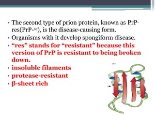
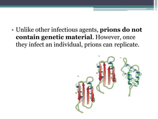
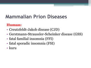
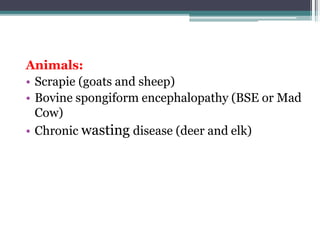
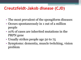
Prions are abnormal infectious protein particles that cause neurodegenerative diseases. They can occur through infection, genetic mutation, or spontaneously. Prions are unique in that they can replicate without nucleic acids by converting normal prion proteins (PrP-sen) into an abnormal form (PrP-res). Accumulation of PrP-res causes neuronal death and symptoms like dementia. Common prion diseases include Creutzfeldt-Jakob disease in humans and mad cow/scrapie in animals. There is no cure for prion diseases and most result in death within a year of symptoms appearing.
This is my report on our cell biology. I hope it could help you.
Objectives: Identify infectious proteins (PrPsc), difference of PrPc and PrPsc, list of neurodegenerative diseases that caused by prions.
The slides explain history of Prion diseases, proposed mechanisms of pathogenesis, investigations and proposed treatment options. Pl watch after downloading as the slides are mostly animated.
Prepared in June 2014
Proteins are fundamental components of living cells and come in many forms like hemoglobin, hormones, antibodies, enzymes, and structural proteins. Proteins have a primary structure defined by their amino acid sequence and commonly form secondary structures like alpha helices and beta sheets. For a protein to function properly it must fold into its correct three-dimensional tertiary structure. Misfolded proteins can aggregate and poison cells. Prions are abnormal infectious protein particles that cause neurodegenerative diseases by converting normal cellular prion proteins into the abnormal misfolded prion form.
Prions are proteinaceous infectious particles that lack nucleic acid and cause fatal neurodegenerative diseases. They exist in two forms - a normal cellular form (PrPc) and an abnormal disease-causing form (PrPsc). PrPsc has a different structure than PrPc which allows it to accumulate and convert PrPc into more PrPsc, ultimately causing neuropathology. Prion diseases affect both humans and animals and manifest as infectious, genetic, and sporadic disorders with varied clinical presentations. Common features include neurological deficits, dementia, and spongiform changes in the brain. Prions are extremely resistant to decomposition and differ from bacteria and viruses by being protein-only entities that propagate through conversion of
- In the 1950s, a disease called kuru was observed among the Fore people of New Guinea that caused trembling, loss of motor control, and ultimately death.
- Kuru and other transmissible spongiform encephalopathies (TSEs) are caused by prion proteins, which are unique infectious proteins that can reproduce on their own and convert normal prion proteins into the abnormal infectious form.
- Prions are extremely resistant to heat and chemicals and can survive in the environment for many years. They infect the central nervous system and cause neurodegeneration through a chain reaction that multiplies copies of the infectious prion protein.
Prion diseases are caused by misfolded prion proteins (PrPSc) that recruit and convert normal prion proteins (PrPC) into more infectious PrPSc. Prion diseases can be transmitted and occur in sporadic, genetic, or infectious forms. PrPSc has a different conformation than PrPC but the same amino acid sequence. PrPSc is resistant to breakdown, aggregates, and causes neurodegeneration through currently unknown mechanisms. Prions violate traditional understandings of infectious disease by lacking nucleic acids and instead being composed solely of misfolded protein.
Prions are infectious proteins that can cause neurodegenerative diseases such as mad cow disease and CJD in humans. Prions lack nucleic acids and are composed solely of abnormal versions of host-encoded proteins. They propagate by converting normal protein molecules into the abnormal prion form. This causes disease by disrupting normal protein folding in the brain. While prion diseases are generally untreatable and fatal, research on yeast prions has provided insights into prion transmission and conversion mechanisms at the molecular level.
Prions are infectious protein particles that cause fatal neurodegenerative diseases in humans and animals. Prions are composed solely of protein and lack DNA or RNA. They are the cause of transmissible spongiform encephalopathies (TSEs), diseases which destroy brain tissue and cause large vacuoles, giving the brain a spongy appearance. Prions convert normal cellular prion proteins (PrPc) into an abnormal disease-causing form (PrPsc) which aggregates to form plaques that damage brain tissue. Common symptoms of TSEs include dementia, loss of motor coordination, insomnia, and eventually death.
Transmissible spongiform encephalopathies (TSEs) are a group of fatal neurodegenerative disorders caused by prions. Prions are abnormally folded forms of a normal cellular protein that are highly resistant to destruction and can transmit their abnormal shape to other normal proteins. TSEs affect both humans and animals. Some examples include Creutzfeldt-Jakob disease (CJD) in humans, bovine spongiform encephalopathy (BSE, or "mad cow disease") in cattle, and chronic wasting disease in deer. Prion diseases have long incubation periods, are always fatal, and currently have no treatment or cure.
This document summarizes prion diseases, which are a group of progressive neurodegenerative disorders known as transmissible spongiform encephalopathies. The diseases affect both animals like sheep and deer, as well as humans. They are caused by misfolded prion proteins that form plaques in the central nervous system, disrupting normal tissue structure and causing holes or vacuoles in neurons. Prion diseases have a long incubation period, cause neurologic symptoms, and are ultimately fatal. Key features include spongiosis, gliosis, and neuronal loss. The diseases can be sporadic, genetic, or acquired through infection or medical procedures.
Prion diseases are rapidly progressive neurodegenerative disorders caused by abnormal prion proteins (PrP) that can be sporadic, inherited, or transmitted. They include Creutzfeldt-Jakob disease (CJD) in humans and related diseases in other animals. Prion diseases are characterized by brain vacuoles and dementia. Normal PrP converts to an abnormal beta-sheet form (PrPsc) that accumulates in the brain and causes neurodegeneration through an unknown mechanism. Variant CJD results from exposure to bovine spongiform encephalopathy. There are no cures, so prevention focuses on infection control and screening high-risk populations.
Scrapie (/ˈskreɪpi/) is a fatal, degenerative disease affecting the nervous systems of sheep and goats.[1] It is one of several transmissible spongiform encephalopathies (TSEs), and as such it is thought to be caused by a prion.[2][3] Scrapie has been known since at least 1732 and does not appear to be transmissible to humans.[4][5] However, new studies suggest a link between scrapie and sporadic CJD.[6]
The name scrapie is derived from one of the clinical signs of the condition, wherein affected animals will compulsively scrape off their fleeces against rocks, trees or fences. The disease apparently causes an itching sensation in the animals. Other clinical signs include excessive lip smacking, altered gaits and convulsive collapse.[7]
Scrapie is infectious and transmissible among conspecifics, so one of the most common ways to contain it (since it is incurable) is to quarantine and kill those affected. However, scrapie tends to persist in flocks and can also arise apparently spontaneously in flocks that have not previously had cases of the disease. The mechanism of transmission between animals and other aspects of the biology of the disease are only poorly understood, and are active areas of research. Recent studies suggest prions may be spread through urine and persist in the environment for decade
Prions are infectious agents that are composed of misfolded protein aggregates called PrP, lacking nucleic acid. They can cause fatal neurodegenerative diseases in humans and animals by converting normal PrP proteins into additional misfolded prions. Common prion diseases include Creutzfeldt-Jakob disease in humans, mad cow disease in cattle, and scrapie in sheep. Prions are highly resistant to sterilization by heat, chemicals, radiation, and are difficult to inactivate.
Misfolded proteins which catalyses the refolding of the native protein into the misfolded pathogenic forms are called as Prions. They are responsible for fatal neurodegenerative diseases. Some of the neurodegenerative diseases such as Scrapie, BSE, CWD of animals and Kuru, CJD, Fatal familial insomnia of human beings are included in the presentation.
- Prion diseases are rapidly progressive dementias caused by infectious proteins called prions. They include Creutzfeldt-Jakob disease (CJD), Gerstmann-Sträussler-Scheinker syndrome (GSS), fatal familial insomnia (FFI), and kuru.
- Prion diseases occur sporadically in 85% of cases, are inherited in 15% of cases, and are infectious in less than 1% of cases. They result from the accumulation of abnormal prion protein (PrPsc) which differs from its normal cellular form (PrPc).
- Clinically, prion diseases present with dementia, myoclonus, and death usually within a year
This document discusses slow virus diseases, which are infections characterized by very long incubation periods, slow but relentless courses, and fatal outcomes. It describes several examples of slow virus diseases in humans and animals caused by viruses from families including retroviruses, paramyxoviruses, togaviruses, polyomaviruses, and prions. These diseases have long latency periods ranging from months to decades before symptoms appear. They progressively affect the central nervous system and have no effective treatment or immune response, inevitably leading to death.
Prions are proteinaceous infectious particles that lack nucleic acid and cause neurodegenerative diseases. They exist in two forms - the normal cellular PrPc form containing alpha helical structure, and the pathogenic PrPsc form containing beta pleated sheet structure. PrPsc is able to convert the normal PrPc protein into the pathogenic form, reproducing through this mechanism without requiring nucleic acids. Prion diseases in humans include Creutzfeldt-Jakob disease and its variants, as well as kuru and Gerstmann-Straussler-Scheinker disease. Neuropathological features include spongiform degeneration, gliosis, and amyloid plaques.
Largest viruses that infect vertebrates
Can be seen under light microscope
Poxvirus diseases are characterized by skin lesions – localized or generalized
Important diseases caused by poxviruses are-
Smallpox
Monkeypox
Cowpox
Tanapox
Molluscum contagiosum
Parvoviruses are the smallest DNA viruses, including human parvovirus B19. B19 is pathogenic in humans, infecting erythroid progenitor cells and causing fifth disease in children characterized by a rash. It can also cause aplastic crisis, hydrops fetalis in fetuses, and chronic anemia in immunocompromised patients. Diagnosis involves detecting IgG and IgM antibodies by ELISA or PCR to detect the virus. There is no treatment, though a vaccine is in clinical trials.
Poxviruses are a family of large, complex enveloped viruses that contain double-stranded DNA. They include viruses that infect humans and other vertebrates. Smallpox and molluscum contagiosum are human poxviruses, while viruses like vaccinia, cowpox and monkeypox can infect humans incidentally from animal hosts. Poxviruses replicate in the cytoplasm and have complex virion structures. Important human poxviruses include variola (smallpox virus), which was eradicated in the 1970s through vaccination, and molluscum contagiosum, which causes a generally mild skin infection.
This document discusses the Reoviridae family of viruses. It provides information on Rotavirus, including that it is the most common cause of severe diarrhea in infants and children worldwide, with about 500,000 child deaths annually. Rotavirus has a double-stranded RNA genome and infects the intestinal tract. It spreads easily between young children and causes symptoms like vomiting, diarrhea, and dehydration. While there is no specific treatment, prevention through vaccination is available.
Satellite viruses are sub-viral agents that depend on a helper virus for replication. The first reported satellite virus was Tobacco necrosis satellite virus. Satellite viruses contain nucleic acids enclosed in a protein coat and lack genes for replication. Satellite genomes can be single-stranded RNA, DNA, or circular RNA.
Satellite RNAs are small, linear or circular RNA strands found in certain multicomponent virus particles. They do not encode their own coat protein and depend on a helper virus for replication and encapsidation.
Viroids were discovered in 1971 and are small, circular, naked RNA molecules that replicate independently using host polymerases. Well-studied viroids include potato spindle tuber viroid and av
Picornaviruses are a family of small RNA viruses that includes enteroviruses like poliovirus and rhinoviruses that cause the common cold. They are spherical and non-enveloped, around 30nm in diameter, and contain a single strand of positive-sense RNA genome around 7-8kb in size. Picornaviruses infect the cytoplasm and their replication results in cell lysis and spread to other cells. Important human pathogens include the polioviruses, coxsackieviruses, echoviruses and rhinoviruses. Both live attenuated and inactivated vaccines have been developed to prevent diseases like polio.
This document discusses various concepts related to bacterial infection and virulence factors. It begins by defining key terms like infection, disease, signs and symptoms. It then describes the different types of hosts and modes of transmission of infectious agents. It discusses the concepts of reservoirs, carriers and zoonotic infections. The major sections cover the epidemiology of infections, modes of transmission including direct and indirect, and routes of entry of pathogens. The last section provides details on various bacterial virulence factors that enhance pathogenicity like adherence factors, invasion factors, toxins and mechanisms of biofilm formation.
Picorna viruses include poliovirus, coxsackie virus, echovirus, rhinovirus, and others. They are small, non-enveloped viruses with positive-sense RNA genomes. Poliovirus causes the disease poliomyelitis. It is transmitted fecally-orally and can infect the central nervous system, causing paralysis. Vaccines including both live attenuated (OPV) and killed (IPV) versions have been highly effective in polio's global eradication efforts. Rhinoviruses are the most common cause of the common cold.
The document summarizes key information about influenza virus. It belongs to the Orthomyxoviridae family and is a segmented, single-stranded RNA virus. It causes the highly contagious disease influenza, or flu. There are three main types - A, B, and C - with Type A being the most virulent and causing pandemics through antigenic drift and shift. Symptoms include fever, cough, and fatigue. Treatment involves antiviral drugs and vaccination, while prevention focuses on hand washing and avoiding contact with infected individuals.
The document discusses slow virus diseases and prion diseases. It describes how Björn Sigurðsson first introduced the slow virus concept and studied diseases like maedi, visna, and scrapie in sheep. These diseases have long incubation periods and involve the central nervous system. Prion diseases like Kuru, Creutzfeldt-Jakob disease, and mad cow disease are also described, noting they involve misfolded prion proteins and have characteristics like spongiform changes in the brain. Measles virus diseases like subacute sclerosing panencephalitis are also slow virus diseases that occur due to mutations in the measles virus.
Prions: structure diseases associated with and clinical picuture.pptxKhalidJahir1
a lecture that describes what are prions, what diseases they cause, and how they cause disease, incubation period and symptoms and signs of the disease they cause.
This document provides an overview of prion diseases, also known as transmissible spongiform encephalopathies (TSEs). It defines prion diseases as progressive neurodegenerative disorders that can affect both humans and animals. The causative agent is an abnormal prion protein (PrPsc) which induces abnormal folding of normal prion proteins in neurons. Several human and animal prion diseases are described, including their signs, symptoms, transmission methods, and neuropathological features. The mechanism of prion propagation and the role of the normal (PrPc) and abnormal (PrPsc) prion proteins are explained.
Prions are infectious protein particles that cause neurodegenerative diseases like Creutzfeldt-Jakob disease in humans and bovine spongiform encephalopathy (mad cow disease) in cattle. Prions are composed solely of protein and contain no nucleic acids. They propagate by converting the normal cellular prion protein (PrPC) into an abnormal disease-causing form (PrPSc). The normal prion protein is involved in the replication of the mutated form. Prion diseases are difficult to treat as prions are highly resistant to heat and chemical inactivation.
This document provides an overview of diseases and the immune response. It discusses different types of pathogens including viruses, bacteria, fungi, protozoans, and prions. Viruses are non-cellular agents that can only replicate inside host cells. Bacteria are prokaryotic cells that can cause disease through toxins or disrupting host cells. Fungi infections include ringworm and athletes foot. Protozoans include the malaria parasite which has a complex lifecycle alternating between mosquitos and humans. Prions cause serious diseases like mad cow disease and have an unusual transmission through abnormal protein folding.
This document provides information on transmissible spongiform encephalopathies (TSEs), also known as prion diseases. It discusses several TSEs that can infect humans and animals, including the causative agents, affected species, transmission routes, clinical signs, and regulations/guidelines regarding prevention and control. Specific TSEs summarized include scrapie, bovine spongiform encephalopathy (BSE), chronic wasting disease (CWD), Creutzfeldt-Jakob disease (CJD), and variant CJD (vCJD). Risk factors for transmission via pharmaceuticals or through food/animal products are also addressed.
Presentation on prions diseases and its preventionomtiwaricv20
Prions are infectious agents composed of misfolded protein that can induce normal proteins to take the same misfolded shape. They were discovered in 1982 by Stanley Prusiner and are believed to cause fatal neurodegenerative diseases like Creutzfeldt-Jakob disease in humans and mad cow disease. Prions exist in two forms - a normal cellular form called PrPc and an infectious disease-causing form called PrPSc. PrPSc is able to recruit PrPc and convert it into more PrPSc through a chain reaction. As PrPSc accumulates in the brain, it causes damage that interferes with normal brain function. Currently there is no cure for prion diseases.
Prions are misfolded proteins that cause fatal neurodegenerative diseases in humans and animals. They are not living organisms and do not contain DNA or RNA. Prions work by converting normal proteins into additional misfolded prions, accumulating in the brain and causing spongiform changes. Some diseases caused by prions include kuru, Creutzfeldt-Jakob disease in humans, and mad cow disease in cattle. Prions are highly resistant to heat and chemicals and can survive in the environment for many years. Currently there is no cure for prion diseases, so prevention is key.
India.involved in various industries that include Engineering, construction ,...OM PRAKASH SINGH
The document provides an overview of prions, including their structure, functions, how they infect cells and replicate without nucleic acids, and the diseases they cause. Prions are misfolded protein aggregates that can induce normal cellular prion proteins to also misfold. They have two forms - PrPC found normally in cells, and the infectious misfolded form PrPSc. PrPSc spreads between cells through direct contact or release into the environment. Upon infection of a new cell, PrPSc acts as a template to convert more PrPC into the abnormal form. This allows prions to replicate without nucleic acids and cause neurodegenerative diseases like Creutzfeldt-Jakob disease in humans.
A prion is a protein that can fold in multiple, structurally distinct ways, at least one of which is transmissible to other prion proteins. It is this form of replication that leads to disease that is similar to viral infection. The word prion, coined in 1982 by Stanley B. Prusiner, is short for “proteinaceous infectious particle” derived from the words protein and infection, in reference to a prion's ability to self-propagate and transmit its conformation to other prions. While several yeast proteins have been identified as having prionogenic properties, the first prion protein was discovered in mammals and is referred to as the major prion protein (PrP). This infectious agent causes mammalian transmissible spongiform encephalopathies, including bovine spongiform encephalopathy (BSE, also known as "mad cow disease") and scrapie in sheep. In humans, PrP causes Creutzfeldt-Jakob Disease (CJD), variant Creutzfeldt-Jakob Disease (vCJD), Gerstmann–Sträussler–Scheinker syndrome, Fatal Familial Insomnia and kuru.
The document discusses prion proteins, which are infectious particles that cause neurodegenerative diseases like Creutzfeldt-Jakob disease. It covers the PRNP gene that encodes prion proteins, their normal cellular function, and how abnormal prion proteins cause diseases by converting normal prions into an abnormal shape. The document also discusses diagnosing prion diseases through tests like MRI, spinal taps, and brain scans, as well as developing new diagnostic techniques like protein misfolding amplification. Currently, there are no cures for prion diseases but some drugs may help slow their progression.
This document provides information on Bovine Spongiform Encephalopathy (BSE), also known as mad cow disease. It discusses that BSE is a fatal neurological disease affecting cattle that results in a spongy appearance of the brain. The disease first emerged in the UK in 1986 and has since spread to several other countries. Cattle primarily become infected through consuming protein supplements containing meat and bone meal from infected animals. The document also provides details on the clinical signs, transmission, diagnosis, control and prevention of BSE.
Scrapie is a fatal, neurological disease that affects sheep and goats. It is caused by prions, which are misfolded proteins, and results in the accumulation of prions in the nervous system, leading to neuronal death. Symptoms include weight loss, behavioral changes, and tremors. It is transmitted from infected females to offspring at birth or through contact with birth materials. There is no treatment for scrapie. Control methods focus on selective breeding of resistant sheep genotypes and restricting the spread of infected tissues and milk.
This document summarizes a study that investigated the ability of prions to infect and propagate in primary neuronal and astrocyte cell cultures. The study found that:
1) Primary cerebellar cultures from transgenic mice expressing ovine prion protein accumulated prion protein and infectivity when exposed to sheep scrapie agent.
2) Both astrocytes and neurons in these primary cultures were capable of sustaining prion propagation independently, producing prion protein with the same characteristics as in diseased brain tissue.
3) Infected neuronal cultures consistently showed late-occurring apoptosis, in contrast to chronically infected cell lines that do not show cytotoxicity. This suggests primary cultured neural cells may be useful for studying
Prions are abnormal protein particles that can cause neurodegenerative diseases. They have a normal function in the body but can transform into an infectious form. When this happens, they induce other normal prion proteins to adopt the abnormal shape. Common prion diseases include scrapie in sheep, mad cow disease, and Creutzfeldt-Jakob disease in humans. There are currently no effective treatments for prion diseases, so prevention and diagnosis are important.
The document discusses prion diseases, which are neurodegenerative disorders caused by abnormal prion proteins. It covers the discovery of prions, the different forms of human and animal prion diseases, and the molecular pathogenesis involving the conversion of normal prion protein (PrPc) to abnormal disease-causing prion protein (PrPsc). Clinical features of various human prion diseases like CJD are described. Diagnosis involves neuroimaging, EEG, CSF analysis and detection of PrPsc in brain tissue. Histopathology shows vacuolation, gliosis and prion plaques. Variant CJD results from eating meat from cows with BSE.
The human PRNP gene is located on chromosome 20 and encodes the prion protein (PrP). Mutations in this gene can cause neurodegenerative diseases like Creutzfeldt-Jakob disease. The protein's exact function is unknown but it may be involved in copper transport, neuroprotection, and communication between neurons. Alternatively spliced variants have been identified for this gene and the mouse protein shares high sequence identity with the human protein. Clinically relevant genetic variants in PRNP have been associated with prion diseases.
Prion diseases are rapidly progressive neurodegenerative disorders caused by abnormal prion proteins. There are several types including sporadic, familial, variant, and acquired from animals. Prion diseases are untreatable and fatal due to prion protein conversion and deposition in the brain leading to neurodegeneration. Diagnosis involves tests of brain imaging, EEG, CSF and genetic analysis while treatment is largely supportive due to lack of effective disease-modifying therapies.
Prion diseases are caused by abnormal prion proteins that accumulate in the brain and cause neurodegeneration. After infection, prions are transported to secondary lymphoid organs like lymph nodes where they infect immune cells, facilitating transmission to the brain. While prion infections do not elicit an adaptive immune response, the innate immune system is activated in the brain, and immune cells can influence disease progression either by protecting the host or accelerating neurodegeneration. The immune system plays an important role in prion disease pathogenesis through its interaction with prions.
Prions are misfolded proteins that can cause fatal neurodegenerative diseases in humans and animals. Prions are highly resistant to breakdown in the body and can transmit their misfolded shape to normal proteins. Stanley Prusiner discovered prions and won the 1997 Nobel Prize in Physiology or Medicine for his research. Prion diseases include scrapie in sheep, mad cow disease in cattle, and Creutzfeldt-Jakob disease in humans. Prions are composed solely of protein and are able to self-replicate by inducing normal proteins to take on their misfolded shape.
This document summarizes the key characteristics of 18 orders of bony fishes (class Osteichthyes) based on their anatomical features and examples. It describes traits such as fin structure, scale type, presence of swim bladders and their connections to other organs, body shape adaptations, and example fish from each order. The orders discussed include Crossopterygii, Dipnoi, Polypteriformes, Acipenseriformes, and others.
The document discusses the origin and evolution of mammals. It describes two main theories for the ancestry of mammals - descent from amphibians or reptiles. While Huxley proposed an amphibian ancestry, most evidence supports a reptilian ancestry, with mammals evolving from mammal-like reptiles called therapsids. True mammals first appeared in the Jurassic period and were small, nocturnal creatures. After the extinction of dinosaurs, mammals underwent an adaptive radiation, diversifying to fill new ecological niches. Limbs and teeth evolved adaptations for different diets and modes of locomotion like burrowing, swimming and climbing. Distantly related groups sometimes converged on similar forms when occupying similar habitats.
The document discusses the origin and evolution of mammals. It describes two main theories for the ancestry of mammals - through amphibians or reptiles. While amphibian ancestry was proposed, reptilian ancestry is now widely accepted based on fossil evidence. Many characteristics of early mammal-like reptiles called therapsids were mammalian. True mammals first appeared in the Jurassic period but remained small until after the extinction of dinosaurs. When ecological niches opened up in the Cenozoic era, mammals underwent adaptive radiations into various forms through modifications of limbs, teeth and other features for different habitats like trees, ground, burrows, water and air. Convergent evolution also led to similarities between unrelated mammals adapting to the same nic
The Draco volans, or flying lizard, has a dorso-ventrally compressed body up to 25cm long with a 12cm tail. It has wing-like patagia membranes between its limbs that it uses for gliding. Males have an orange gular pouch and nuchal crest while females have a blue pouch. It is found in Indonesia and feeds on ants and possibly termites.
1. The document describes the digestive system of vertebrates, including the development and anatomy of key structures like the mouth, pharynx, esophagus, stomach, intestines, liver and gallbladder.
2. It notes that the digestive tract develops from the embryonic archenteron and surrounding mesoderm. Structures vary between aquatic and terrestrial vertebrates.
3. Accessory organs like the tongue, teeth and salivary glands are described. The stomach and intestines show diversity between species related to diet. The liver and gallbladder function to produce and store bile.
1. The document discusses the common peafowl, specifically the Indian peafowl. It describes the classification, appearance, habitat, and distribution of male peacocks and female peahens.
2. Male peacocks have iridescent blue-green plumage and elongated tail feathers called a "train" with eye-like patterns. Females are duller in coloration and lack the elongated tail feathers.
3. Peafowl are found in forests, farmland, bushlands and rainforests, preferring areas with open spaces for displaying and dust bathing. Their historic range was northern India but they are now protected in several national parks and reserves in Nepal.
The document summarizes the development and comparative anatomy of the vertebrate brain. It describes how the embryonic brain divides into three primary vesicles - the prosencephalon, mesencephalon, and rhombencephalon. It then discusses the specific structures that develop from each of these regions across different vertebrate groups, including differences in brain structure between cyclostomes, fish, amphibians, reptiles, birds, and mammals. Key differences include the size of regions like the cerebrum, cerebellum, and olfactory bulbs across the groups.
This document discusses the comparative anatomy of the integument and its derivatives in vertebrates. It describes the general structure of skin, which consists of an outer epidermis and inner dermis layers. The epidermis is stratified squamous epithelium that provides protection, while the dermis contains connective tissue, blood vessels, and sensory structures. Major derivatives of the integument discussed include epidermal glands, scales, claws/nails/hooves, horns, feathers, and hair. The integument and its derivatives have evolved to suit the environment and provide functions like protection, thermoregulation, sensation, and identification.
Water is essential for life and exists in three states: liquid, solid, and gas. It has unique properties like being a universal solvent and exhibiting surface tension due to hydrogen bonding between molecules. Water is used for many important purposes including household use, agriculture, industry, recreation, hydropower, and more. As the global population increases, proper management of water resources and conservation efforts will be needed to ensure adequate fresh water supplies for the future.
Translation is the process by which the genetic code in mRNA is used to direct the synthesis of proteins. It involves three main steps - initiation, elongation, and termination. Initiation requires the small and large ribosomal subunits to assemble around an mRNA molecule along with initiator tRNA and other initiation factors. Elongation then adds amino acids one by one to the growing polypeptide chain according to the mRNA codons. Termination occurs when a stop codon is reached, causing the ribosome to dissociate and release the complete protein.
The document discusses the structure of DNA and genome organization. It describes that DNA is usually composed of two polynucleotide chains twisted around each other in a double helix structure. The backbone of each strand is made up of alternating sugar and phosphate residues, while the bases project inward. The two strands are linked by hydrogen bonds between complementary bases, with adenine pairing with thymine and guanine pairing with cytosine. Together, the sugar, phosphate, and base components make up the nucleotides that serve as the fundamental building blocks of DNA.
This document discusses the life cycle and pathogenesis of Plasmodium vivax, the parasite that causes vivax malaria. P. vivax has a digenetic life cycle involving two hosts. In humans, it undergoes asexual reproduction in the liver and red blood cells. Mosquitoes ingest the parasite's gametocyte stages, where sexual reproduction occurs leading to sporozoite formation. Sporozoites are then transmitted to humans to continue the cycle, causing symptoms of malaria including fever and anemia. Diagnosis involves blood smear microscopy and treatment involves chloroquine and primaquine to prevent relapses.
The document describes the development and anatomy of the human eye and compares it to eyes in other organisms. It discusses that in the third week of human development, optic vesicles form and later develop into the optic cup, which will become the retina. By week six, tissues surrounding the eye differentiate into the sclera and choroid. The document then compares key features of eyes across vertebrate groups like fish, amphibians, reptiles, birds, and mammals.
Friedrich Miescher first isolated nucleic acids in 1869 and called them "nuclein" due to their acidic properties. The discovery of DNA's double helix structure in 1953 by Watson and Crick was monumental. Nucleic acids function to store and transmit genetic information through DNA and RNA. DNA is made of two strands bound together by complementary nucleotide bases, with adenine pairing with thymine and guanine pairing with cytosine. The discovery of DNA's structure explained the mechanism of heredity.
This document provides information about honey bees. It begins with the classification of honey bees, then discusses their social behavior and advantages. It provides examples of social insects including honey bees, wasps, termites and ants. The document focuses on honey bees, describing their appearance, distribution, and the different types of bees within a hive: the queen, workers, and drones. It explains the key roles and characteristics of each type of honey bee.
This document discusses genetic engineering of humans through various methods such as germline genetic modification and somatic genetic modification. It notes both the promising applications for improving health through gene therapy but also the ethical concerns regarding enhancing traits and the possibility of genetic inequality. Specific examples discussed include using CRISPR to genetically modify human embryos to potentially treat disease as well as concerns about "designer babies". The document examines issues around testing for non-disease traits, gene doping in athletes, and the potential benefits and risks of human gene editing technologies.
Genetic engineering and genetically modified organisms have potential risks and downsides according to the document. While genetic engineering has applications in agriculture and medicine, it can also have detrimental effects on humanity. Introducing foreign genes into other species can have uncontrolled and unpredictable consequences. Studies have shown genetically modified foods can be toxic and cause health issues in animals. Genetically engineered crops also have negative impacts on biodiversity and the environment by promoting the growth of pesticide-resistant superweeds and superbugs. The long-term effects of genetic engineering are uncertain due to its new and crude nature, making it a controversial technology that manipulates nature in ways bypassing normal evolution.
The document describes isolating and identifying Azotobacter species from a soil sample. It discusses enriching the soil in Ashby's Mannitol Broth for a week to activate nitrogen-fixing bacteria. Colonies were then streaked on the broth and incubated for 4-6 days. Gram staining identified gram-negative cells with a red color and capsule staining showed transparent capsules around violet cells, indicating motility. The colonies were creamy white, raised, circular and mucoid, consistent with Azotobacter.
The document summarizes the fluid mosaic model of the cell membrane. It describes the main components of the membrane - phospholipids, cholesterol, proteins, and carbohydrates. The phospholipid bilayer forms the main structure of the membrane, with phospholipids arranged in two layers with their hydrophobic tails facing each other and hydrophilic heads facing outwards. Proteins embedded within or attached to the membrane carry out important functions like transport and cell signaling. Carbohydrates attached to proteins and lipids on the outer surface of the membrane help with cell recognition. The fluid nature of the membrane allows it to maintain selective permeability and flexibility. Passive and active transport mechanisms allow movement of substances across the membrane.
1) Atomic radius is the distance from the nucleus to the outermost shell of electrons or the point where electron density is highest.
2) There are three types of atomic radii: covalent, van der Waals, and metallic.
3) Covalent radius is half the distance between bonded atoms. Van der Waals radius is half the distance between adjacent non-bonded atoms. Metallic radius is half the distance between metal ions in a lattice.
More from St. Xavier's college, maitighar,Kathmandu (20)
NAVIGATING THE HORIZONS OF TIME LAPSE EMBRYO MONITORING.pdfRahul Sen
Time-lapse embryo monitoring is an advanced imaging technique used in IVF to continuously observe embryo development. It captures high-resolution images at regular intervals, allowing embryologists to select the most viable embryos for transfer based on detailed growth patterns. This technology enhances embryo selection, potentially increasing pregnancy success rates.
PGx Analysis in VarSeq: A User’s PerspectiveGolden Helix
Since our release of the PGx capabilities in VarSeq, we’ve had a few months to gather some insights from various use cases. Some users approach PGx workflows by means of array genotyping or what seems to be a growing trend of adding the star allele calling to the existing NGS pipeline for whole genome data. Luckily, both approaches are supported with the VarSeq software platform. The genotyping method being used will also dictate what the scope of the tertiary analysis will be. For example, are your PGx reports a standalone pipeline or would your lab’s goal be to handle a dual-purpose workflow and report on PGx + Diagnostic findings.
The purpose of this webcast is to:
Discuss and demonstrate the approaches with array and NGS genotyping methods for star allele calling to prep for downstream analysis.
Following genotyping, explore alternative tertiary workflow concepts in VarSeq to handle PGx reporting.
Moreover, we will include insights users will need to consider when validating their PGx workflow for all possible star alleles and options you have for automating your PGx analysis for large number of samples. Please join us for a session dedicated to the application of star allele genotyping and subsequent PGx workflows in our VarSeq software.
Breast cancer: Post menopausal endocrine therapyDr. Sumit KUMAR
Breast cancer in postmenopausal women with hormone receptor-positive (HR+) status is a common and complex condition that necessitates a multifaceted approach to management. HR+ breast cancer means that the cancer cells grow in response to hormones such as estrogen and progesterone. This subtype is prevalent among postmenopausal women and typically exhibits a more indolent course compared to other forms of breast cancer, which allows for a variety of treatment options.
Diagnosis and Staging
The diagnosis of HR+ breast cancer begins with clinical evaluation, imaging, and biopsy. Imaging modalities such as mammography, ultrasound, and MRI help in assessing the extent of the disease. Histopathological examination and immunohistochemical staining of the biopsy sample confirm the diagnosis and hormone receptor status by identifying the presence of estrogen receptors (ER) and progesterone receptors (PR) on the tumor cells.
Staging involves determining the size of the tumor (T), the involvement of regional lymph nodes (N), and the presence of distant metastasis (M). The American Joint Committee on Cancer (AJCC) staging system is commonly used. Accurate staging is critical as it guides treatment decisions.
Treatment Options
Endocrine Therapy
Endocrine therapy is the cornerstone of treatment for HR+ breast cancer in postmenopausal women. The primary goal is to reduce the levels of estrogen or block its effects on cancer cells. Commonly used agents include:
Selective Estrogen Receptor Modulators (SERMs): Tamoxifen is a SERM that binds to estrogen receptors, blocking estrogen from stimulating breast cancer cells. It is effective but may have side effects such as increased risk of endometrial cancer and thromboembolic events.
Aromatase Inhibitors (AIs): These drugs, including anastrozole, letrozole, and exemestane, lower estrogen levels by inhibiting the aromatase enzyme, which converts androgens to estrogen in peripheral tissues. AIs are generally preferred in postmenopausal women due to their efficacy and safety profile compared to tamoxifen.
Selective Estrogen Receptor Downregulators (SERDs): Fulvestrant is a SERD that degrades estrogen receptors and is used in cases where resistance to other endocrine therapies develops.
Combination Therapies
Combining endocrine therapy with other treatments enhances efficacy. Examples include:
Endocrine Therapy with CDK4/6 Inhibitors: Palbociclib, ribociclib, and abemaciclib are CDK4/6 inhibitors that, when combined with endocrine therapy, significantly improve progression-free survival in advanced HR+ breast cancer.
Endocrine Therapy with mTOR Inhibitors: Everolimus, an mTOR inhibitor, can be added to endocrine therapy for patients who have developed resistance to aromatase inhibitors.
Chemotherapy
Chemotherapy is generally reserved for patients with high-risk features, such as large tumor size, high-grade histology, or extensive lymph node involvement. Regimens often include anthracyclines and taxanes.
Histololgy of Female Reproductive System.pptxAyeshaZaid1
Dive into an in-depth exploration of the histological structure of female reproductive system with this comprehensive lecture. Presented by Dr. Ayesha Irfan, Assistant Professor of Anatomy, this presentation covers the Gross anatomy and functional histology of the female reproductive organs. Ideal for students, educators, and anyone interested in medical science, this lecture provides clear explanations, detailed diagrams, and valuable insights into female reproductive system. Enhance your knowledge and understanding of this essential aspect of human biology.
How to Control Your Asthma Tips by gokuldas hospital.Gokuldas Hospital
Respiratory issues like asthma are the most sensitive issue that is affecting millions worldwide. It hampers the daily activities leaving the body tired and breathless.
The key to a good grip on asthma is proper knowledge and management strategies. Understanding the patient-specific symptoms and carving out an effective treatment likewise is the best way to keep asthma under control.
These lecture slides, by Dr Sidra Arshad, offer a simplified look into the mechanisms involved in the regulation of respiration:
Learning objectives:
1. Describe the organisation of respiratory center
2. Describe the nervous control of inspiration and respiratory rhythm
3. Describe the functions of the dorsal and respiratory groups of neurons
4. Describe the influences of the Pneumotaxic and Apneustic centers
5. Explain the role of Hering-Breur inflation reflex in regulation of inspiration
6. Explain the role of central chemoreceptors in regulation of respiration
7. Explain the role of peripheral chemoreceptors in regulation of respiration
8. Explain the regulation of respiration during exercise
9. Integrate the respiratory regulatory mechanisms
10. Describe the Cheyne-Stokes breathing
Study Resources:
1. Chapter 42, Guyton and Hall Textbook of Medical Physiology, 14th edition
2. Chapter 36, Ganong’s Review of Medical Physiology, 26th edition
3. Chapter 13, Human Physiology by Lauralee Sherwood, 9th edition
Nano-gold for Cancer Therapy chemistry investigatory projectSIVAVINAYAKPK
chemistry investigatory project
The development of nanogold-based cancer therapy could revolutionize oncology by providing a more targeted, less invasive treatment option. This project contributes to the growing body of research aimed at harnessing nanotechnology for medical applications, paving the way for future clinical trials and potential commercial applications.
Cancer remains one of the leading causes of death worldwide, prompting the need for innovative treatment methods. Nanotechnology offers promising new approaches, including the use of gold nanoparticles (nanogold) for targeted cancer therapy. Nanogold particles possess unique physical and chemical properties that make them suitable for drug delivery, imaging, and photothermal therapy.
Travel vaccination in Manchester offers comprehensive immunization services for individuals planning international trips. Expert healthcare providers administer vaccines tailored to your destination, ensuring you stay protected against various diseases. Conveniently located clinics and flexible appointment options make it easy to get the necessary shots before your journey. Stay healthy and travel with confidence by getting vaccinated in Manchester. Visit us: www.nxhealthcare.co.uk
Osvaldo Bernardo Muchanga-GASTROINTESTINAL INFECTIONS AND GASTRITIS-2024.pdfOsvaldo Bernardo Muchanga
GASTROINTESTINAL INFECTIONS AND GASTRITIS
Osvaldo Bernardo Muchanga
Gastrointestinal Infections
GASTROINTESTINAL INFECTIONS result from the ingestion of pathogens that cause infections at the level of this tract, generally being transmitted by food, water and hands contaminated by microorganisms such as E. coli, Salmonella, Shigella, Vibrio cholerae, Campylobacter, Staphylococcus, Rotavirus among others that are generally contained in feces, thus configuring a FECAL-ORAL type of transmission.
Among the factors that lead to the occurrence of gastrointestinal infections are the hygienic and sanitary deficiencies that characterize our markets and other places where raw or cooked food is sold, poor environmental sanitation in communities, deficiencies in water treatment (or in the process of its plumbing), risky hygienic-sanitary habits (not washing hands after major and/or minor needs), among others.
These are generally consequences (signs and symptoms) resulting from gastrointestinal infections: diarrhea, vomiting, fever and malaise, among others.
The treatment consists of replacing lost liquids and electrolytes (drinking drinking water and other recommended liquids, including consumption of juicy fruits such as papayas, apples, pears, among others that contain water in their composition).
To prevent this, it is necessary to promote health education, improve the hygienic-sanitary conditions of markets and communities in general as a way of promoting, preserving and prolonging PUBLIC HEALTH.
Gastritis and Gastric Health
Gastric Health is one of the most relevant concerns in human health, with gastrointestinal infections being among the main illnesses that affect humans.
Among gastric problems, we have GASTRITIS AND GASTRIC ULCERS as the main public health problems. Gastritis and gastric ulcers normally result from inflammation and corrosion of the walls of the stomach (gastric mucosa) and are generally associated (caused) by the bacterium Helicobacter pylor, which, according to the literature, this bacterium settles on these walls (of the stomach) and starts to release urease that ends up altering the normal pH of the stomach (acid), which leads to inflammation and corrosion of the mucous membranes and consequent gastritis or ulcers, respectively.
In addition to bacterial infections, gastritis and gastric ulcers are associated with several factors, with emphasis on prolonged fasting, chemical substances including drugs, alcohol, foods with strong seasonings including chilli, which ends up causing inflammation of the stomach walls and/or corrosion. of the same, resulting in the appearance of wounds and consequent gastritis or ulcers, respectively.
Among patients with gastritis and/or ulcers, one of the dilemmas is associated with the foods to consume in order to minimize the sensation of pain and discomfort.
- Video recording of this lecture in English language: https://youtu.be/Pt1nA32sdHQ
- Video recording of this lecture in Arabic language: https://youtu.be/uFdc9F0rlP0
- Link to download the book free: https://nephrotube.blogspot.com/p/nephrotube-nephrology-books.html
- Link to NephroTube website: www.NephroTube.com
- Link to NephroTube social media accounts: https://nephrotube.blogspot.com/p/join-nephrotube-on-social-media.html
“Psychiatry and the Humanities”: An Innovative Course at the University of Mo...Université de Montréal
“Psychiatry and the Humanities”: An Innovative Course at the University of Montreal Expanding the medical model to embrace the humanities. Link: https://www.psychiatrictimes.com/view/-psychiatry-and-the-humanities-an-innovative-course-at-the-university-of-montreal
The biomechanics of running involves the study of the mechanical principles underlying running movements. It includes the analysis of the running gait cycle, which consists of the stance phase (foot contact to push-off) and the swing phase (foot lift-off to next contact). Key aspects include kinematics (joint angles and movements, stride length and frequency) and kinetics (forces involved in running, including ground reaction and muscle forces). Understanding these factors helps in improving running performance, optimizing technique, and preventing injuries.
3. Introduction
• Prion diseases can occur through
▫ infection (Infectious) ,
▫ as a dominantly inherited genetic disorder
(Familial), or
▫ as a consequence of a spontaneous mutation
(Sporadic).
4. History
• In 1920s, Creutzfeldt and Jakob described first
cases of progressive mental, motor and
neurological deficits in young patients - the
eponym 'Creutzfeldt-Jakob disease’(CJD) was
first used to describe degenerative CNS diseases
• In 1950s, disease known as 'kuru', in Papua New
Guinea discovered; neuropathological
similarity between kuru, CJD.
5. • In 1960s, the term transmissible spongiform
encephalopathy (TSE) applied after discovery of
the transmissible ability of both kuru and CJD
diseases to chimpanzees.
• In 1976, Gajdusek awarded Nobel prize for his
work on 'slow virus' infections theory
6. • In 1980s, the 'protein-only' hypothesis was
developed by Prusiner; he was awarded Nobel
Prize in Medicine in 1997 for his work on
prions; theory remains controversial
• In 1990s, outbreak of BSE in cattle in UK raised
public concern and spawned vigorous research
efforts to understand mechanism underlying
prion diseases
7. Prion
• Prions (PREE-ons) are proteins that are
unique in their ability to reproduce on their own
and become infectious.
• They can occur in two forms
PrP-sen : Sensitive or PrPC (cellular)
PrP-res : Resistant or PrPSc (scrapie)
8. PrP gene structure and expression
• Entire open reading frame (ORF) of all known
mammalian PrP genes resides within a single
exon
• PrP mRNA is constitutively expressed in brain;
highest levels found in the central
nervous system although protein is found in
most tissues, especially cells of the immune
system
9. • Scientists don’t know the exact function of PrP-sen,
but there is evidence that it may be involved in
communication between neurons, cell death, and
controlling sleep patterns.
• Besides:
▫ It may have a role in cell adhesion or signaling
processes
▫ PrP is involved in maintaining myelin formation,
development of T cells and is critical in stem cells
that make blood cells and M cells of the intestine
immune centers.
10.
11. • Normal form of the protein, PrPC, is a
35kDa cell surface glycoprotein that is
anchored to specific areas of the
membrane (caveolae or lipid rafts)by a
glycosyl phosphatidyl inositol (GPI)
lipid linker
• Half-life on the cell surface is 5 h, after
which protein is internalized by a
caveolae-dependent mechanism
12. • The acidic pH of the endosomal
compartment may facilitate the
conformational change
• PrP-sen is produced by normal healthy cells.
• The sen stands for “sensitive” because this
version of the protein is sensitive to being
broken down.
13. • PrP-sen featuring more alpha-helices
• soluble
• protease-sensitive
• α-helix rich
• Normal PrP is soluble in det
and readily digested by prot
ergents
eases
14. • Both PrP-sen and PrP-res are made up of the
exact same string of amino acids, the building
blocks that make up proteins.
• However, the two forms have different shapes
15. • The second type of prion protein, known as PrP-
res(PrP-sc), is the disease-causing form.
• Organisms with it develop spongiform disease.
• “res” stands for “resistant” because this
version of PrP is resistant to being broken
down.
• insoluble filaments
• protease-resistant
• β-sheet rich
16. • Unlike other infectious agents, prions do not
contain genetic material. However, once
they infect an individual, prions can replicate.
17. • Scientists are still working out the details, but
evidence supports the idea that when PrP-sen
comes into contact with PrP-res it is converted to
PrP-res.
• The result is a chain reaction that multiplies
copy after copy of the infectious prion.
18. Prion proteins can exist in multiple structures
called conformers. These different shapes are created
by sigma bonds in their chemical structures that allow
the rotation of parts of the protein.
19. • Because of their abnormal shape, PrP-res
proteins tend to stick to each other.
• Over time, the PrP-res molecules stack up to form
long chains called “amyloid fibers”.
20. • The accumulation of prions in the brain causes
neuronal cells to die and, in some types of TSE, a
type of protein called amyloid accumulates in
plaques, or flat areas, and causes degeneration of
brain tissue.
• Recent research suggests prions disrupt the
normal cell process of protein recycling which
causes a buildup of faulty proteins and causes the
death of the cell.
21. • Amyloid fibers are toxic to cells, and
ultimately kill them.
• Cells called astrocytes crawl through the
brain digesting the dead neurons, leaving
holes where neurons used to be.
• The amyloid fibers remain.
22. • Astrocytes comes into action
• The destruction of neural cells causes tiny
holes in the brain tissue and a sponge-like
appearance under the microscope, thus
giving rise to the term spongiform disease.
23. • As this process progresses, we begin to see
the unique features that characterize all
spongiform diseases:
* Clumps of amyloid fibers
* Holes in the brain
* Large numbers of astrocytes
24. • There are three ways an individual
can end up with a spongiform
disease:
PrP-res
gene that
*Eating tissue infected with
*An inherited mutation in the
codes for PrP-sen
* PrP-res forms spontaneously
25.
26. • Prions cannot be destroyed by boiling, alcohol,
acid, standard autoclaving methods, or
radiation.
• In fact, infected brains that have been sitting in
formaldehyde for decades can still transmit
spongiform disease.
27. Molecular characterization of the PrP
gene.
• PrP gene is highly conserved gene among all vertebrates.
Prion protein gene structure
• PrP is encoded by a single open reading frame (ORF)
encoding 253 amino acids in all known mammalian and avian
PrP genes.
• Alignment of the 44 known PrP sequences shows a striking
degree of conservation between the mammalian sequences,
suggestion the retention of an important function through
evolution.
• However, PrP-null mice are fine.
• Therefore, PrP is not an essential protein.
28. Species barrier
• It is very difficult to establish prion disease in one
species using innoculum from another species ,where
interspecific prion diseases can be established,
incubation times are long.
• However, once prion disease is established in a species,
intraspecific infections are easy to establish, and
incubation times are short.
• Bovine spongiform encephalopathy (BSE) has
been transmitted from cattle to humans to cause a new
variant of Creutzfeldt-Jakob syndrome.
29. Therapeutic approaches for prion
disorders
• Various compounds which bind to PrPSc, including
polyene antibiotics, dextran sulfate, and ß-sheet
breaker peptides have limited effects in animal
models of prion disease. They show a significant
effect only if administered long before clinical onset.
▫ Any ligand that selectively stabilizes the PrPC state will
prevent its rearrangement and block prion replication
(or the production of any toxic intermediate forms of
PrP on the pathway to PrPSc formation)
30. Prions can enter the body through
food and injection
• Injection of prions has occurred during corneal
transplants.
• Prions can, also, enter through blood transfusions,
which demonstrates that blood allows prions entry
into the brain.
• If prions are on the skin or in aerosols, they also end
up in lymph tissue
• It was originally discovered in Kuru, a prion disease
that was self injected by people using sticks for
crushing animal brains to eat. They sharpened their
sticks by stabbing themselves injecting the prion into
their blood.
31. • Prions can enter orally and cross the wall of the GI
tract.
• They survive in gastric acid and intestinal enzymes.
• The prion seems to enter through specialized immune
tissue in the intestine called the Peyer’s patch.
▫ In experiments with animals, inflammation of the
bowel increases the number of prions entering.
▫ M immune cells in the Peyer’s patch take samples of
the material floating by for immune surveillance. M
cells pick up prions from and bring them into the
body. There are other possible mechanisms where
intestinal cells actively transport the proteins into
the cell.
32. Immune System Harbors Prions
• In naturally occurring diseases of many wild
animals, prions multiply in the lymph tissues
before entering the brain.
• Tonsil biopsies can pick up prion disease.
• The spleen is a major place for prions, especially
the stromal region. The spleen appears to be
critical for early multiplication.
33. • Dendritic cells can, also, pick up prions and
transfer them into the lymph system to the
lymph nodes.
• Once in the brain, the higher concentration of
cellular prion protein speeds up the replication
process.
• Prions also may enter lesions or wounds in the
oral cavity and access the Vagus as a pathway to
the brain.
34. • T cells are not responsible for prion multiplication.
• B cells are necessary for prions to enter the brain, but
once they are in the brain they are not dependent on B
cells to spread.
•
• Because prions start as natural proteins they do not
appear to trigger the ordinary innate immune response
that responds to unusual shapes including cancer cells.
• They also don’t trigger the adaptive immunity of T cells
and antibodies.
36. Animals:
• Scrapie (goats and sheep)
• Bovine spongiform encephalopathy (BSE or Mad
Cow)
• Chronic wasting disease (deer and elk)
37.
38. Creutzfeldt-Jakob disease (CJD)
• The most prevalent of the spongiform diseases
• Occurs spontaneously in 1 out of a million
people
• 10% of cases are inherited mutations in the
PRPN gene
• Usually strikes people age 50 to 75
• Symptoms: dementia, muscle twitching, vision
problem
39. • CJD occurs in three different forms:
▫ sporadic
▫ familial
▫ iatrogenic
40. iCJD (iatrogenic)
Patients have been infected iatrogenically from
injections of human growth hormone derived
from human pituitary gland extracts,dura mater
graft, cornea transplantation, use of
neurosurgical instruments and EEG depth
electrodes
41. sCJD (spontaneous)
▫ CJD can occur in the absence of a familial history or
infection.
▫ Can be due to a spontaneous mutation on the PrP
gene, or even in the presence of only wild-type genes
• fCJD: (familial)
▫ Dominantly inherited trait, i.e. heterozygous
individuals develop fCJD.
▫ Lybian jews. Had been thought to be due to the
consumption of lightly cooked/raw sheep brain and
eyeballs. Later shown to be due to a specific mutation.
42. Gerstmann-Steussler-Scheinker disease
(GSS)
• The first of the spongiform encephalopathy to be
described as a genetically inherited trait.
• Linkage analysis from GSS families
demonstrated that PrP was the responsible gene.
• GSS may also arise spontaneously.
43. Fatal Familial Insommnia
• FFI: characterized by adults generally over age
50 who present with a progressive sleep disorder
who die within about a year of onset. More than
30 families worldwide have been identified.
• FSI (fatal sporadic insomnia). The sporadic
form of FFI.All cases are inherited mutations in
the PrP gene.Usually strikes people age 36 to
61.Disruption of sleep/wake cycle leads to coma,
then death
44. Kuru
• Affects the Fore people of New Guniea.
• Struck members of the Fore tribe in the 1950s and
1960s
• The Fore people called this sickness kuru, which
means "trembling in fear."
• After intially becoming unable to walk, victims of
kuru lost the ability to swallow or chew.
• Drastic weight loss would inevitably lead to death.
• 1976 Carleton Gajdusek became co-recipient of
the Nobel Prize in Medicine for his "discoveries
concerning new mechanisms for the origin and
dissemination of infectious diseases.
45. • Today we know that kuru is one of several
diseases in humans and animals caused by prion
(PREE-on) proteins.
• Muscle weakness, loss of coordination, tremors,
inappropriate episodes of laughter or crying
• Transmitted by ritual cannibalism as part of
funeral ceremonies
46. Animal
• Scrapie
▫ The classic spongiform encephalopathy of sheep
characterized by progressive loss of motor control
and the propensity of infected animals to
obsessively rub or scrape themselves against
things (fence posts, sides of enclosures, etc) to the
point of scraping off all their hair and rubbing
their skin raw.
▫ Horizontally transmissible through the herd.
▫ There is no evidence of spread to humans
47. Mad Cow disease (Bovine Spongiform
Encephalopathy or BSE)
• Transmissible, neuro-degenerative fatal brain
disease of cattle.
• Long incubation period of 4-5 years
• It is fatal for cattle within weeks to months of its
onset.
• It is transmitted through the consumption of
BSE-contaminated meat and bone meal
supplements in cattle feed.
49. Route of infection
• Infective sheep and cattle tissue in meat and
bone meal, a component of feed for cattle and
other domestic livestock.
• The traditional includes an extraction step with
organic solvents, which is thought to have
extracted out the prion protein.
• In Great Britian in the early 1980's a new cost
effective method was implemented that omitted
the organic extraction step.
50. • Scientists speculate that the disease has spread
among cattle primarily through "animal recycling" --
the use of bone meal and other ground animal parts
in feeds
• The result was the BSE epidemic of the mid- to late-
80‘s.
• Following the introduction of the ruminant feed ban
in July1988, and the wholesale slaughter of infected
herds, BSE is declining in Great Britain.
51. the human variant of Mad
Cow Disease
Risks to human health
• a new variant of CJD (vCJD) began to appear in
humans 8 years after the first known case of BSE
in cattle.
• Although there have only been 21 confirmed
cases of vCJD to date, the young ages of the
patients and the lack of mutant PrP alleles
suggests that they developed disease via an
infectious route.
52. • GSS is distinct from CJD characterized by
cerebellar ataxia and concomitant motor
problems, dementia is less common (disease
course lasts several years before ultimate death)
• Fatal insomnias present with an untreatable
insomnia and dysautonomia; pathological
changes are characterized by severe selective
atrophy of the thalamus
53. Prognosis:
Death within one year after the onset of symptoms
in 90% (further 5% of patients die within the
next year