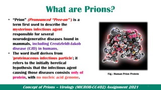
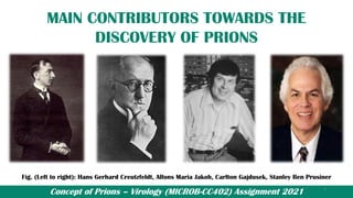
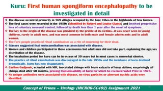
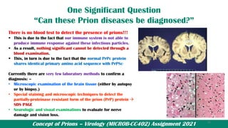
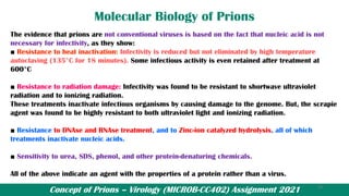
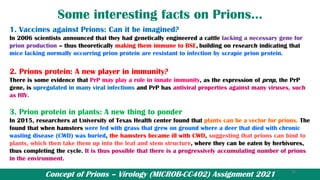
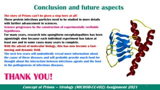
Abhilash Jeas George – B. Sc (H) Microbiology, Sem-IV 2021;
Virology Assignment – Ram Lal Anand College, University of Delhi
A presentation on
“CONCEPT OF
PRIONS”
1
“Just as nucleic acids can carry out enzymatic
reactions, proteins can be genes”— Reed Wickner
Concept of Prions – Virology (MICROB-CC402) Assignment 2021
OBJECTIVES OF THIS STUDY
The main objectives of this study are: -
• To know “What are prions actually?”
• When, How and Who discovered them?
• Are they something related to viruses or are they something
different?
• How are “Prions” formed and what is their significance among the
living organisms?
• To know the diseases and complications caused by these “Prions”
in both humans and other animals.
• Whether these agents can be diagnosed or not?
• To take a look on some lesser known facts regarding “Prions”.
2
Concept of Prions – Virology (MICROB-CC402) Assignment 2021
What are Prions?
• “Prion” (Pronounced “Pree-on” ) is a
term first used to describe the
mysterious infectious agent
responsible for several
neurodegenerative diseases found in
mammals, including Creutzfeldt-Jakob
disease (CJD) in humans.
• The word itself derives from
'proteinaceous infectious particle'; it
refers to the initially heretical
hypothesis that the infectious agent
causing those diseases consists only of
protein, with no nucleic acid genome.
Fig.: Human Prion Protein
3
Concept of Prions – Virology (MICROB-CC402) Assignment 2021
HISTORY OF PRIONS
1730s: Scrapie first appears: The disease affecting sheep and goats are called scrapie because the
animals scraped their fleece or skin against rocks, trees or fences. Reason being that the disease
apparently causes an itching sensation in the animals.
In 1920, Hans Creutzfeldt and Alfons Maria Jakob had described the disease in man
known as Creutzfeldt-Jakob disease.
- Experimental transmission of scrapie by Cuille and Chelle in 1936.
In 1967, Tikvah Alper was the first to suggest that the agent of scrapie might replicate without nucleic
acid.
Carlton Gajdusek received Nobel Prize in 1976 for proving transmissibility of Kuru.
Stanley Ben Prusiner
- American neurologist and biochemist at University of California, San Francisco
- Prusiner discovered prions, a class of infectious self-reproducing pathogens primarily or solely
composed of protein from "proteinaceous infections particle that lacks nucleic acid."
- Got Albert Lasker Award for Basic Medical Research in 1994 and the Nobel Prize in Physiology and
Medicine in 1997 for his prion research.
4
Concept of Prions – Virology (MICROB-CC402) Assignment 2021
MAIN CONTRIBUTORS TOWARDS THE
DISCOVERY OF PRIONS
Fig. (Left to right): Hans Gerhard Creutzfeldt, Alfons Maria Jakob, Carlton Gajdusek, Stanley Ben Prusiner
5
Concept of Prions – Virology (MICROB-CC402) Assignment 2021
MORE DETAILS ON PRIONS
• Prions are transmissible particles that
are devoid of nucleic acid and seem to
be composed exclusively of a modified
protein (PrPSc).
• The normal, cellular PrP (PrPC) is
converted into PrPSc through a
posttranslational process during which
it acquires a high β-sheet content.
• Here ‘c’ refers to cellular or common
PrP.
• ‘Sc’ refers to Scrapie, a prion disease
occuring in sheep.
• Structure of PrP is well defined but of
PrPSc is polydispersed and defined at a
relatively poor level.
Fig.: Normal Prion Protein v/s Diseased Prion
Protein
Ref: A.J. Cann – Principles of Molecular Virology
6
Concept of Prions – Virology (MICROB-CC402) Assignment 2021
Cellular Prion Protein (PrPc)
• PrPC is a normal protein found on the membranes of cells.
• It has 209 amino acids (in humans), one disulfide bond, a
molecular mass of 35–36 kDa and a mainly α--helical
structure.
• The normal protein is not sedimentable ; meaning that it
cannot be separated by centrifuging techniques.
• Its function is not yet known properly.
• PrPC binds copper (II) ions with high affinity.
• PrPC is readily digested by proteinase K.
• PrP has been reported to play important roles in cell-cell
adhesion and intracellular signaling in-vivo, and may
therefore be involved in cell-cell communication in the
brain.
Fig. PrP Protein
7
Concept of Prions – Virology (MICROB-CC402) Assignment 2021
Diseased Prion Protein (PrPsc)
• The infectious isoform of PrP, known as PrPSc, is able to convert
normal PrPc proteins into the infectious isoform by changing
their conformation, or shape; this, in turn, alters the way the
proteins interconnect.
• PrPSc always causes prion disease, hence infectious in nature.
• Although the exact 3D structure of PrPSc is not known, it has a
higher proportion of β-sheet structure in place of the normal α-
helix structure.
• Aggregations of these abnormal isoforms form highly structured
amyloid fibers, which accumulate to form plaques. It is also
insoluble and forms aggregates.
• The end of each fiber acts as a template onto which free protein
molecules may attach, allowing the fiber to grow.
• Under most circumstances, only PrP molecules with an identical
amino acid sequence to the infectious PrPSc are incorporated
into the growing fiber. Fig. PrPSc Protein
8
Concept of Prions – Virology (MICROB-CC402) Assignment 2021
Table 1: Differences between the infectious and the non-
infectious forms of Prion Protein
PrPc PrPSc
Globular Protein Fibrous Protein
Composed of α-helix mostly Composed of β-sheets mostly
Protease Sensitive Protease Insensitive
Non-Infectious Infectious
Soluble: doesn’t forms aggregates Insoluble: forms aggregates, form highly
structured amyloid fibers, which accumulate to
form plaques.
Monomeric Multimeric
209 amino acid long (in humans), one disulfide
bond, a molecular mass of 35–36 kDa
209 amino acid long (in humans), one disulfide
bond, a molecular mass of 35–36 kDa
Heat resistance: low Heat resistance: high
Normal cellular protein involved in cell-cell
adhesion and intracellular signaling
Role in TSEs (Transmissible spongiform
encephalopathies)
9
Concept of Prions – Virology (MICROB-CC402) Assignment 2021
Prion Hypothesis (Prion Replication)
HETERODIMER MODEL FOR PRION
REPLICATION
• This hypothesis that tried to explain how prions replicate
in a protein-only manner was the heterodimer model.
• This model assumed that a single PrPSc molecule binds
to a single PrPC molecule and catalyzes its conversion
into PrPSc.
• The two PrPSc molecules then come apart and can go on
to convert more PrPC.
• Manfred Eigen showed that the heterodimer model
requires PrPSc to be an extraordinarily effective catalyst,
increasing the rate of the conversion reaction by a factor
of around 1015.
• When enough PrPSc proteins have been made they form
long filamentous aggregates that gradually damage
neuronal tissue.
• An alternative model assumes that PrPSc exists only as
fibrils, and that fibril ends bind PrPC and convert it into
PrPSc. Fig. Heterodimer model for prion replication.
10
Concept of Prions – Virology (MICROB-CC402) Assignment 2021
Why are Prions (PrPSc) disease causing?
(Pathology of Prion diseases)
Since PrPSc is majorly composed of the β-
sheets, it is highly resistant to the action
of the enzyme Proteinase – K, which, in-
turn is able to readily digest PrPc and
hence can be liberated from the cell
surface in vitro by enzyme
phosphoinositide phospholipase (PI-PLC).
But, due to its resistance to cleavage,
PrPSc, brain tissue collects PrPSc causing
protein accumulation in high
concentration, which is distinguished by
nerve cell death causing large vacuoles
and plaques in brain tissue.
Fig. (A) Normal healthy brain tissue. (B) Brain tissue from the
temporal lobe of a patient with Creutzfeldt-Jakob’s disease. Note
the large spaces in the tissue where the prion proteins have
caused damage.
Image credits: Nephron via Wikimedia Commons and Sherif Zaki
11
Concept of Prions – Virology (MICROB-CC402) Assignment 2021
Pathology of Prion diseases (continued)
• All prion diseases share a similar underlying
pathology, although there are significant differences
between various conditions.
• Various diseases are characterized by the deposition
of abnormal protein deposits in various organs (e.g.,
kidney, spleen, liver or brain). These ‘amyloid’
deposits consist of accumulations of various proteins
in the form of plaques or fibrils depending on their
origin; for example, Alzheimer’s disease is
characterized by the deposition of plaques and
‘tangles’ composed of beta-amyloid protein.
• Amyloid deposits appear to be inherently cytotoxic.
• Although the molecular mechanisms involved in
cell death are unclear, it is this effect that gives
the ‘spongiform encephalopathies’ their name
owing to the characteristic holes in thin
sections of affected brain tissue viewed under
the microscope; these holes are caused by
neuronal loss and gliosis.
• These holes are due to digestion of dead
neurons by astrocytes.
• Thus, deposition of amyloid is end stage, linking
conventional amyloidosis and TSEs and
explaining the tissue damage seen in both types
of disease, but it does not reveal anything about
their underlying causes.
12
Concept of Prions – Virology (MICROB-CC402) Assignment 2021
Pathology of Prion diseases (continued)
vacuole
Fig. Brain damage from Spongiform Encephalopathy (A prion disease)
Source: UC Davis School of Veterinary Medicine
13
Concept of Prions – Virology (MICROB-CC402) Assignment 2021
Characteristics of Prion diseases
Characteristics of infection:
• Loss of motor control & Paralysis (Development of myoclonus (involuntary muscular
contractions) in the majority of patients)
• Dementia (Development of forgetfulness and memory disturbance progressing to severe
cognitive impairment)
• Predominantly psychiatric presentation including anxiety, depression, withdrawal, and
behavioral changes rather than neurological symptoms
• Encephalitis
• Widespread neuronal loss (Neuropathologic spongiform change, neuronal loss, and astrocytic
gliosis, most evident in the basal ganglia and thalamus)
Mode of transmission:
• Infectious (including diet, after surgical procedures, corneal transplants etc.)
• Hereditary (autosomal and dominant mutations)
14
Concept of Prions – Virology (MICROB-CC402) Assignment 2021
Prion diseases
Prion diseases or transmissible spongiform encephalopathies (TSEs) are a family of rare
progressive neurodegenerative disorders that affect both humans and animals.
They are distinguished by long incubation periods, characteristic spongiform changes (hence
the name) associated with neuronal loss, and a failure to induce inflammatory response.
Types of TSE’s
Sporadic
No clear exposure to
infectious TSE agent
No PrP mutation
E.g. Creutzfeldt-Jakob
disease (CJD), sporadic
fatal familial insomnia
(FFI)
Infectious/ Iatrogenic
This occurs due to
recognized risks (e.g.
neurosurgery,
transplantation),
consumption of infected
material, transfusion etc.
E.g. Kuru, BSE, Scrapie
Familial
Occurs due to autosomal
dominant mutation of PrP
Inherited → at least 10 -
15% of total human TSE
cases
E.g. Familial CJD
15
Concept of Prions – Virology (MICROB-CC402) Assignment 2021
Transmissible Spongiform
Encephalopathies
Animals
➢ Bovine spongiform
encephalopathy (BSE)
➢ Feline Spongiform
encephalopathy (FSE)
➢ Scrapie in sheep and goats
➢ Transmissible mink
encephalopathy (TME)
➢ Chronic wasting disease of
ungulates (deer, kudu etc.)
Humans
❖ Kuru (Means ‘trembling’)
❖ Creutzfeldt-Jacob disease
(CJD)
❖ Fatal familial insomnia
(FFI)
❖ Gerstmann–Straussler
Scheinker disease (GSS)
16
Concept of Prions – Virology (MICROB-CC402) Assignment 2021
Details of some of the Animal TSE’s
SCRAPIE
• First described more than 200 years ago, scrapie is a naturally occurring disease of
sheep found in many parts of the world, although it can also affect goats.
• Infected sheep show severe and progressive neurological symptoms such as abnormal
gait ; they often repeatedly scrape against fences or posts.
• The incidence of the disease increases with the age of the animals.
• The natural mode of transmission between sheep is unclear. Lambs of scrapie-infected
sheep are more likely to develop the disease, but the reason for this is unclear.
• # Symptoms of scrapie are not seen in sheep less than one and a half years old which
indicates that the incubation period of scrapie is at least this long.
• The first traces of infectivity can be detected in the tonsils, mesenteric lymph nodes, and
intestines of sheep 10 to 14 months old which suggests an oral route of infection.
17
Concept of Prions – Virology (MICROB-CC402) Assignment 2021
Bovine Spongiform Encephalopathy (BSE)
❖ First recognized in dairy cattle the United Kingdom in 1986 as a typical spongiform
encephalopathy.
❖ Affected cattle showed altered behaviour and a staggering gait, giving the disease its name in the
press of ‘mad cow disease.’ On microscopic examination, the brains of affected cattle showed
extensive spongiform degeneration.
❖ Because scrapie is endemic in Britain, it was assumed that this was the source of the infectious
agent in the feed. Hence, the hydrocarbon - solvent extraction of meat and bone meal (MBM) used
for cattle feed traditionally was banned for use.
❖ In 1989, human consumption of bovine CNS tissue (thought to have the highest prion
concentration) banned based on fears of transmission to humans.
❖ A similar ban on consumption of offals from sheep, goats, and deer was finally announced in July
1996 to counter concerns about transmission of BSE to sheep.
❖ Characteristics of BSE: The time between infection and onset of symptoms is generally four to five
years, Time from onset of symptoms to death is generally weeks to months.
❖ Symptoms include abnormal behavior, trouble walking, and weight loss. Later in the course of the
disease the cow becomes unable to function normally.
18
Concept of Prions – Virology (MICROB-CC402) Assignment 2021
Bovine Spongiform Encephalopathy (BSE)
(A)
(B)
Fig. (A) Brain tissue of a cow with BSE showing the typical microscopic "holes" in the grey matter
(B) Reported incidence of bovine spongiform encephalopathy (BSE) in the United Kingdom
(Ref. AJ Cann, Principles of Molecular Virology)
19
Concept of Prions – Virology (MICROB-CC402) Assignment 2021
Human TSE’s
There are 4 known human TSE’s which can be summarized as follows
Table 2: Transmissible spongiform encephalopathies (TSEs) in human
Disease Comments
CJD (Creutzfeldt–Jakob disease) Three forms: sporadic, iatrogenic (recognised risk e.g.,
neurosurgery), familial (same disease in first degree
relative).
Familial Fatal Insomnia (FFI) The area most commonly affected region is the thalamus,
which interferes with a person’s sleep-wake cycle,
preventing progression past a sleep stage described as
stage 1 sleep. This makes it impossible to achieve restful
sleep.
Gerstmann–Straussler Scheinker disease (GSS) Symptoms start with slowly developing dysarthria
(difficulty speaking) and cerebellar truncal ataxia
(unsteadiness) and then the progressive dementia
becomes more evident. Loss of memory can be the first
symptom of GSS.
Kuru Occurs in the Fore population of New Guinea due to ritual
cannibalism, now eliminated.
20
Concept of Prions – Virology (MICROB-CC402) Assignment 2021
Kuru: First human spongiform encephalopathy to be
investigated in detail
❖ The disease occurred primarily in 169 villages occupied by the Fore tribes in the highlands of New Guinea.
❖ The first cases were recorded in the 1950s (identified by Robert and Louise Glasse) and involved progressive
loss of voluntary neuronal control, followed by death less than 1 year after the onset of symptoms.
❖ The key to the origin of the disease was provided by the profile of its victims—it was never seen in young
children, rarely in adult men, and was most common in both male and female adolescents and in adult
women.
❖ The Fore people practiced ritual cannibalism as a rite of mourning for their dead.
❖ Glasses suggested that endocannibalism was associated with disease.
❖ Women and children participated in these ceremonies but adult men did not take part, explaining the age/sex
distribution of the disease.
❖ The incubation period for kuru can be in excess of 30 years but in most cases is somewhat shorter.
❖ The practice of ritual cannibalism was discouraged in the late 1950s and the incidence of kuru declined
dramatically. Kuru has now disappeared.
❖ Carlton Gadjusek, scientist with NIH, inoculated chimps with brain extracts of kuru victims; surprisingly all
chimps died after 50 months, proving transmissibility of Kuru for which he received Nobel Prize in 1976.
❖ No unique antibodies were associated with disease, no virus particles or aberrant nucleic acids were
identified.
21
Concept of Prions – Virology (MICROB-CC402) Assignment 2021
One Significant Question
“Can these Prion diseases be diagnosed?”
There is no blood test to detect the presence of prions!!!
▪ This is due to the fact that our immune system is not able to
produce immune response against these infectious particles.
▪ As a result, nothing significant cannot be detected through a
blood examination.
▪ This, in turn is due to the fact that the normal PrPc protein
shares identical primary amino acid sequence with PrPSc.
Currently there are very few laboratory methods to confirm a
diagnosis: –
• Microscopic examination of the brain tissue (either by autopsy
or by biopsy.)
• Special staining and microscopic techniques to detect the
partially-proteinase resistant form of the prion (PrP) protein →
SDS PAGE
• Neurologic and visual examinations to evaluate for nerve
damage and vision loss.
22
Concept of Prions – Virology (MICROB-CC402) Assignment 2021
Molecular Biology of Prions
The evidence that prions are not conventional viruses is based on the fact that nucleic acid is not
necessary for infectivity, as they show:
■ Resistance to heat inactivation: Infectivity is reduced but not eliminated by high temperature
autoclaving (135°C for 18 minutes). Some infectious activity is even retained after treatment at
600°C
■ Resistance to radiation damage: Infectivity was found to be resistant to shortwave ultraviolet
radiation and to ionizing radiation.
These treatments inactivate infectious organisms by causing damage to the genome. But, the scrapie
agent was found to be highly resistant to both ultraviolet light and ionizing radiation.
■ Resistance to DNAse and RNAse treatment, and to Zinc-ion catalyzed hydrolysis, all of which
treatments inactivate nucleic acids.
■ Sensitivity to urea, SDS, phenol, and other protein-denaturing chemicals.
All of the above indicate an agent with the properties of a protein rather than a virus.
23
Concept of Prions – Virology (MICROB-CC402) Assignment 2021
Some interesting facts on Prions…
1. Vaccines against Prions: Can it be imagined?
In 2006 scientists announced that they had genetically engineered a cattle lacking a necessary gene for
prion production – thus theoretically making them immune to BSE, building on research indicating that
mice lacking normally occurring prion protein are resistant to infection by scrapie prion protein.
2. Prions protein: A new player in immunity?
There is some evidence that PrP may play a role in innate immunity, as the expression of prnp, the PrP
gene, is upregulated in many viral infections and PrP has antiviral properties against many viruses, such
as HIV.
3. Prion protein in plants: A new thing to ponder
In 2015, researchers at University of Texas Health center found that plants can be a vector for prions. The
found that when hamsters were fed with grass that grew on ground where a deer that died with chronic
wasting disease (CWD) was buried, the hamsters became ill with CWD, suggesting that prions can bind to
plants, which then take them up into the leaf and stem structure, where they can be eaten by herbivores,
thus completing the cycle. It is thus possible that there is a progressively accumulating number of prions
in the environment.
24
Concept of Prions – Virology (MICROB-CC402) Assignment 2021
Conclusion and future aspects
The story of Prions can’t be given a stop here at all!
These protein infectious particles need to be studied in more details
with further advancement in sciences.
Science progresses by the construction of experimentally verifiable
hypotheses.
For many years, research into spongiform encephalopathies has been
agonizingly slow because each individual experiment has taken at
least one and in some cases many years to complete.
With the advent of molecular biology, this has now become a fast-
moving and dynamic field.
The next few years will undoubtedly reveal more information about
the cause of these diseases and will probably provide much food for
thought about the interaction between infectious agents and the host
in the pathogenesis of infectious diseases.
THANK YOU!
25
Concept of Prions – Virology (MICROB-CC402) Assignment 2021
References
1. A.J. Cann, Principles of Molecular Virology, Sixth Edition - Elsevier Academic Press (2015).
2. Edward K. Wagner, Martinez J. Hewlett, David C. Bloom, David Camerini - Basic Virology, 3rd
Edition-Wiley - Blackwell (2007).
3. John Carter, Venetia Saunders – Virology: Principles and Applications - Wiley (2013).
4. Belay, E.D. and Schonberger, L.B., 2005. The Public Health Impact of Prion Diseases.
5. Collinge, J., (2001). Prion Diseases of Humans and Animals: Their Causes and Molecular Basis.
6. Zabel MD, Avery AC (2015) Prions—Not Your Immunologist’s Pathogen. PLoS Pathog 11(2):
e1004624. https://doi.org/10.1371/journal.ppat.1004624
7. Lathe R, Darlix JL (December 2017). "Prion Protein PRNP: A New Player in Innate Immunity? The
Aβ Connection". Journal of Alzheimer's Disease Reports. 1 (1): 263–275. doi:10.3233/ADR-170037
8. Büeler H, Aguzzi A, Sailer A, Greiner RA, Autenried P, Aguet M, Weissmann C (July 1993). "Mice
devoid of PrP are resistant to scrapie". Cell. 73 (7): 1339–47. doi:10.1016/0092-8674(93)90360-3
9. Pritzkow S, Morales R, Moda F, Khan U, Telling GC, Hoover E, Soto C (May 2015). "Grass plants
bind, retain, uptake, and transport infectious prions". Cell Reports. 11 (8): 1168–75.
doi:10.1016/j.celrep.2015.04.036
10. wikipedia.org
26

PRIONS

  • 1.
    Abhilash Jeas George– B. Sc (H) Microbiology, Sem-IV 2021; Virology Assignment – Ram Lal Anand College, University of Delhi A presentation on “CONCEPT OF PRIONS” 1 “Just as nucleic acids can carry out enzymatic reactions, proteins can be genes”— Reed Wickner
  • 2.
    Concept of Prions– Virology (MICROB-CC402) Assignment 2021 OBJECTIVES OF THIS STUDY The main objectives of this study are: - • To know “What are prions actually?” • When, How and Who discovered them? • Are they something related to viruses or are they something different? • How are “Prions” formed and what is their significance among the living organisms? • To know the diseases and complications caused by these “Prions” in both humans and other animals. • Whether these agents can be diagnosed or not? • To take a look on some lesser known facts regarding “Prions”. 2
  • 3.
    Concept of Prions– Virology (MICROB-CC402) Assignment 2021 What are Prions? • “Prion” (Pronounced “Pree-on” ) is a term first used to describe the mysterious infectious agent responsible for several neurodegenerative diseases found in mammals, including Creutzfeldt-Jakob disease (CJD) in humans. • The word itself derives from 'proteinaceous infectious particle'; it refers to the initially heretical hypothesis that the infectious agent causing those diseases consists only of protein, with no nucleic acid genome. Fig.: Human Prion Protein 3
  • 4.
    Concept of Prions– Virology (MICROB-CC402) Assignment 2021 HISTORY OF PRIONS 1730s: Scrapie first appears: The disease affecting sheep and goats are called scrapie because the animals scraped their fleece or skin against rocks, trees or fences. Reason being that the disease apparently causes an itching sensation in the animals. In 1920, Hans Creutzfeldt and Alfons Maria Jakob had described the disease in man known as Creutzfeldt-Jakob disease. - Experimental transmission of scrapie by Cuille and Chelle in 1936. In 1967, Tikvah Alper was the first to suggest that the agent of scrapie might replicate without nucleic acid. Carlton Gajdusek received Nobel Prize in 1976 for proving transmissibility of Kuru. Stanley Ben Prusiner - American neurologist and biochemist at University of California, San Francisco - Prusiner discovered prions, a class of infectious self-reproducing pathogens primarily or solely composed of protein from "proteinaceous infections particle that lacks nucleic acid." - Got Albert Lasker Award for Basic Medical Research in 1994 and the Nobel Prize in Physiology and Medicine in 1997 for his prion research. 4
  • 5.
    Concept of Prions– Virology (MICROB-CC402) Assignment 2021 MAIN CONTRIBUTORS TOWARDS THE DISCOVERY OF PRIONS Fig. (Left to right): Hans Gerhard Creutzfeldt, Alfons Maria Jakob, Carlton Gajdusek, Stanley Ben Prusiner 5
  • 6.
    Concept of Prions– Virology (MICROB-CC402) Assignment 2021 MORE DETAILS ON PRIONS • Prions are transmissible particles that are devoid of nucleic acid and seem to be composed exclusively of a modified protein (PrPSc). • The normal, cellular PrP (PrPC) is converted into PrPSc through a posttranslational process during which it acquires a high β-sheet content. • Here ‘c’ refers to cellular or common PrP. • ‘Sc’ refers to Scrapie, a prion disease occuring in sheep. • Structure of PrP is well defined but of PrPSc is polydispersed and defined at a relatively poor level. Fig.: Normal Prion Protein v/s Diseased Prion Protein Ref: A.J. Cann – Principles of Molecular Virology 6
  • 7.
    Concept of Prions– Virology (MICROB-CC402) Assignment 2021 Cellular Prion Protein (PrPc) • PrPC is a normal protein found on the membranes of cells. • It has 209 amino acids (in humans), one disulfide bond, a molecular mass of 35–36 kDa and a mainly α--helical structure. • The normal protein is not sedimentable ; meaning that it cannot be separated by centrifuging techniques. • Its function is not yet known properly. • PrPC binds copper (II) ions with high affinity. • PrPC is readily digested by proteinase K. • PrP has been reported to play important roles in cell-cell adhesion and intracellular signaling in-vivo, and may therefore be involved in cell-cell communication in the brain. Fig. PrP Protein 7
  • 8.
    Concept of Prions– Virology (MICROB-CC402) Assignment 2021 Diseased Prion Protein (PrPsc) • The infectious isoform of PrP, known as PrPSc, is able to convert normal PrPc proteins into the infectious isoform by changing their conformation, or shape; this, in turn, alters the way the proteins interconnect. • PrPSc always causes prion disease, hence infectious in nature. • Although the exact 3D structure of PrPSc is not known, it has a higher proportion of β-sheet structure in place of the normal α- helix structure. • Aggregations of these abnormal isoforms form highly structured amyloid fibers, which accumulate to form plaques. It is also insoluble and forms aggregates. • The end of each fiber acts as a template onto which free protein molecules may attach, allowing the fiber to grow. • Under most circumstances, only PrP molecules with an identical amino acid sequence to the infectious PrPSc are incorporated into the growing fiber. Fig. PrPSc Protein 8
  • 9.
    Concept of Prions– Virology (MICROB-CC402) Assignment 2021 Table 1: Differences between the infectious and the non- infectious forms of Prion Protein PrPc PrPSc Globular Protein Fibrous Protein Composed of α-helix mostly Composed of β-sheets mostly Protease Sensitive Protease Insensitive Non-Infectious Infectious Soluble: doesn’t forms aggregates Insoluble: forms aggregates, form highly structured amyloid fibers, which accumulate to form plaques. Monomeric Multimeric 209 amino acid long (in humans), one disulfide bond, a molecular mass of 35–36 kDa 209 amino acid long (in humans), one disulfide bond, a molecular mass of 35–36 kDa Heat resistance: low Heat resistance: high Normal cellular protein involved in cell-cell adhesion and intracellular signaling Role in TSEs (Transmissible spongiform encephalopathies) 9
  • 10.
    Concept of Prions– Virology (MICROB-CC402) Assignment 2021 Prion Hypothesis (Prion Replication) HETERODIMER MODEL FOR PRION REPLICATION • This hypothesis that tried to explain how prions replicate in a protein-only manner was the heterodimer model. • This model assumed that a single PrPSc molecule binds to a single PrPC molecule and catalyzes its conversion into PrPSc. • The two PrPSc molecules then come apart and can go on to convert more PrPC. • Manfred Eigen showed that the heterodimer model requires PrPSc to be an extraordinarily effective catalyst, increasing the rate of the conversion reaction by a factor of around 1015. • When enough PrPSc proteins have been made they form long filamentous aggregates that gradually damage neuronal tissue. • An alternative model assumes that PrPSc exists only as fibrils, and that fibril ends bind PrPC and convert it into PrPSc. Fig. Heterodimer model for prion replication. 10
  • 11.
    Concept of Prions– Virology (MICROB-CC402) Assignment 2021 Why are Prions (PrPSc) disease causing? (Pathology of Prion diseases) Since PrPSc is majorly composed of the β- sheets, it is highly resistant to the action of the enzyme Proteinase – K, which, in- turn is able to readily digest PrPc and hence can be liberated from the cell surface in vitro by enzyme phosphoinositide phospholipase (PI-PLC). But, due to its resistance to cleavage, PrPSc, brain tissue collects PrPSc causing protein accumulation in high concentration, which is distinguished by nerve cell death causing large vacuoles and plaques in brain tissue. Fig. (A) Normal healthy brain tissue. (B) Brain tissue from the temporal lobe of a patient with Creutzfeldt-Jakob’s disease. Note the large spaces in the tissue where the prion proteins have caused damage. Image credits: Nephron via Wikimedia Commons and Sherif Zaki 11
  • 12.
    Concept of Prions– Virology (MICROB-CC402) Assignment 2021 Pathology of Prion diseases (continued) • All prion diseases share a similar underlying pathology, although there are significant differences between various conditions. • Various diseases are characterized by the deposition of abnormal protein deposits in various organs (e.g., kidney, spleen, liver or brain). These ‘amyloid’ deposits consist of accumulations of various proteins in the form of plaques or fibrils depending on their origin; for example, Alzheimer’s disease is characterized by the deposition of plaques and ‘tangles’ composed of beta-amyloid protein. • Amyloid deposits appear to be inherently cytotoxic. • Although the molecular mechanisms involved in cell death are unclear, it is this effect that gives the ‘spongiform encephalopathies’ their name owing to the characteristic holes in thin sections of affected brain tissue viewed under the microscope; these holes are caused by neuronal loss and gliosis. • These holes are due to digestion of dead neurons by astrocytes. • Thus, deposition of amyloid is end stage, linking conventional amyloidosis and TSEs and explaining the tissue damage seen in both types of disease, but it does not reveal anything about their underlying causes. 12
  • 13.
    Concept of Prions– Virology (MICROB-CC402) Assignment 2021 Pathology of Prion diseases (continued) vacuole Fig. Brain damage from Spongiform Encephalopathy (A prion disease) Source: UC Davis School of Veterinary Medicine 13
  • 14.
    Concept of Prions– Virology (MICROB-CC402) Assignment 2021 Characteristics of Prion diseases Characteristics of infection: • Loss of motor control & Paralysis (Development of myoclonus (involuntary muscular contractions) in the majority of patients) • Dementia (Development of forgetfulness and memory disturbance progressing to severe cognitive impairment) • Predominantly psychiatric presentation including anxiety, depression, withdrawal, and behavioral changes rather than neurological symptoms • Encephalitis • Widespread neuronal loss (Neuropathologic spongiform change, neuronal loss, and astrocytic gliosis, most evident in the basal ganglia and thalamus) Mode of transmission: • Infectious (including diet, after surgical procedures, corneal transplants etc.) • Hereditary (autosomal and dominant mutations) 14
  • 15.
    Concept of Prions– Virology (MICROB-CC402) Assignment 2021 Prion diseases Prion diseases or transmissible spongiform encephalopathies (TSEs) are a family of rare progressive neurodegenerative disorders that affect both humans and animals. They are distinguished by long incubation periods, characteristic spongiform changes (hence the name) associated with neuronal loss, and a failure to induce inflammatory response. Types of TSE’s Sporadic No clear exposure to infectious TSE agent No PrP mutation E.g. Creutzfeldt-Jakob disease (CJD), sporadic fatal familial insomnia (FFI) Infectious/ Iatrogenic This occurs due to recognized risks (e.g. neurosurgery, transplantation), consumption of infected material, transfusion etc. E.g. Kuru, BSE, Scrapie Familial Occurs due to autosomal dominant mutation of PrP Inherited → at least 10 - 15% of total human TSE cases E.g. Familial CJD 15
  • 16.
    Concept of Prions– Virology (MICROB-CC402) Assignment 2021 Transmissible Spongiform Encephalopathies Animals ➢ Bovine spongiform encephalopathy (BSE) ➢ Feline Spongiform encephalopathy (FSE) ➢ Scrapie in sheep and goats ➢ Transmissible mink encephalopathy (TME) ➢ Chronic wasting disease of ungulates (deer, kudu etc.) Humans ❖ Kuru (Means ‘trembling’) ❖ Creutzfeldt-Jacob disease (CJD) ❖ Fatal familial insomnia (FFI) ❖ Gerstmann–Straussler Scheinker disease (GSS) 16
  • 17.
    Concept of Prions– Virology (MICROB-CC402) Assignment 2021 Details of some of the Animal TSE’s SCRAPIE • First described more than 200 years ago, scrapie is a naturally occurring disease of sheep found in many parts of the world, although it can also affect goats. • Infected sheep show severe and progressive neurological symptoms such as abnormal gait ; they often repeatedly scrape against fences or posts. • The incidence of the disease increases with the age of the animals. • The natural mode of transmission between sheep is unclear. Lambs of scrapie-infected sheep are more likely to develop the disease, but the reason for this is unclear. • # Symptoms of scrapie are not seen in sheep less than one and a half years old which indicates that the incubation period of scrapie is at least this long. • The first traces of infectivity can be detected in the tonsils, mesenteric lymph nodes, and intestines of sheep 10 to 14 months old which suggests an oral route of infection. 17
  • 18.
    Concept of Prions– Virology (MICROB-CC402) Assignment 2021 Bovine Spongiform Encephalopathy (BSE) ❖ First recognized in dairy cattle the United Kingdom in 1986 as a typical spongiform encephalopathy. ❖ Affected cattle showed altered behaviour and a staggering gait, giving the disease its name in the press of ‘mad cow disease.’ On microscopic examination, the brains of affected cattle showed extensive spongiform degeneration. ❖ Because scrapie is endemic in Britain, it was assumed that this was the source of the infectious agent in the feed. Hence, the hydrocarbon - solvent extraction of meat and bone meal (MBM) used for cattle feed traditionally was banned for use. ❖ In 1989, human consumption of bovine CNS tissue (thought to have the highest prion concentration) banned based on fears of transmission to humans. ❖ A similar ban on consumption of offals from sheep, goats, and deer was finally announced in July 1996 to counter concerns about transmission of BSE to sheep. ❖ Characteristics of BSE: The time between infection and onset of symptoms is generally four to five years, Time from onset of symptoms to death is generally weeks to months. ❖ Symptoms include abnormal behavior, trouble walking, and weight loss. Later in the course of the disease the cow becomes unable to function normally. 18
  • 19.
    Concept of Prions– Virology (MICROB-CC402) Assignment 2021 Bovine Spongiform Encephalopathy (BSE) (A) (B) Fig. (A) Brain tissue of a cow with BSE showing the typical microscopic "holes" in the grey matter (B) Reported incidence of bovine spongiform encephalopathy (BSE) in the United Kingdom (Ref. AJ Cann, Principles of Molecular Virology) 19
  • 20.
    Concept of Prions– Virology (MICROB-CC402) Assignment 2021 Human TSE’s There are 4 known human TSE’s which can be summarized as follows Table 2: Transmissible spongiform encephalopathies (TSEs) in human Disease Comments CJD (Creutzfeldt–Jakob disease) Three forms: sporadic, iatrogenic (recognised risk e.g., neurosurgery), familial (same disease in first degree relative). Familial Fatal Insomnia (FFI) The area most commonly affected region is the thalamus, which interferes with a person’s sleep-wake cycle, preventing progression past a sleep stage described as stage 1 sleep. This makes it impossible to achieve restful sleep. Gerstmann–Straussler Scheinker disease (GSS) Symptoms start with slowly developing dysarthria (difficulty speaking) and cerebellar truncal ataxia (unsteadiness) and then the progressive dementia becomes more evident. Loss of memory can be the first symptom of GSS. Kuru Occurs in the Fore population of New Guinea due to ritual cannibalism, now eliminated. 20
  • 21.
    Concept of Prions– Virology (MICROB-CC402) Assignment 2021 Kuru: First human spongiform encephalopathy to be investigated in detail ❖ The disease occurred primarily in 169 villages occupied by the Fore tribes in the highlands of New Guinea. ❖ The first cases were recorded in the 1950s (identified by Robert and Louise Glasse) and involved progressive loss of voluntary neuronal control, followed by death less than 1 year after the onset of symptoms. ❖ The key to the origin of the disease was provided by the profile of its victims—it was never seen in young children, rarely in adult men, and was most common in both male and female adolescents and in adult women. ❖ The Fore people practiced ritual cannibalism as a rite of mourning for their dead. ❖ Glasses suggested that endocannibalism was associated with disease. ❖ Women and children participated in these ceremonies but adult men did not take part, explaining the age/sex distribution of the disease. ❖ The incubation period for kuru can be in excess of 30 years but in most cases is somewhat shorter. ❖ The practice of ritual cannibalism was discouraged in the late 1950s and the incidence of kuru declined dramatically. Kuru has now disappeared. ❖ Carlton Gadjusek, scientist with NIH, inoculated chimps with brain extracts of kuru victims; surprisingly all chimps died after 50 months, proving transmissibility of Kuru for which he received Nobel Prize in 1976. ❖ No unique antibodies were associated with disease, no virus particles or aberrant nucleic acids were identified. 21
  • 22.
    Concept of Prions– Virology (MICROB-CC402) Assignment 2021 One Significant Question “Can these Prion diseases be diagnosed?” There is no blood test to detect the presence of prions!!! ▪ This is due to the fact that our immune system is not able to produce immune response against these infectious particles. ▪ As a result, nothing significant cannot be detected through a blood examination. ▪ This, in turn is due to the fact that the normal PrPc protein shares identical primary amino acid sequence with PrPSc. Currently there are very few laboratory methods to confirm a diagnosis: – • Microscopic examination of the brain tissue (either by autopsy or by biopsy.) • Special staining and microscopic techniques to detect the partially-proteinase resistant form of the prion (PrP) protein → SDS PAGE • Neurologic and visual examinations to evaluate for nerve damage and vision loss. 22
  • 23.
    Concept of Prions– Virology (MICROB-CC402) Assignment 2021 Molecular Biology of Prions The evidence that prions are not conventional viruses is based on the fact that nucleic acid is not necessary for infectivity, as they show: ■ Resistance to heat inactivation: Infectivity is reduced but not eliminated by high temperature autoclaving (135°C for 18 minutes). Some infectious activity is even retained after treatment at 600°C ■ Resistance to radiation damage: Infectivity was found to be resistant to shortwave ultraviolet radiation and to ionizing radiation. These treatments inactivate infectious organisms by causing damage to the genome. But, the scrapie agent was found to be highly resistant to both ultraviolet light and ionizing radiation. ■ Resistance to DNAse and RNAse treatment, and to Zinc-ion catalyzed hydrolysis, all of which treatments inactivate nucleic acids. ■ Sensitivity to urea, SDS, phenol, and other protein-denaturing chemicals. All of the above indicate an agent with the properties of a protein rather than a virus. 23
  • 24.
    Concept of Prions– Virology (MICROB-CC402) Assignment 2021 Some interesting facts on Prions… 1. Vaccines against Prions: Can it be imagined? In 2006 scientists announced that they had genetically engineered a cattle lacking a necessary gene for prion production – thus theoretically making them immune to BSE, building on research indicating that mice lacking normally occurring prion protein are resistant to infection by scrapie prion protein. 2. Prions protein: A new player in immunity? There is some evidence that PrP may play a role in innate immunity, as the expression of prnp, the PrP gene, is upregulated in many viral infections and PrP has antiviral properties against many viruses, such as HIV. 3. Prion protein in plants: A new thing to ponder In 2015, researchers at University of Texas Health center found that plants can be a vector for prions. The found that when hamsters were fed with grass that grew on ground where a deer that died with chronic wasting disease (CWD) was buried, the hamsters became ill with CWD, suggesting that prions can bind to plants, which then take them up into the leaf and stem structure, where they can be eaten by herbivores, thus completing the cycle. It is thus possible that there is a progressively accumulating number of prions in the environment. 24
  • 25.
    Concept of Prions– Virology (MICROB-CC402) Assignment 2021 Conclusion and future aspects The story of Prions can’t be given a stop here at all! These protein infectious particles need to be studied in more details with further advancement in sciences. Science progresses by the construction of experimentally verifiable hypotheses. For many years, research into spongiform encephalopathies has been agonizingly slow because each individual experiment has taken at least one and in some cases many years to complete. With the advent of molecular biology, this has now become a fast- moving and dynamic field. The next few years will undoubtedly reveal more information about the cause of these diseases and will probably provide much food for thought about the interaction between infectious agents and the host in the pathogenesis of infectious diseases. THANK YOU! 25
  • 26.
    Concept of Prions– Virology (MICROB-CC402) Assignment 2021 References 1. A.J. Cann, Principles of Molecular Virology, Sixth Edition - Elsevier Academic Press (2015). 2. Edward K. Wagner, Martinez J. Hewlett, David C. Bloom, David Camerini - Basic Virology, 3rd Edition-Wiley - Blackwell (2007). 3. John Carter, Venetia Saunders – Virology: Principles and Applications - Wiley (2013). 4. Belay, E.D. and Schonberger, L.B., 2005. The Public Health Impact of Prion Diseases. 5. Collinge, J., (2001). Prion Diseases of Humans and Animals: Their Causes and Molecular Basis. 6. Zabel MD, Avery AC (2015) Prions—Not Your Immunologist’s Pathogen. PLoS Pathog 11(2): e1004624. https://doi.org/10.1371/journal.ppat.1004624 7. Lathe R, Darlix JL (December 2017). "Prion Protein PRNP: A New Player in Innate Immunity? The Aβ Connection". Journal of Alzheimer's Disease Reports. 1 (1): 263–275. doi:10.3233/ADR-170037 8. Büeler H, Aguzzi A, Sailer A, Greiner RA, Autenried P, Aguet M, Weissmann C (July 1993). "Mice devoid of PrP are resistant to scrapie". Cell. 73 (7): 1339–47. doi:10.1016/0092-8674(93)90360-3 9. Pritzkow S, Morales R, Moda F, Khan U, Telling GC, Hoover E, Soto C (May 2015). "Grass plants bind, retain, uptake, and transport infectious prions". Cell Reports. 11 (8): 1168–75. doi:10.1016/j.celrep.2015.04.036 10. wikipedia.org 26