Downloaded 209 times
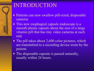


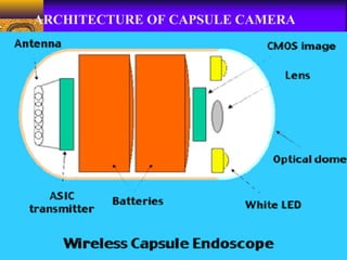
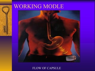
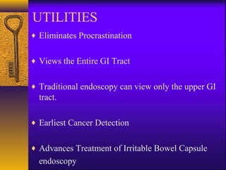
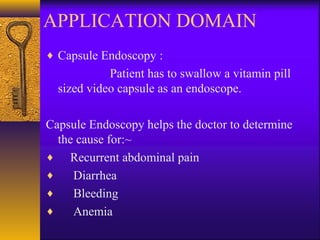

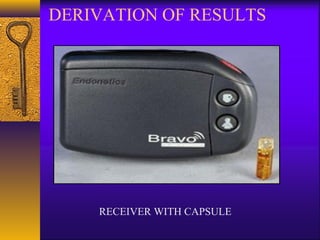


This document discusses capsule endoscopy, a medical procedure where patients swallow a pill-sized capsule containing a camera. The capsule passes naturally through the digestive tract, taking over 2,600 images which are transmitted to a recorder. This allows physicians to noninvasively examine the entire small intestine. The technology has advanced from basic endoscopy in the 1960s to now include capsule cameras, which provide benefits over traditional endoscopy like viewing the entire GI tract and earlier cancer detection. The capsule uses ultra-low power wireless transmission of images to help diagnose conditions like bleeding, Crohn's disease, and small bowel tumors.