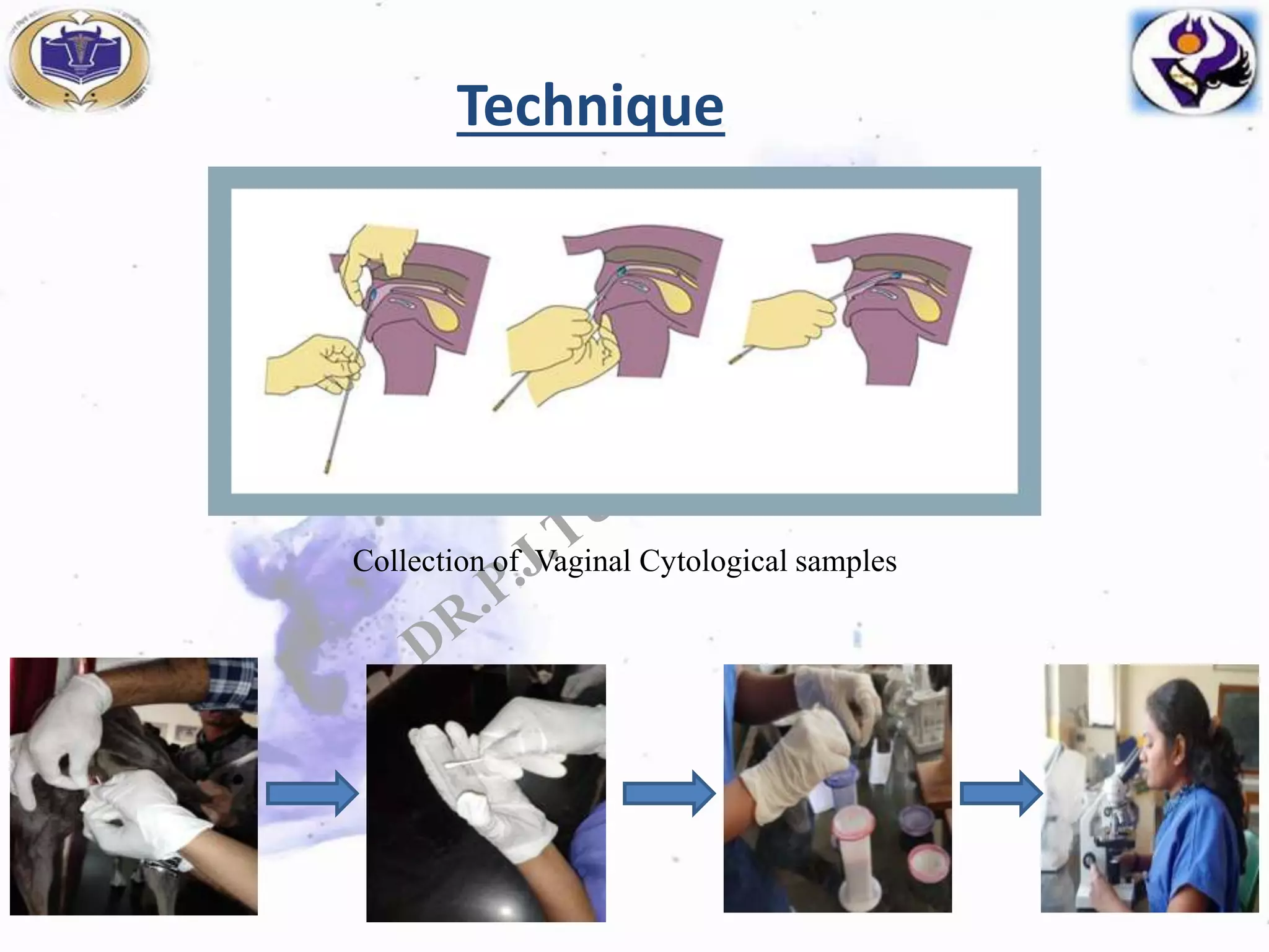
VAGINAL EXFOLIATIVE CYTOLOGY
IN SHE DOG
Presented by,
Dr.Tumbare Prajakta Jalinder
M.V.Sc (ARGO)
Department of Animal Reproduction, Gynaecology and Obstetrics
KNP College of Veterinary Science, Shirwal, Dist:Satara
Principle
Vaginal epithelium is sensitive to levels of estrodiol
and it changes from a bistratified cuboidal
epithelium to stratified squamous epithelium of
greater than 30 cell layers.
Thereafter these cells slowly mature, keratinized and
fall in vaginal lumen( exfoliate).
Case study
• A total of 70 cases of vaginal cytological examination
were presented to the TVCC of KNPCVS, Shirwal
during the period from April 2017 to December
2019.
• Total 13 Artificial insemination was done at the TVCC
of KNPCVS, Shirwal.
• And other are advised for natural mating.
• Conception rate – 92.30% .
Technique
Collection of Vaginal Cytological samples
Epithelial cells found in Normal canine vaginal smears
Cells Characteristics
Parabasal
Smallest and healthiest
Rounded with a large vesiculated nucleus
Small amount of cytoplasm
Intermediate
Slightly larger
Oval , round or irregularly round
Vesiculated nucleus slightly larger than parabasal
cell
Superficial
Largest cell
Sharp, flat, angular cytoplasmic border
Small, pyknotic, fading or ghost nucleus
Cornified/
Anuclear
keratinized
Large
Dead irregular cells
Nucleus are absent
Interpretation of vaginal smear
Cell type
Stages
Parabasal Intermediate Superficial
Intermediate
Superficial Anuclear
keratinized
Red
blood
cells
Neutrophil Bacteria
Proestrus
Early
Mid
Late
+++
+
+/-
++
+
+/-
+
++
+++
+/-
+
++
+/-
+/-
+
+++
++
+/-
++
+
+
++
+
+
Estrus
Early
Late
-
-
-
-
+
-
+++
+
++
+++
+/-
-
-
-
-
-
Diestrus
Early
Late
++
+
+++
+++
-
-
+
-
+
-
-
-
+
-
-
-
Anestrus +++ + - - - - - -
6
(Sridevi,2005)
Clinical situations wherein vaginal
cytology could be used as a diagnostic
tool
• Evaluation of infertility in bitches and those with
abnormal reproductive cycle.
• In prediction of approximate date of whelping.
• Diagnosis of follicular cyst.
• Diagnosis of vaginities.
• Identification of Transmissible venereal tumour.
• Diagnosis of transitional cell carcinomas of the
bladder
• Identification of sperms in vaginal smear within 48
hrs of mismating which helps to determining
whether an abortificient should be administered or
not.
1.TVT 2.Superficial cells with sperm
Conclusion
• Exfoliative vaginal cytology is best tool determine the
stage of estrus cycle
• As well as on the basis cornification index gives idea
about rising level of estrogen which predicts the
tentative date ovulation in she dogs.
• Exfoliative vaginal cytology is very simple, cost-
effective and comparatively accurate test for
predicting the she dogs fit for breeding.
Vaginal exfoliative cytology

Vaginal exfoliative cytology

  • 1.
    VAGINAL EXFOLIATIVE CYTOLOGY INSHE DOG Presented by, Dr.Tumbare Prajakta Jalinder M.V.Sc (ARGO) Department of Animal Reproduction, Gynaecology and Obstetrics KNP College of Veterinary Science, Shirwal, Dist:Satara
  • 2.
    Principle Vaginal epithelium issensitive to levels of estrodiol and it changes from a bistratified cuboidal epithelium to stratified squamous epithelium of greater than 30 cell layers. Thereafter these cells slowly mature, keratinized and fall in vaginal lumen( exfoliate).
  • 3.
    Case study • Atotal of 70 cases of vaginal cytological examination were presented to the TVCC of KNPCVS, Shirwal during the period from April 2017 to December 2019. • Total 13 Artificial insemination was done at the TVCC of KNPCVS, Shirwal. • And other are advised for natural mating. • Conception rate – 92.30% .
  • 4.
  • 5.
    Epithelial cells foundin Normal canine vaginal smears Cells Characteristics Parabasal Smallest and healthiest Rounded with a large vesiculated nucleus Small amount of cytoplasm Intermediate Slightly larger Oval , round or irregularly round Vesiculated nucleus slightly larger than parabasal cell Superficial Largest cell Sharp, flat, angular cytoplasmic border Small, pyknotic, fading or ghost nucleus Cornified/ Anuclear keratinized Large Dead irregular cells Nucleus are absent
  • 6.
    Interpretation of vaginalsmear Cell type Stages Parabasal Intermediate Superficial Intermediate Superficial Anuclear keratinized Red blood cells Neutrophil Bacteria Proestrus Early Mid Late +++ + +/- ++ + +/- + ++ +++ +/- + ++ +/- +/- + +++ ++ +/- ++ + + ++ + + Estrus Early Late - - - - + - +++ + ++ +++ +/- - - - - - Diestrus Early Late ++ + +++ +++ - - + - + - - - + - - - Anestrus +++ + - - - - - - 6 (Sridevi,2005)
  • 7.
    Clinical situations whereinvaginal cytology could be used as a diagnostic tool • Evaluation of infertility in bitches and those with abnormal reproductive cycle. • In prediction of approximate date of whelping. • Diagnosis of follicular cyst. • Diagnosis of vaginities.
  • 8.
    • Identification ofTransmissible venereal tumour. • Diagnosis of transitional cell carcinomas of the bladder • Identification of sperms in vaginal smear within 48 hrs of mismating which helps to determining whether an abortificient should be administered or not. 1.TVT 2.Superficial cells with sperm
  • 9.
    Conclusion • Exfoliative vaginalcytology is best tool determine the stage of estrus cycle • As well as on the basis cornification index gives idea about rising level of estrogen which predicts the tentative date ovulation in she dogs. • Exfoliative vaginal cytology is very simple, cost- effective and comparatively accurate test for predicting the she dogs fit for breeding.