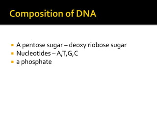
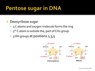
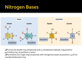
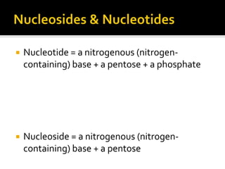
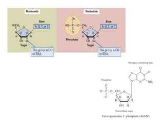
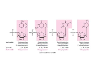
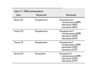
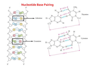
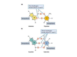
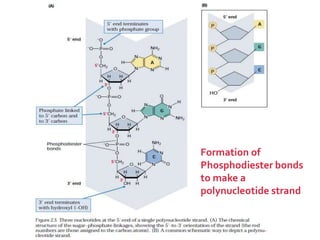
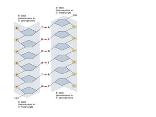
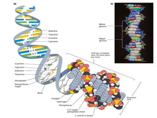
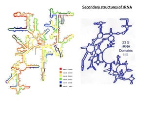
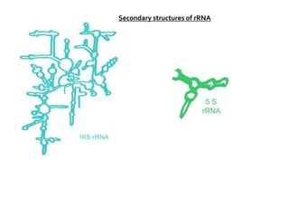
Rashmi Vijay
   A pentose sugar – deoxy riobose sugar
   Nucleotides – A,T,G,C
   a phosphate
   Deoxyribose sugar
     4 C atoms and oxygen molecule forms the ring
     5th C atom is outside the, part of CH2 group
     3 OH groups at positions 1,3,5
Purines are double ring compounds with 5 membered imidazole ring joined to
pyrimidine ring at positions 4’ and 5’
Pyrimidines are single ring compounds with nitrogenous bases at positions 1,3 of a 6
membered benzene ring
   Alternate with deoxyribose sugars joined by
    3’-C atom of one deoxyribose to 5’-C atom of
    the other
   Nucleotide = a nitrogenous (nitrogen-
    containing) base + a pentose + a phosphate


   Nucleoside = a nitrogenous (nitrogen-
    containing) base + a pentose
Nucleotide Base Pairing
Formation of
Phosphodiester bonds
to make a
polynucleotide strand
 The discovery of the structure of DNA by Watson
  and Crick in 1953 was a momentous event in
  science, an event that gave rise to entirely new
  disciplines and influenced the course of many
  established ones.
 Won Nobel Prize
1.    Right handed double helix, wound around central axis with
      plectonemic coiling.
2.    Two polynucloetide strands run antiparallel
3.    The offset pairing of the two strands creates a major groove
      and minor groove on the surface of the duplex
4.    Phosphate and dRibose forms the back bone of each
      polynucloetide strand
5.    Nitrogen bases are projected inward
6.    Two polynucloetide strands held together by H- bonds , AT
      and GC
7.    Each base pair tilts 360 and hence has 3600, and each of this
      turn has 10 nucleotide base pair
8.    Bases are place at a distance of 3.4 A0
9.    Diameter of the helix 20 A0
10.   Molecular weight / unit length = 2x10 6/ micrometr
   To shed more light on the structure
    of DNA, Rosalind Franklin and
    Maurice Wilkins used the powerful
    method of x-ray diffraction to
    analyze DNA fibers.

   They showed in the early 1950s that
    DNA produces a characteristic x-ray
    diffraction pattern.

   In 1953 Watson and Crick postulated
    a three dimensional model of DNA
    structure that accounted for all the
    available data.
   Erwin Chargaff developed a chemical
    technique to measure the amount of each
    base present in DNA.
   Chargaff also observed certain regular
    relationships among the molar
    concentrations of the different bases.
   These relationships are now called Chargaff’s
    rules
Chargaff’s rules


    He found that the base composition of the DNA, defined as
     the percent G+C, differs among species but is constant in
     all cells of an organism and within a species.
   The biochemical investigation of DNA began with Friedrich
    Miescher, who in the year 1868 isolated a phosphorus-
    containing substance, which he called “nuclein,” from the
    nuclei of pus cells (leukocytes) obtained from discarded
    surgical bandages.

   Miescher and many others suspected this substance is in
    some way with cell inheritance, but the first direct evidence
    that DNA is the bearer of genetic information came in 1944
    through a discovery made by Oswald T. Avery, Colin
    MacLeod, and Maclyn McCarty.
   These investigators found that DNA
    extracted from a virulent (disease-causing)
    strain of the bacterium Streptococcus
    pneumoniae, also known as pneumococcus,
    genetically transformed a nonvirulent strain
    of this organism into a virulent form.
   When injected into mice, the encapsulated strain of pneumococcus is
    lethal, whereas the nonencapsulated strain, like the heat-killed
    encapsulated strain, is harmless.
   Earlier research by the bacteriologist Frederick Griffith had shown
    that adding heat-killed virulent bacteria (harmless to mice) to a live
    nonvirulent strain permanently transformed the latter into lethal,
    virulent, encapsulated bacteria.
   Avery and his colleagues extracted the DNA from heat-killed virulent
    pneumococci, removing the protein as completely as possible, and
    added this DNA to non-virulent bacteria.
   The DNA gained entrance into the non-virulent bacteria, which were
    permanently transformed into a virulent strain.
   Avery and his colleagues concluded that the DNA extracted from the
    virulent strain carried the inheritable genetic message for virulence.
   A second important experiment provided independent
    evidence that DNA carries genetic information.


   In 1952 Alfred D. Hershey and Martha Chase used radioactive
    phosphorus (32P) and radioactive sulfur (35S) tracers to show
    that when the bacterial virus (bacteriophage) T2 infects its
    host cell, Escherichia coli, it is the phosphorus-containing
    DNA of the viral particle, not the sulfur-containing protein of
    the viral coat, that enters the host cell and furnishes the
    genetic information for viral replication
   DNA is a remarkably flexible molecule.
   Many significant deviations from the Watson-
    Crick DNA structure are found in cellular DNA,
    some or all of which may play important roles in
    DNA metabolism.
   These structural variations generally do not
    affect the key properties of DNA defined by
    Watson and Crick: strand complementarity,
    antiparallel strands, and the requirement for A=T
    and G C base pairs.
   Structural variation in DNA
    reflects three things:
     the different possible
      conformations of the deoxyribose,
     rotation about the contiguous
      bonds that make up the
      phosphodeoxyribose backbone,
      and
     Free rotation about the C-1–N-
      glycosyl bond
Comparison of A, B, and Z forms of DNA.
Each structure shown here has 36 base pairs.
The bases are shown in gray, the
phosphate atoms in yellow, and the riboses
and phosphate oxygens in blue. Blue is the
color used to represent DNA strands in later
chapters.
   The Watson-Crick structure is        Whether A-DNA occurs in
    also referred to as B-form            cells is uncertain, but there is
    DNA, or B-DNA, which is               evidence for some short
    the most stable structure for         stretches (tracts) of Z-DNA in
    a random-sequence DNA                 both prokaryotes and
    molecule under physiological          eukaryotes.
    conditions and is therefore
    the standard point of                These Z-DNA tracts may play
    reference in any study of the         a role (as yet undefined) in
    properties of DNA.                    regulating the expression of
                                          some genes or in genetic
   Two structural variants that          recombination.
    have been well characterized
    in crystal structures are the A
    and Z forms.
   Single stranded polymer of nucleotide monomers made
    of Ribose sugar, Nitrogenous bases and Phosphate
    group
   The structure of RNA is similar to, but not identical with,
    that of DNA.
   There is a difference in the sugar (RNA contains the
    sugar ribose instead of deoxyribose), RNA is usually
    single-stranded (not a duplex), and
   RNA contains the base uracil (U) instead of thymine (T),
    which is present in DNA.
   Serves as genetic material in some Viruses
   Present in 3 predominant forms rRNA, tRNA
    and mRNA
   Normally doesn't replicate or transcribe
   Made of fewer nucleotides (max 12000)
   Chain starts with adenine or guanine
   Contents expressed in terms of
    sedimentation coefficients ‘S’ – Svedberg
    constant
   rRNA = forms 80% total cellular RNA
   tRNA = 10-20% total cellular RNA
   mRNA = 3-5% total cellular RNA
   Found in Ribosomes
   most abundant RNA in cells (75%)
   most stable RNA in cells
   four species rRNA in eukaryotes
       28S rRNA (large subunit)
       5.8S rRNA (large subunit)
       5S rRNA (large subunit)
       18S rRNA (small subunit)
   28, 18 & 5.8S rRNA are synthesized as a single transcript, then
    processed -DNA has one promoter, one termination sequence for all
    three pieces of rRNA -DNA has multiple copies of the whole
    transcription unit.
     cut into pieces yielding four spacers & 3 rRNAs.
     spacers are broken down to nucleotides.
     rRNA pieces bind ribosomal proteins and begin to self assemble into
        ribosomal subunits
   5S rRNA transcribed from another gene as a separate transcription unit
    three species of rRNA in prokaryotes 23S in large subunit, 5S in the
    large subunit and 16S in the small subunit
Types of rRNA
   Made up of nucleotides twisted around itself at
    some region forming helical structure
   The strand assumes a shape of rod, coil or extended
    strand depending on ionic strength, temp, pH.
   In helical region, most of the bases are
    complementary and are joined by H – bonds
   The unpaired/unfolded single strand will have bases,
    and are not complementary, hence don’t show
    purine-pyrimidine equality
   rRNA strand unfold upon heating and refold on
    cooling
Secondary structures of rRNA
   Jacob and Monod coined the term mRNA
   least abundant (about 5%) of cellular RNAs
   mRNA may be mono or poly cistronic.
   bacterial mRNA is not processed
   eukaryotic mRNA is processed
   initial eukaryotic transcripts are quite large
     allows for posttranscriptional regulation of gene expression
     introns, exons and splicing
       (1) eukaryotic mRNAs contain "introns“
       (2) introns = intervening sequences
       (3) exons = expressed sequences
       (4) introns must be excised from mRNA before translation
       (5) RNA splicing = process of excising introns from mRNA and splicing the
          mRNA back together
   Single stranded linear molecule without any
    base pairing (base pairing destroys its nature)
   They have complementary sequence to the
    segment of DNA on which they are
    transcribed
 Cap: region at 5’ end. Important for protein synthesis, otherwise mRNA
  binds poorly to ribosomes. cap protects mRNA from 5'-endonucleases
 Non coding region - I (leader): region next to cap with 10-100 nucleotides.
  Rich in A & U residues and doesn't translate into proteins
 Initiation codon: region next NC-I. Common in both Prokaryotes and
  Eukaryotes
 The coding region: consists an average of 1500 nucleotides, which
  translates in to amino acids
 Termination Codon: do not code for any amino acids and thus brings
  termination of translation
 Non coding region – II (trailer): consists about 50-150 nucleotides. doesn't
  translate into proteins. Contains AAUAA in all sequenced examples
 Poly ‘A’ sequence: present at 3’ end. Contains about 200-250 nucleotides,
  which become shorter with age of the organism. Poly ‘A’ is added in
  nucleus before m-RNA reaches the cytoplasm from nucleus.
mRNA has rapid turnover.
 rRNA & tRNA are stable (days, months) -
  rRNA deeply buried in structure of ribosomes
 both rRNA and tRNA have many modified
  bases -modified bases help protect against
  nuclease attack
 mRNA turns over fast
  bacterial mRNA half life = minutes
  eukaryotic mRNA half life = hours
   nucleotide composition of mRNA : mRNA base
    composition like total genomic DNA
   base sequence complementarity of mRNA:
    measure complementarity by doing RNA-DNA
    hybridization
   size heterogeneity: mRNA varies greatly in size
    relative to rRNA & tRNA. mRNA varies in size
    depending on protein for which it codes
   gene amplification: one gene (DNA) gives rise
    to many transcripts (mRNA). each transcript can
    be translated to many proteins. get more
    proteins faster
   2nd most abundant (20%) RNA in cells
   synthesized in precursor form and then processed
   16 nucleotide leader sequence removed from 5' end
    and terminal UU is removed from 3' end ,UU replaced
    by CCA
   CCA found at the 3' end of all functional tRNAs
   intron "loop" is removed
   many bases methylated
   many bases modified –
     uracil converted to dihydrouracil, ribothymine or
      pseudouridine
     adenine converted to inosine

   Transfer RNAs vary in length from 73 to 93 nucleotides.
Nucleotide sequence of yeast tRNA Ala.
This structure was deduced in 1965 by Robert W.
Holley and his colleagues;
it is shown in the cloverleaf conformation in
which intrastrand base pairing is maximal.
The following symbols are used for the modified
nucleotides (shaded pink): ,
     Ψ, pseudouridine;
     I, inosine;
     T, ribothymidine;
     D, 5,6-dihydrouridine;
     mI I, 1-methylinosine;
     m1G, 1-methylguanosine;
     m2G, N2-dimethylguanosine
Blue lines between parallel sections indicate
Watson-Crick base pairs.
The anticodon can recognize three codons for
alanine (GCA, GCU, and GCC).
Note the presence of two G=U base pairs,
signified by a blue dot to indicate non-Watson-Crick
pairing.
In RNAs, guanosine is often basepaired with
uridine, although the G=U pair is not as stable as the
Watson- Crick G=C pair
Extra nucleotides occur in the extra arm or in
the D arm.
Two of the arms of a tRNA are critical for its
adaptor function.
The amino acid arm can carry a specific
amino acid esterified by its carboxyl group to
the 2- or 3-hydroxyl group of the amino acid
residue at the 3 end of the tRNA.
The anticodon arm contains the anticodon.
The other major arms are the D arm (DHU
arm), which contains the unusual nucleotide
dihydrouridine (D), and the T Ψ C arm, which
contains ribothymidine (T), not usually present
in RNAs, and pseudouridine (Ψ), which has an
unusual carbon–carbon bond between the base
and ribose.
The D and T Ψ C arms contribute important
interactions for the overall folding of tRNA
molecules,
and the T Ψ C arm interacts with the large-
subunit rRNA.

Structure and forms of dna&rna

  • 1.
  • 2.
    A pentose sugar – deoxy riobose sugar  Nucleotides – A,T,G,C  a phosphate
  • 3.
    Deoxyribose sugar  4 C atoms and oxygen molecule forms the ring  5th C atom is outside the, part of CH2 group  3 OH groups at positions 1,3,5
  • 4.
    Purines are doublering compounds with 5 membered imidazole ring joined to pyrimidine ring at positions 4’ and 5’ Pyrimidines are single ring compounds with nitrogenous bases at positions 1,3 of a 6 membered benzene ring
  • 5.
    Alternate with deoxyribose sugars joined by 3’-C atom of one deoxyribose to 5’-C atom of the other
  • 6.
    Nucleotide = a nitrogenous (nitrogen- containing) base + a pentose + a phosphate  Nucleoside = a nitrogenous (nitrogen- containing) base + a pentose
  • 10.
  • 12.
    Formation of Phosphodiester bonds tomake a polynucleotide strand
  • 14.
     The discoveryof the structure of DNA by Watson and Crick in 1953 was a momentous event in science, an event that gave rise to entirely new disciplines and influenced the course of many established ones.  Won Nobel Prize
  • 16.
    1. Right handed double helix, wound around central axis with plectonemic coiling. 2. Two polynucloetide strands run antiparallel 3. The offset pairing of the two strands creates a major groove and minor groove on the surface of the duplex 4. Phosphate and dRibose forms the back bone of each polynucloetide strand 5. Nitrogen bases are projected inward 6. Two polynucloetide strands held together by H- bonds , AT and GC 7. Each base pair tilts 360 and hence has 3600, and each of this turn has 10 nucleotide base pair 8. Bases are place at a distance of 3.4 A0 9. Diameter of the helix 20 A0 10. Molecular weight / unit length = 2x10 6/ micrometr
  • 19.
    To shed more light on the structure of DNA, Rosalind Franklin and Maurice Wilkins used the powerful method of x-ray diffraction to analyze DNA fibers.  They showed in the early 1950s that DNA produces a characteristic x-ray diffraction pattern.  In 1953 Watson and Crick postulated a three dimensional model of DNA structure that accounted for all the available data.
  • 21.
    Erwin Chargaff developed a chemical technique to measure the amount of each base present in DNA.  Chargaff also observed certain regular relationships among the molar concentrations of the different bases.  These relationships are now called Chargaff’s rules
  • 22.
    Chargaff’s rules  He found that the base composition of the DNA, defined as the percent G+C, differs among species but is constant in all cells of an organism and within a species.
  • 23.
    The biochemical investigation of DNA began with Friedrich Miescher, who in the year 1868 isolated a phosphorus- containing substance, which he called “nuclein,” from the nuclei of pus cells (leukocytes) obtained from discarded surgical bandages.  Miescher and many others suspected this substance is in some way with cell inheritance, but the first direct evidence that DNA is the bearer of genetic information came in 1944 through a discovery made by Oswald T. Avery, Colin MacLeod, and Maclyn McCarty.
  • 24.
    These investigators found that DNA extracted from a virulent (disease-causing) strain of the bacterium Streptococcus pneumoniae, also known as pneumococcus, genetically transformed a nonvirulent strain of this organism into a virulent form.
  • 25.
    When injected into mice, the encapsulated strain of pneumococcus is lethal, whereas the nonencapsulated strain, like the heat-killed encapsulated strain, is harmless.  Earlier research by the bacteriologist Frederick Griffith had shown that adding heat-killed virulent bacteria (harmless to mice) to a live nonvirulent strain permanently transformed the latter into lethal, virulent, encapsulated bacteria.  Avery and his colleagues extracted the DNA from heat-killed virulent pneumococci, removing the protein as completely as possible, and added this DNA to non-virulent bacteria.  The DNA gained entrance into the non-virulent bacteria, which were permanently transformed into a virulent strain.  Avery and his colleagues concluded that the DNA extracted from the virulent strain carried the inheritable genetic message for virulence.
  • 26.
    A second important experiment provided independent evidence that DNA carries genetic information.  In 1952 Alfred D. Hershey and Martha Chase used radioactive phosphorus (32P) and radioactive sulfur (35S) tracers to show that when the bacterial virus (bacteriophage) T2 infects its host cell, Escherichia coli, it is the phosphorus-containing DNA of the viral particle, not the sulfur-containing protein of the viral coat, that enters the host cell and furnishes the genetic information for viral replication
  • 28.
    DNA is a remarkably flexible molecule.  Many significant deviations from the Watson- Crick DNA structure are found in cellular DNA, some or all of which may play important roles in DNA metabolism.  These structural variations generally do not affect the key properties of DNA defined by Watson and Crick: strand complementarity, antiparallel strands, and the requirement for A=T and G C base pairs.
  • 29.
    Structural variation in DNA reflects three things:  the different possible conformations of the deoxyribose,  rotation about the contiguous bonds that make up the phosphodeoxyribose backbone, and  Free rotation about the C-1–N- glycosyl bond
  • 30.
    Comparison of A,B, and Z forms of DNA. Each structure shown here has 36 base pairs. The bases are shown in gray, the phosphate atoms in yellow, and the riboses and phosphate oxygens in blue. Blue is the color used to represent DNA strands in later chapters.
  • 33.
    The Watson-Crick structure is  Whether A-DNA occurs in also referred to as B-form cells is uncertain, but there is DNA, or B-DNA, which is evidence for some short the most stable structure for stretches (tracts) of Z-DNA in a random-sequence DNA both prokaryotes and molecule under physiological eukaryotes. conditions and is therefore the standard point of  These Z-DNA tracts may play reference in any study of the a role (as yet undefined) in properties of DNA. regulating the expression of some genes or in genetic  Two structural variants that recombination. have been well characterized in crystal structures are the A and Z forms.
  • 34.
    Single stranded polymer of nucleotide monomers made of Ribose sugar, Nitrogenous bases and Phosphate group  The structure of RNA is similar to, but not identical with, that of DNA.  There is a difference in the sugar (RNA contains the sugar ribose instead of deoxyribose), RNA is usually single-stranded (not a duplex), and  RNA contains the base uracil (U) instead of thymine (T), which is present in DNA.
  • 35.
    Serves as genetic material in some Viruses  Present in 3 predominant forms rRNA, tRNA and mRNA  Normally doesn't replicate or transcribe  Made of fewer nucleotides (max 12000)  Chain starts with adenine or guanine  Contents expressed in terms of sedimentation coefficients ‘S’ – Svedberg constant
  • 36.
    rRNA = forms 80% total cellular RNA  tRNA = 10-20% total cellular RNA  mRNA = 3-5% total cellular RNA
  • 37.
    Found in Ribosomes  most abundant RNA in cells (75%)  most stable RNA in cells
  • 38.
    four species rRNA in eukaryotes  28S rRNA (large subunit)  5.8S rRNA (large subunit)  5S rRNA (large subunit)  18S rRNA (small subunit)  28, 18 & 5.8S rRNA are synthesized as a single transcript, then processed -DNA has one promoter, one termination sequence for all three pieces of rRNA -DNA has multiple copies of the whole transcription unit.  cut into pieces yielding four spacers & 3 rRNAs.  spacers are broken down to nucleotides.  rRNA pieces bind ribosomal proteins and begin to self assemble into ribosomal subunits  5S rRNA transcribed from another gene as a separate transcription unit  three species of rRNA in prokaryotes 23S in large subunit, 5S in the large subunit and 16S in the small subunit
  • 39.
  • 40.
    Made up of nucleotides twisted around itself at some region forming helical structure  The strand assumes a shape of rod, coil or extended strand depending on ionic strength, temp, pH.  In helical region, most of the bases are complementary and are joined by H – bonds  The unpaired/unfolded single strand will have bases, and are not complementary, hence don’t show purine-pyrimidine equality  rRNA strand unfold upon heating and refold on cooling
  • 41.
  • 42.
  • 43.
    Jacob and Monod coined the term mRNA  least abundant (about 5%) of cellular RNAs  mRNA may be mono or poly cistronic.  bacterial mRNA is not processed  eukaryotic mRNA is processed  initial eukaryotic transcripts are quite large  allows for posttranscriptional regulation of gene expression  introns, exons and splicing (1) eukaryotic mRNAs contain "introns“ (2) introns = intervening sequences (3) exons = expressed sequences (4) introns must be excised from mRNA before translation (5) RNA splicing = process of excising introns from mRNA and splicing the mRNA back together
  • 45.
    Single stranded linear molecule without any base pairing (base pairing destroys its nature)  They have complementary sequence to the segment of DNA on which they are transcribed
  • 46.
     Cap: regionat 5’ end. Important for protein synthesis, otherwise mRNA binds poorly to ribosomes. cap protects mRNA from 5'-endonucleases  Non coding region - I (leader): region next to cap with 10-100 nucleotides. Rich in A & U residues and doesn't translate into proteins  Initiation codon: region next NC-I. Common in both Prokaryotes and Eukaryotes  The coding region: consists an average of 1500 nucleotides, which translates in to amino acids  Termination Codon: do not code for any amino acids and thus brings termination of translation  Non coding region – II (trailer): consists about 50-150 nucleotides. doesn't translate into proteins. Contains AAUAA in all sequenced examples  Poly ‘A’ sequence: present at 3’ end. Contains about 200-250 nucleotides, which become shorter with age of the organism. Poly ‘A’ is added in nucleus before m-RNA reaches the cytoplasm from nucleus.
  • 47.
    mRNA has rapidturnover.  rRNA & tRNA are stable (days, months) - rRNA deeply buried in structure of ribosomes  both rRNA and tRNA have many modified bases -modified bases help protect against nuclease attack  mRNA turns over fast  bacterial mRNA half life = minutes  eukaryotic mRNA half life = hours
  • 48.
    nucleotide composition of mRNA : mRNA base composition like total genomic DNA  base sequence complementarity of mRNA: measure complementarity by doing RNA-DNA hybridization  size heterogeneity: mRNA varies greatly in size relative to rRNA & tRNA. mRNA varies in size depending on protein for which it codes  gene amplification: one gene (DNA) gives rise to many transcripts (mRNA). each transcript can be translated to many proteins. get more proteins faster
  • 49.
    2nd most abundant (20%) RNA in cells  synthesized in precursor form and then processed  16 nucleotide leader sequence removed from 5' end and terminal UU is removed from 3' end ,UU replaced by CCA  CCA found at the 3' end of all functional tRNAs  intron "loop" is removed  many bases methylated  many bases modified –  uracil converted to dihydrouracil, ribothymine or pseudouridine  adenine converted to inosine  Transfer RNAs vary in length from 73 to 93 nucleotides.
  • 50.
    Nucleotide sequence ofyeast tRNA Ala. This structure was deduced in 1965 by Robert W. Holley and his colleagues; it is shown in the cloverleaf conformation in which intrastrand base pairing is maximal. The following symbols are used for the modified nucleotides (shaded pink): , Ψ, pseudouridine; I, inosine; T, ribothymidine; D, 5,6-dihydrouridine; mI I, 1-methylinosine; m1G, 1-methylguanosine; m2G, N2-dimethylguanosine Blue lines between parallel sections indicate Watson-Crick base pairs. The anticodon can recognize three codons for alanine (GCA, GCU, and GCC). Note the presence of two G=U base pairs, signified by a blue dot to indicate non-Watson-Crick pairing. In RNAs, guanosine is often basepaired with uridine, although the G=U pair is not as stable as the Watson- Crick G=C pair
  • 51.
    Extra nucleotides occurin the extra arm or in the D arm. Two of the arms of a tRNA are critical for its adaptor function. The amino acid arm can carry a specific amino acid esterified by its carboxyl group to the 2- or 3-hydroxyl group of the amino acid residue at the 3 end of the tRNA. The anticodon arm contains the anticodon. The other major arms are the D arm (DHU arm), which contains the unusual nucleotide dihydrouridine (D), and the T Ψ C arm, which contains ribothymidine (T), not usually present in RNAs, and pseudouridine (Ψ), which has an unusual carbon–carbon bond between the base and ribose. The D and T Ψ C arms contribute important interactions for the overall folding of tRNA molecules, and the T Ψ C arm interacts with the large- subunit rRNA.