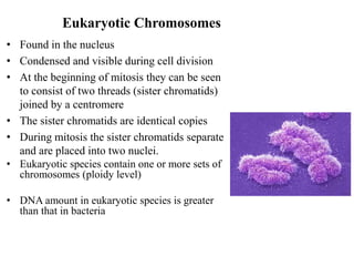
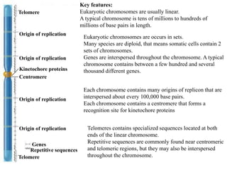
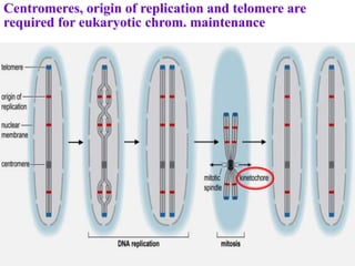
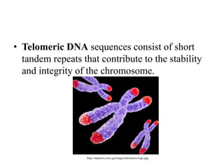
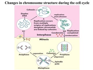
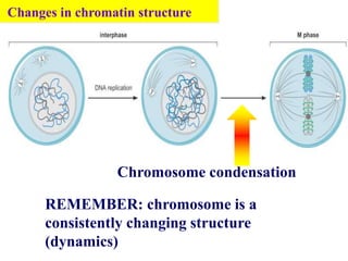
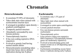
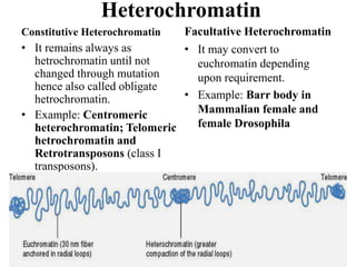
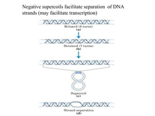
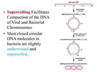
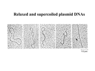
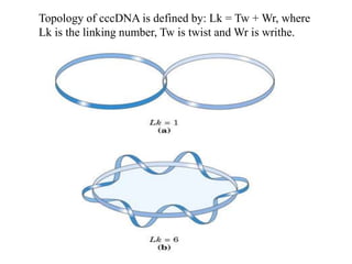
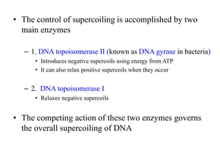
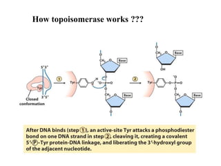
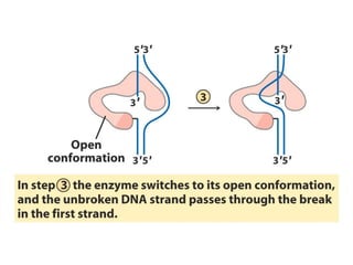
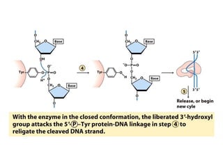
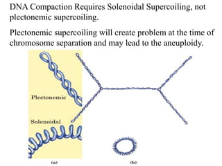
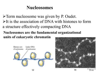
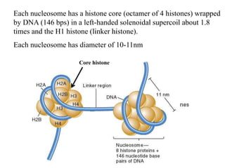
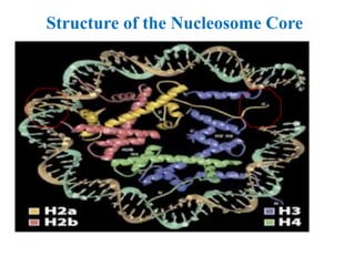
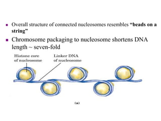
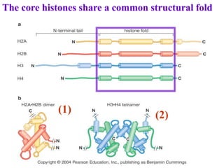
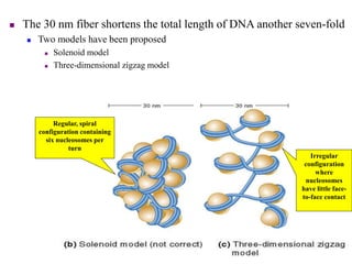
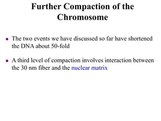
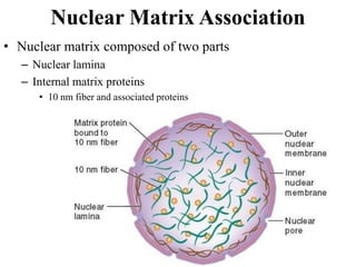
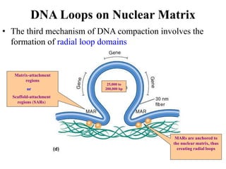
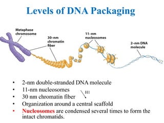
CHROMOSOME
ARTI YADAV
Dept. of Botany
Dayalbagh Educational Institute, Agra
Gene ???
• Gene: A segment of DNA which codes for
protein.
• Each gene has two alleles present on the same
loci in homologous chromosome.
• Thousands of genes are present in an
eukaryotic cell which are divided among the
chromosomes.
• Thus chromosomes act as vehicle for gene
(DNA).
• Chromosome are composed of DNA and associated proteins.
 Viral genomic DNA may be associated with capsid proteins
 Prokaryotic DNA is associated with proteins in the nucleoid
 Eukaryotic DNA is organized with proteins into a complex structure called chromatin
Chromosome
Chromosomes in eukaryotes and prokaryotes are different
PROKARYOTES EUKARYOTES
single chromosome plus plasmids many chromosomes
circular chromosome linear chromosomes
made only of DNA made of chromatin, a nucleoprotein
(DNA coiled around histone proteins)
found in cytoplasm found in a nucleus
copies its chromosome and divides
immediately afterwards
copies chromosomes, then the cell
grows, then goes through mitosis to
organise chromosomes in two equal
groups
Chromosome in Prokaryotes and
Viruses
• The DNA molecules of prokaryotes and
viruses are organized into negatively
supercoiled domains.
• Most viruses and prokaryotes have a single set of
genes stored in a single chromosome, which
contains a single molecule of nucleic acid.
• Prokaryotes are monoploid and are part of the
nucleoids.
• Bacterial chromosomes are double-stranded DNA
and are compacted into a nucleoid.
• Bacterial chromosomes contain circular molecules
of DNA segregated into 50 to 100 domains.
• Bacterial chromosomal DNA is usually a circular
molecule that is of few million nucleotides in
length
– Escherichia coli  ~ 4.6 million base pairs
– Haemophilus influenzae  ~ 1.8 million base
pairs
• A typical bacterial chromosome contains a few
thousand different genes
– Structural gene sequences (encoding proteins)
account for the majority of bacterial DNA
– The non-transcribed DNA between adjacent
genes are termed intergenic regions
A few hundred
nucleotides in length
These play roles in DNA
folding, DNA replication,
and gene expression
The E. coli Chromosome
Folded genome: is the functional state of a isolated bacterial
chromosome
Mild conditions
(no ionic detergents)
1M salt
Polyamines(-)
Model of E. coli Chromosome
folded=coiled
protein
Nicked=single strand
Some Virus Structures
Phage
Capsid
protein
TMV
• Chromosomes of Viruses consist of single- or
double-stranded DNA or RNA.
Eukaryotic Chromosomes
• Found in the nucleus
• Condensed and visible during cell division
• At the beginning of mitosis they can be seen
to consist of two threads (sister chromatids)
joined by a centromere
• The sister chromatids are identical copies
• During mitosis the sister chromatids separate
and are placed into two nuclei.
• Eukaryotic species contain one or more sets of
chromosomes (ploidy level)
• DNA amount in eukaryotic species is greater
than that in bacteria
Centromere
Kinetochore proteins
Origin of replication
Telomere
Telomere
Genes
Repetitive sequences
Key features:
Eukaryotic chromosomes are usually linear.
A typical chromosome is tens of millions to hundreds of
millions of base pairs in length.
Eukaryotic chromosomes are occurs in sets.
Many species are diploid, that means somatic cells contain 2
sets of chromosomes.
Genes are interspersed throughout the chromosome. A typical
chromosome contains between a few hundred and several
thousand different genes.
Each chromosome contains many origins of replicon that are
interspersed about every 100,000 base pairs.
Each chromosome contains a centromere that forms a
recognition site for kinetochore proteins
Telomeres contains specialized sequences located at both
ends of the linear chromosome.
Repetitive sequences are commonly found near centromeric
and telomeric regions, but they may also be interspersed
throughout the chromosome.
Chromosome Organization
• Genes located between centromere & telomeres
hundreds to thousands of genes are present
In lower eukaryotes (i.e. yeast)
• Genes are relatively small
• Very few introns
In higher eukaryotes (i.e. mammals)
• Genes are long
• Have many introns
• Non-gene sequences
– Repetitive DNA
• Telomere
• Centromere
• Satellite
Chromosome Structure
• Sister chromatid
– One of two attached
members of a duplicated
eukaryotic chromosome
• Centromere
– Constricted region in a
eukaryotic chromosome
where sister chromatids
are attached
Important Structural Elements of
the Eukaryotic Chromosome
• Centromere are the primary
constrictions along
eukaryotic chromosomes
• Centromere mediate
chromosomal migration
during mitosis and meiosis
• Centromere functions in cell
division; where the two daughter
chromosomes are held together
during mitosis (after DNA
replication but before cell division)
• Mitotic segregation of
chromosomes. Simple-sequence
DNA is located at centromere in
higher eukaryotes.
Required for the correct segregation of the
chromosomes after replication
 Direct the formation of kinetochore (an
elaborate protein complex) essential for chrom.
segregation
 One chromosome, one centromere
 The size varies (200 bp- >40 kb)
 Composed of largely repetitive DNA
sequences
Centromeres
Centromeres, origin of replication and telomere are
required for eukaryotic chrom. maintenance
Telomeres
• Telomeres cap the ends of linear chromosomes and are
needed for successful cell division
• Functions of telomeres
– Protect the ends of linear DNA molecules from
deoxyribonucleases
– Prevent fusion of chromosomes
– Facilitate complete replication of the ends of linear DNA
molecules
• Most telomeres contain repetitive sequences and a
distinct structure.
Telomere Structure
-TTAGGG
-500 to 3000 repeats
-G-rich overhang
-T-loop (D-loop)
-Telomeres specific
Proteins ( protection)
POT1
TRF1 and 2
TIN2 and TPP1
Telomeres and Cellular Aging
• In many tissues, telomeres are shortened after each round
of replication (end-replication problem of linear DNA); the
cellular DNA ages
• Normal human cells divide about 52 times before losing
ability to divide again (Hayflick limit)
• Sequence complexity refers to the number of
times a particular base sequence appears in the
genome
• Eukaryotic chromosomes contain repetitive DNA
( 15 to 80 %), Human (~50%)
• 3 main types of sequences
– Non-repetitive
– Moderately repetitive
– Highly repetitive (low complexity)
Repetitive Sequences
• Unique or non-repetitive sequences
– Found once or a few times in the genome
– Includes structural genes as well as intergenic areas
• Moderately repetitive
– Found a few hundred to a few thousand times
– Includes
• Genes for rRNA and histones
• Origins of replication
• Transposable elements
• Highly repetitive
– Found tens of thousands to millions of times
– Each copy is relatively short (a few nucleotides to several hundred in
length)
– Some sequences are interspersed throughout the genome
• Example: Alu family in humans
– Other sequences are clustered together in tandem arrays
• Example: AATAT and AATATAT sequences in Drosophila
• These are commonly found in the centromeric regions
Repetitive Sequences
DNA repeats
Centromeric: specific repeated regions (non-coding DNA sequences=
heterochromatin) of chromosome for attachment of spindle microtubules
( 5000 to 15000 bp).
Satellite sequences:
--Tandemly repeating
--Non-coding DNA
Alpha-------171 ( unit repeat as base pair)
Beta----------68
Satellite 1---48
Satellite 2-----5
Satellite 3-----5
Most satellite DNA is localized to the telomeric or
the centromeric region of the chromosome
• Telomeric DNA sequences consist of short
tandem repeats that contribute to the stability
and integrity of the chromosome.
http://topnews.com.sg/images/telomeres-logo.jpg
• Short interspersed elements (SINES) and
long interspersed elements (LINES) are
dispersed throughout the genome rather than
tandemly repeated, and constitute over 1/3 of
the human genome.
• These transposable elements are generated via
an RNA intermediate and are referred to as
retrotransposons.
• The Vast Majority of a Eukaryotic Genome
Does Not Encode Functional Genes
• Only a small portion of the eukaryotic genome
(2%–10%) constitute protein-encoding genes.
• There are also a large number of single-copy
noncoding regions, some of which are
pseudogenes.
M phase: condensed state, completely
disentangled from each other
G1, S, G2 phases: diffused, significantly less
compact. The structure of chrom. changes,
e.g.
DNA replication requires the nearly complete
disassembly and reassembly of the proteins
associated with each chromosome
Chromosome structure changes as
eukaryotic cells divide
Changes in chromosome structure during the cell cycle
Chromosome condensation
Changes in chromatin structure
REMEMBER: chromosome is a
consistently changing structure
(dynamics)
Numbers of chromosomes
• Constant for each cell in the body (except sex
cells which only have half sets).
• Constant throughout the life of an individual
(you don’t lose or gain chromosomes)
• Constant for all members of a species
Chromosome Number
• A eukaryotic cell’s DNA is
divided into a characteristic
number of chromosomes
• Chromosome number
– Sum of all chromosomes in a
cell of a given type
– A human body cell has 23
pairs of chromosomes
• Diploid
– Cells having two of each
type of chromosome
characteristic of the species
(2n)
Types of Chromosomes
• There are two types of eukaryotic chromosomes:
autosomes and sex chromosomes
• Autosomes
– Paired chromosomes with the same length, shape,
centromere location, and genes
– Any chromosome other than a sex chromosome
• Sex chromosomes
– Members of a pair of chromosomes that differ between
males and females
Other types of Eukaryotic chromosomes
• Polytene chromosome
• Lampbrush chromosome
Both these chromosome increases transcriptional
activity of gene
• Polytene chromosomes:
– have distinctive banding
patterns
– represent paired homologs
– are composed of many DNA
strands
Polytene chromosomes and lampbrush chromosomes are
very large and can be visualized by light microscopy.
Polytene chromosomes have puff regions where the DNA has
uncoiled and are visible manifestations of a high level of gene
activity.
• Lampbrush
chromosomes -
large and have
extensive DNA
looping.
• Formed through
DNA replication
without separation
or cell division
• Found in oocytes
in the diplotene
stage of meiosis.
Karyotype
• Karyotyping reveals characteristics of an individual’s chromosomes
• Karyotype
– Image of an individual’s complement of chromosomes arranged by size,
length, shape, and centromere location
Constructing a Karyotype
Half of the molecular mass of eukaryotic chromosome is protein
Two types of proteins are associated with DNA in eukaryotes:
1. Histones
2. Non-histones
Chromosome along with these proteins called Chromatin.
Chromatin is formed through end to end non-covalent attachment of
chromosome.
 The majority of the associated proteins are small, basic proteins
called histones.
 Other proteins associated with the chromosome are referred to as
non-histone proteins, including numerous DNA binding proteins
that regulate the transcription, replication, repair and recombination
of DNA.
Proteins in chromosome
Chromatin Composition
(+)
(-)
Histones:
H1, H2a, H2b, H3, H4
Structural
Nonhistone proteins:
Non structural
Regulation
Nucleosomes: DNA + histones
except H1
• Chromatin formation is an adaptation to increase
the stability of nucleus due to decrease in entropy.
• Telomere ends of chromosome allows only non-
covalent fusion to form chromatin.
• Covalent fusion is not possible due to triple
helical structure of telomere cap hence free ends
are not available for covalent bonding.
• Triple helical telomere cap is formed as a result of
Hoogstein base pairing in which homopurine of
guanine pairs with cytosine of its complementary
strands.
Chromatin
Heterochromatin
• It constitute 97-99% of chromatin
• Takes dark stain when stained with
acetocarmine (nuclear dye)
• It arranged at periphery upon density
gradient centrifugation
• Compactly packed structure
• Abundantly surrounded by non-
histone proteins
• Transcriptionaly inactive
• It has non-coding repetetive DNA
sequences
• Evolved as genetic load having
neutral mutant alleles
• Heterochromatin remodeling takes
much time hence shows delayed
replication during late S-phase
Euchromatin
• It constitute only 1-3% part of
chromatin
• Takes light stain when stained with
acetocarmine
• It arranged at centre upon centrifugation
• Loosely packed structure
• Few non-histone protein surrounds
euchromatin
• Transcriptionaly active
• It has protein coding non-repetetive
DNA sequences (*exceptionally histone
genes are moderately repetetive)
• Evolved as housekeeping genes and
remain switch on or off depending upon
requirement.
• Euchromatin remodeling occurs faster
hence it replicate during early S-phase.
Heterochromatin
Constitutive Heterochromatin
• It remains always as
hetrochromatin until not
changed through mutation
hence also called obligate
hetrochromatin.
• Example: Centromeric
heterochromatin; Telomeric
hetrochromatin and
Retrotransposons (class I
transposons).
Facultative Heterochromatin
• It may convert to
euchromatin depending
upon requirement.
• Example: Barr body in
Mammalian female and
female Drosophila
• Barr body (Facultative heterochromatin)
• Barr body was reported by Barr in cat
• To explain barr body Lyon proposed Lyon hypothesis
which depicts that one X chromosome in mammalian
female and female Drosophila becomes
heterochromatic to achieve dosage compensation.
• This heterochromatic X chromosome is called barr
body.
• Heterochromatic X chromosome have 85% qualitative
genes while 15% quantitative genes (escape genes).
• Heterochromatization occurs during early embryonic
stage (gastrula stage).
How is DNA packed in the
chromosomes
• DNA Supercoiling.
• Proteins assisted packaging (nucleosomes)
1. DNA Supercoiling
• DNA in the cell must be organized to allow:
– Packing of large DNA molecules within the cells
– Access of proteins to read the information in DNA sequence
• There are several levels of organization, one of which is
the supercoiling of the double-stranded DNA helix
What is coil and Supercoils ??
Supercoiling of DNA can only occur in closed-
circular DNA or linear DNA where the ends
are fixed.
Underwinding produces negative supercoils,
wheres overwinding produces positive supercoils.
Negative and positive supercoils .
Topoisomerases catalyze changes in the linking number of DNA.
Supercoiling induced by separating the strands
of duplex DNA (eg., during DNA replication)
Negative supercoils facilitate separation of DNA
strands (may facilitate transcription)
• Supercoiling Facilitates
Compaction of the DNA
of Viral and Bacterial
Chromosomes
• Most closed circular
DNA molecules in
bacteria are slightly
underwound and
supercoiled.
Relaxed and supercoiled plasmid DNAs
Topology of cccDNA is defined by: Lk = Tw + Wr, where
Lk is the linking number, Tw is twist and Wr is writhe.
• The control of supercoiling is accomplished by two
main enzymes
– 1. DNA topoisomerase II (known as DNA gyrase in bacteria)
• Introduces negative supercoils using energy from ATP
• It can also relax positive supercoils when they occur
– 2. DNA topoisomerase I
• Relaxes negative supercoils
• The competing action of these two enzymes governs
the overall supercoiling of DNA
How topoisomerase works ???
DNA Compaction Requires Solenoidal Supercoiling, not
plectonemic supercoiling.
Plectonemic supercoiling will create problem at the time of
chromosome separation and may lead to the aneuploidy.
DNA has negative charge due to the phosphate backbone and this
negative charge of DNA is neutralized by positevely charged histone
proteins.
Histones are small, basic proteins (rich in positively charged amino
acids Lysine and arginine).
Histone proteins are most conservative proteins due to conservative
topological charge of DNA.
Hence histone coding genes are treated as conservative genes and not
changed under influence of evolutionary force.
2. Protein assisted packaging of chromosome
Five abundant histones are H1 (linker histone, 20 kd), H2A, H2B, H3 and H4 (core
histones, 11-15 kd).
H2A, H2B, H3 and H4 are the core histones
Two of each make up the octamer
H1 is the linker histone
Binds to linker DNA and also binds to nucleosomes but not as tightly as are the
core histones.
Histone – DNA interaction
• Two non-covalent interactions are key player
of histone DNA interaction
• Electrostatic interaction allows recruitment
of histone proteins on negatively charged
backbone of DNA
• H- Bonding the N-terminal Ser-OH, Thr-OH
or Tyr-OH forms H-bonding with nitrogenous
bases of DNA and favouring stability of
histone DNA interaction.
Term nucleosome was given by P. Oudet.
It is the association of DNA with histones to form
a structure effectively compacting DNA
Nucleosomes are the fundamental organizational
units of eukaryotic chromatin
Nucleosomes
Core histone
Each nucleosome has a histone core (octamer of 4 histones) wrapped
by DNA (146 bps) in a left-handed solenoidal supercoil about 1.8
times and the H1 histone (linker histone).
Each nucleosome has diameter of 10-11nm
• The linker DNA is histone free hence it is called
DNase hypersensitive site.
• Caspase induced DNase cleaves the chromatin
during apoptosis from linker region.
• Eukaryotic promoters are also found in linker
region or H1 occupied region.
• H1 act as sealing protein to nucleosome.
Structure of the Nucleosome Core
Play a role in the organization
and compaction of the
chromosome
The DNA between each nucleosome is called a linker DNA.
Each eukaryote has a characteristic average linker DNA
length (20-60 bp)
 Overall structure of connected nucleosomes resembles “beads on a
string”
 Chromosome packaging to nucleosome shortens DNA
length ~ seven-fold
The core histones share a common structural fold
(1)
(2)
Many DNA sequence-independently make
interaction with the core histones
H-bond is the major stabilizing interaction to a nucleosome structure.
Each nucleosome has around 142 H-bonds
The histone N-terminal tails stabilize
DNA wrapping around the octamer
The histone tails emerge from the core of the nucleosome
at specific positions, serving as the grooves of a screw to
direct the DNA wrapping around the histone core in a
left-handed manner.
Histone variants alter nucleosome
function
Several histone variants are found in
eukaryotes
This variants can replace one of the 4
standard histones to form alternate
nucleosomes
Alteration of chromatin by incorporation of
histone variants
CENP-A is associated with the nucleosomes containing centromeric DNA
 Nucleosomes associate with each other to form a more
compact structure termed the 30 nm fiber or solenoid.
 Term solenoid was given by Finch and Clug.
 Each solenoid has 3 nucleosome at a point and it is the
actual chromatin fibre.
 Histone H1 plays a role in this compaction
 At moderate salt concentrations, H1 is removed
 The result is the classic beads-on-a-string morphology
 At low salt concentrations, H1 remains bound
 Beads associate together into a more compact morphology
Nucleosomes Join to Form a 30 nm Fiber
Histone H1 binds
to the linker DNA
between
nucleosome,
inducing tighter
DNA wrapping
around the
nucleosome
 The 30 nm fiber shortens the total length of DNA another seven-fold
 Two models have been proposed
 Solenoid model
 Three-dimensional zigzag model
Regular, spiral
configuration containing
six nucleosomes per
turn
Irregular
configuration
where
nucleosomes
have little face-
to-face contact
 The two events we have discussed so far have shortened
the DNA about 50-fold
 A third level of compaction involves interaction between
the 30 nm fiber and the nuclear matrix
Further Compaction of the
Chromosome
Nuclear Matrix Association
• Nuclear matrix composed of two parts
– Nuclear lamina
– Internal matrix proteins
• 10 nm fiber and associated proteins
Matrix-attachment
regions
Scaffold-attachment
regions (SARs)
or
MARs are anchored to
the nuclear matrix, thus
creating radial loops
25,000 to
200,000 bp
DNA Loops on Nuclear Matrix
• The third mechanism of DNA compaction involves the
formation of radial loop domains
 The attachment of radial loops to the nuclear matrix is
important in two ways
 1. It plays a role in gene regulation
 2. It serves to organize the chromosomes within the nucleus
An overview of histone DNA interaction
Compaction level
in euchromatin
Compaction level in
heterochromatin
During interphase most
chromosomal regions
are euchromatic
 Condensed chromosomes are referred to as metaphase
chromosomes
 During prophase, the compaction level increases
 As cells enter M phase, the level of compaction changes
dramatically
 By the end of prophase, sister chromatids are entirely
heterochromatic
 Two parallel chromatids have an overall diameter of 1,400 nm
 These highly condensed metaphase chromosomes
undergo little gene transcription
 In metaphase chromosomes, the radial loops are
compacted and anchored to the nuclear matrix scaffold
Metaphase Chromosomes
 In metaphase chromosomes the radial loops are highly
compacted and stay anchored to a scaffold
 The scaffold is formed from the nuclear matrix
 Histones are needed for the compaction of radial loops
Metaphase Chromosomes
 Two multiprotein complexes help to form and organize
metaphase chromosomes
 Condensin
 Plays a critical role in chromosome condensation
 Cohesin
 Plays a critical role in sister chromatid alignment
 Both contain a category of proteins called SMC proteins
 SMC proteins use energy from ATP and catalyze changes in
chromosome structure
The condensation of a metaphase chromosome by condensin
The number of loops has not changed
However, the diameter of each loop is smaller
Condensin travels
into the nucleus
Condensin binds to
chromosomes and
compacts the radial
loops
During interphase,
condensin is in the
cytoplasm
Chromosome Condensation
The alignment of sister chromatids via cohesin
Cohesins along chromosome
arms are released
Cohesin
remains at
centromere
Cohesin at centromer
is degraded
Chromosomes During Mitosis
Levels of DNA Packaging
• 2-nm double-stranded DNA molecule
• 11-nm nucleosomes
• 30 nm chromatin fiber
• Organization around a central scaffold
• Nucleosomes are condensed several times to form the
intact chromatids.
H1
The interaction of DNA with the histone
octamer is dynamic
Regulation of chromatin structure
There are factors acting on the
nucleosome to increase or decrease the
dynamic nature
The dynamic nature of DNA-binding
to the histone core is important for
access of DNA by other proteins
essential genome expression etc.
Nucleosome remodeling complexes
(chromatin remodeling complex)
facilitate nucleosome movement
A large protein complexes facilitate
changes in nucleosome location or
interaction with the DNA using the
energy of ATP hydrolysis.
Modification of the N-terminal tails of the histones alters
chromatin accessibility, and specific enzymes are
responsible for histone modification
• Histone tails are important for histone
modifications such as acetylation,
methylation, and phosphorylation.
• Acetylation it is catalysed by histone acetylase
enzyme. Target site for acetylation is Arg and
Lys residues at N-terminal tail of histone.
• Due to acetylation positive charge of histones
protein is masked and as a result histone
proteins dissociates from DNA due to
electrostatic repulsion.
• Methylation also occurs at N-terminal tail of
histone at Arg residue and modify the histone
protein
• Thus histone protein interaction become week
• Phosphorylation it occurs at Ser-OH, Thr-OH
and Tyr-OH residues at N-terminal and as a
result H-bond broken between nitrogenous
bases and histone.
The importance of packing of DNA into
chromosomes
 Chromosome is a compact form of the DNA that
readily fits inside the cell
 To protect DNA from damage
 DNA in a chromosome can be transmitted
efficiently to both daughter cells during cell
division
 Chromosome confers an overall organization to
each molecule of DNA, which facilitates gene
expression as well as recombination

Chromosome

  • 1.
    CHROMOSOME ARTI YADAV Dept. ofBotany Dayalbagh Educational Institute, Agra
  • 2.
    Gene ??? • Gene:A segment of DNA which codes for protein. • Each gene has two alleles present on the same loci in homologous chromosome. • Thousands of genes are present in an eukaryotic cell which are divided among the chromosomes. • Thus chromosomes act as vehicle for gene (DNA).
  • 3.
    • Chromosome arecomposed of DNA and associated proteins.  Viral genomic DNA may be associated with capsid proteins  Prokaryotic DNA is associated with proteins in the nucleoid  Eukaryotic DNA is organized with proteins into a complex structure called chromatin Chromosome Chromosomes in eukaryotes and prokaryotes are different PROKARYOTES EUKARYOTES single chromosome plus plasmids many chromosomes circular chromosome linear chromosomes made only of DNA made of chromatin, a nucleoprotein (DNA coiled around histone proteins) found in cytoplasm found in a nucleus copies its chromosome and divides immediately afterwards copies chromosomes, then the cell grows, then goes through mitosis to organise chromosomes in two equal groups
  • 4.
    Chromosome in Prokaryotesand Viruses • The DNA molecules of prokaryotes and viruses are organized into negatively supercoiled domains. • Most viruses and prokaryotes have a single set of genes stored in a single chromosome, which contains a single molecule of nucleic acid. • Prokaryotes are monoploid and are part of the nucleoids.
  • 5.
    • Bacterial chromosomesare double-stranded DNA and are compacted into a nucleoid. • Bacterial chromosomes contain circular molecules of DNA segregated into 50 to 100 domains. • Bacterial chromosomal DNA is usually a circular molecule that is of few million nucleotides in length – Escherichia coli  ~ 4.6 million base pairs – Haemophilus influenzae  ~ 1.8 million base pairs • A typical bacterial chromosome contains a few thousand different genes – Structural gene sequences (encoding proteins) account for the majority of bacterial DNA – The non-transcribed DNA between adjacent genes are termed intergenic regions
  • 6.
    A few hundred nucleotidesin length These play roles in DNA folding, DNA replication, and gene expression
  • 7.
    The E. coliChromosome Folded genome: is the functional state of a isolated bacterial chromosome Mild conditions (no ionic detergents) 1M salt Polyamines(-)
  • 8.
    Model of E.coli Chromosome folded=coiled protein Nicked=single strand
  • 9.
  • 10.
    • Chromosomes ofViruses consist of single- or double-stranded DNA or RNA.
  • 11.
    Eukaryotic Chromosomes • Foundin the nucleus • Condensed and visible during cell division • At the beginning of mitosis they can be seen to consist of two threads (sister chromatids) joined by a centromere • The sister chromatids are identical copies • During mitosis the sister chromatids separate and are placed into two nuclei. • Eukaryotic species contain one or more sets of chromosomes (ploidy level) • DNA amount in eukaryotic species is greater than that in bacteria
  • 12.
    Centromere Kinetochore proteins Origin ofreplication Origin of replication Origin of replication Origin of replication Telomere Telomere Genes Repetitive sequences Key features: Eukaryotic chromosomes are usually linear. A typical chromosome is tens of millions to hundreds of millions of base pairs in length. Eukaryotic chromosomes are occurs in sets. Many species are diploid, that means somatic cells contain 2 sets of chromosomes. Genes are interspersed throughout the chromosome. A typical chromosome contains between a few hundred and several thousand different genes. Each chromosome contains many origins of replicon that are interspersed about every 100,000 base pairs. Each chromosome contains a centromere that forms a recognition site for kinetochore proteins Telomeres contains specialized sequences located at both ends of the linear chromosome. Repetitive sequences are commonly found near centromeric and telomeric regions, but they may also be interspersed throughout the chromosome.
  • 13.
    Chromosome Organization • Geneslocated between centromere & telomeres hundreds to thousands of genes are present In lower eukaryotes (i.e. yeast) • Genes are relatively small • Very few introns In higher eukaryotes (i.e. mammals) • Genes are long • Have many introns • Non-gene sequences – Repetitive DNA • Telomere • Centromere • Satellite
  • 14.
    Chromosome Structure • Sisterchromatid – One of two attached members of a duplicated eukaryotic chromosome • Centromere – Constricted region in a eukaryotic chromosome where sister chromatids are attached
  • 15.
    Important Structural Elementsof the Eukaryotic Chromosome • Centromere are the primary constrictions along eukaryotic chromosomes • Centromere mediate chromosomal migration during mitosis and meiosis • Centromere functions in cell division; where the two daughter chromosomes are held together during mitosis (after DNA replication but before cell division) • Mitotic segregation of chromosomes. Simple-sequence DNA is located at centromere in higher eukaryotes.
  • 16.
    Required for thecorrect segregation of the chromosomes after replication  Direct the formation of kinetochore (an elaborate protein complex) essential for chrom. segregation  One chromosome, one centromere  The size varies (200 bp- >40 kb)  Composed of largely repetitive DNA sequences Centromeres
  • 17.
    Centromeres, origin ofreplication and telomere are required for eukaryotic chrom. maintenance
  • 18.
    Telomeres • Telomeres capthe ends of linear chromosomes and are needed for successful cell division • Functions of telomeres – Protect the ends of linear DNA molecules from deoxyribonucleases – Prevent fusion of chromosomes – Facilitate complete replication of the ends of linear DNA molecules • Most telomeres contain repetitive sequences and a distinct structure.
  • 19.
    Telomere Structure -TTAGGG -500 to3000 repeats -G-rich overhang -T-loop (D-loop) -Telomeres specific Proteins ( protection) POT1 TRF1 and 2 TIN2 and TPP1
  • 20.
    Telomeres and CellularAging • In many tissues, telomeres are shortened after each round of replication (end-replication problem of linear DNA); the cellular DNA ages • Normal human cells divide about 52 times before losing ability to divide again (Hayflick limit)
  • 21.
    • Sequence complexityrefers to the number of times a particular base sequence appears in the genome • Eukaryotic chromosomes contain repetitive DNA ( 15 to 80 %), Human (~50%) • 3 main types of sequences – Non-repetitive – Moderately repetitive – Highly repetitive (low complexity) Repetitive Sequences
  • 22.
    • Unique ornon-repetitive sequences – Found once or a few times in the genome – Includes structural genes as well as intergenic areas • Moderately repetitive – Found a few hundred to a few thousand times – Includes • Genes for rRNA and histones • Origins of replication • Transposable elements • Highly repetitive – Found tens of thousands to millions of times – Each copy is relatively short (a few nucleotides to several hundred in length) – Some sequences are interspersed throughout the genome • Example: Alu family in humans – Other sequences are clustered together in tandem arrays • Example: AATAT and AATATAT sequences in Drosophila • These are commonly found in the centromeric regions
  • 23.
  • 24.
    DNA repeats Centromeric: specificrepeated regions (non-coding DNA sequences= heterochromatin) of chromosome for attachment of spindle microtubules ( 5000 to 15000 bp). Satellite sequences: --Tandemly repeating --Non-coding DNA Alpha-------171 ( unit repeat as base pair) Beta----------68 Satellite 1---48 Satellite 2-----5 Satellite 3-----5 Most satellite DNA is localized to the telomeric or the centromeric region of the chromosome
  • 25.
    • Telomeric DNAsequences consist of short tandem repeats that contribute to the stability and integrity of the chromosome. http://topnews.com.sg/images/telomeres-logo.jpg
  • 26.
    • Short interspersedelements (SINES) and long interspersed elements (LINES) are dispersed throughout the genome rather than tandemly repeated, and constitute over 1/3 of the human genome. • These transposable elements are generated via an RNA intermediate and are referred to as retrotransposons.
  • 27.
    • The VastMajority of a Eukaryotic Genome Does Not Encode Functional Genes • Only a small portion of the eukaryotic genome (2%–10%) constitute protein-encoding genes. • There are also a large number of single-copy noncoding regions, some of which are pseudogenes.
  • 28.
    M phase: condensedstate, completely disentangled from each other G1, S, G2 phases: diffused, significantly less compact. The structure of chrom. changes, e.g. DNA replication requires the nearly complete disassembly and reassembly of the proteins associated with each chromosome Chromosome structure changes as eukaryotic cells divide
  • 29.
    Changes in chromosomestructure during the cell cycle
  • 30.
    Chromosome condensation Changes inchromatin structure REMEMBER: chromosome is a consistently changing structure (dynamics)
  • 31.
    Numbers of chromosomes •Constant for each cell in the body (except sex cells which only have half sets). • Constant throughout the life of an individual (you don’t lose or gain chromosomes) • Constant for all members of a species
  • 32.
    Chromosome Number • Aeukaryotic cell’s DNA is divided into a characteristic number of chromosomes • Chromosome number – Sum of all chromosomes in a cell of a given type – A human body cell has 23 pairs of chromosomes • Diploid – Cells having two of each type of chromosome characteristic of the species (2n)
  • 33.
    Types of Chromosomes •There are two types of eukaryotic chromosomes: autosomes and sex chromosomes • Autosomes – Paired chromosomes with the same length, shape, centromere location, and genes – Any chromosome other than a sex chromosome • Sex chromosomes – Members of a pair of chromosomes that differ between males and females
  • 34.
    Other types ofEukaryotic chromosomes • Polytene chromosome • Lampbrush chromosome Both these chromosome increases transcriptional activity of gene
  • 35.
    • Polytene chromosomes: –have distinctive banding patterns – represent paired homologs – are composed of many DNA strands Polytene chromosomes and lampbrush chromosomes are very large and can be visualized by light microscopy. Polytene chromosomes have puff regions where the DNA has uncoiled and are visible manifestations of a high level of gene activity.
  • 36.
    • Lampbrush chromosomes - largeand have extensive DNA looping. • Formed through DNA replication without separation or cell division • Found in oocytes in the diplotene stage of meiosis.
  • 37.
    Karyotype • Karyotyping revealscharacteristics of an individual’s chromosomes • Karyotype – Image of an individual’s complement of chromosomes arranged by size, length, shape, and centromere location Constructing a Karyotype
  • 38.
    Half of themolecular mass of eukaryotic chromosome is protein Two types of proteins are associated with DNA in eukaryotes: 1. Histones 2. Non-histones Chromosome along with these proteins called Chromatin. Chromatin is formed through end to end non-covalent attachment of chromosome.  The majority of the associated proteins are small, basic proteins called histones.  Other proteins associated with the chromosome are referred to as non-histone proteins, including numerous DNA binding proteins that regulate the transcription, replication, repair and recombination of DNA. Proteins in chromosome
  • 39.
    Chromatin Composition (+) (-) Histones: H1, H2a,H2b, H3, H4 Structural Nonhistone proteins: Non structural Regulation Nucleosomes: DNA + histones except H1
  • 40.
    • Chromatin formationis an adaptation to increase the stability of nucleus due to decrease in entropy. • Telomere ends of chromosome allows only non- covalent fusion to form chromatin. • Covalent fusion is not possible due to triple helical structure of telomere cap hence free ends are not available for covalent bonding. • Triple helical telomere cap is formed as a result of Hoogstein base pairing in which homopurine of guanine pairs with cytosine of its complementary strands.
  • 41.
    Chromatin Heterochromatin • It constitute97-99% of chromatin • Takes dark stain when stained with acetocarmine (nuclear dye) • It arranged at periphery upon density gradient centrifugation • Compactly packed structure • Abundantly surrounded by non- histone proteins • Transcriptionaly inactive • It has non-coding repetetive DNA sequences • Evolved as genetic load having neutral mutant alleles • Heterochromatin remodeling takes much time hence shows delayed replication during late S-phase Euchromatin • It constitute only 1-3% part of chromatin • Takes light stain when stained with acetocarmine • It arranged at centre upon centrifugation • Loosely packed structure • Few non-histone protein surrounds euchromatin • Transcriptionaly active • It has protein coding non-repetetive DNA sequences (*exceptionally histone genes are moderately repetetive) • Evolved as housekeeping genes and remain switch on or off depending upon requirement. • Euchromatin remodeling occurs faster hence it replicate during early S-phase.
  • 42.
    Heterochromatin Constitutive Heterochromatin • Itremains always as hetrochromatin until not changed through mutation hence also called obligate hetrochromatin. • Example: Centromeric heterochromatin; Telomeric hetrochromatin and Retrotransposons (class I transposons). Facultative Heterochromatin • It may convert to euchromatin depending upon requirement. • Example: Barr body in Mammalian female and female Drosophila
  • 43.
    • Barr body(Facultative heterochromatin) • Barr body was reported by Barr in cat • To explain barr body Lyon proposed Lyon hypothesis which depicts that one X chromosome in mammalian female and female Drosophila becomes heterochromatic to achieve dosage compensation. • This heterochromatic X chromosome is called barr body. • Heterochromatic X chromosome have 85% qualitative genes while 15% quantitative genes (escape genes). • Heterochromatization occurs during early embryonic stage (gastrula stage).
  • 44.
    How is DNApacked in the chromosomes • DNA Supercoiling. • Proteins assisted packaging (nucleosomes)
  • 45.
    1. DNA Supercoiling •DNA in the cell must be organized to allow: – Packing of large DNA molecules within the cells – Access of proteins to read the information in DNA sequence • There are several levels of organization, one of which is the supercoiling of the double-stranded DNA helix
  • 46.
    What is coiland Supercoils ??
  • 47.
    Supercoiling of DNAcan only occur in closed- circular DNA or linear DNA where the ends are fixed. Underwinding produces negative supercoils, wheres overwinding produces positive supercoils.
  • 48.
    Negative and positivesupercoils . Topoisomerases catalyze changes in the linking number of DNA.
  • 49.
    Supercoiling induced byseparating the strands of duplex DNA (eg., during DNA replication)
  • 50.
    Negative supercoils facilitateseparation of DNA strands (may facilitate transcription)
  • 51.
    • Supercoiling Facilitates Compactionof the DNA of Viral and Bacterial Chromosomes • Most closed circular DNA molecules in bacteria are slightly underwound and supercoiled.
  • 52.
  • 53.
    Topology of cccDNAis defined by: Lk = Tw + Wr, where Lk is the linking number, Tw is twist and Wr is writhe.
  • 54.
    • The controlof supercoiling is accomplished by two main enzymes – 1. DNA topoisomerase II (known as DNA gyrase in bacteria) • Introduces negative supercoils using energy from ATP • It can also relax positive supercoils when they occur – 2. DNA topoisomerase I • Relaxes negative supercoils • The competing action of these two enzymes governs the overall supercoiling of DNA
  • 55.
  • 58.
    DNA Compaction RequiresSolenoidal Supercoiling, not plectonemic supercoiling. Plectonemic supercoiling will create problem at the time of chromosome separation and may lead to the aneuploidy.
  • 59.
    DNA has negativecharge due to the phosphate backbone and this negative charge of DNA is neutralized by positevely charged histone proteins. Histones are small, basic proteins (rich in positively charged amino acids Lysine and arginine). Histone proteins are most conservative proteins due to conservative topological charge of DNA. Hence histone coding genes are treated as conservative genes and not changed under influence of evolutionary force. 2. Protein assisted packaging of chromosome
  • 60.
    Five abundant histonesare H1 (linker histone, 20 kd), H2A, H2B, H3 and H4 (core histones, 11-15 kd). H2A, H2B, H3 and H4 are the core histones Two of each make up the octamer H1 is the linker histone Binds to linker DNA and also binds to nucleosomes but not as tightly as are the core histones.
  • 61.
    Histone – DNAinteraction • Two non-covalent interactions are key player of histone DNA interaction • Electrostatic interaction allows recruitment of histone proteins on negatively charged backbone of DNA • H- Bonding the N-terminal Ser-OH, Thr-OH or Tyr-OH forms H-bonding with nitrogenous bases of DNA and favouring stability of histone DNA interaction.
  • 62.
    Term nucleosome wasgiven by P. Oudet. It is the association of DNA with histones to form a structure effectively compacting DNA Nucleosomes are the fundamental organizational units of eukaryotic chromatin Nucleosomes
  • 63.
    Core histone Each nucleosomehas a histone core (octamer of 4 histones) wrapped by DNA (146 bps) in a left-handed solenoidal supercoil about 1.8 times and the H1 histone (linker histone). Each nucleosome has diameter of 10-11nm
  • 64.
    • The linkerDNA is histone free hence it is called DNase hypersensitive site. • Caspase induced DNase cleaves the chromatin during apoptosis from linker region. • Eukaryotic promoters are also found in linker region or H1 occupied region. • H1 act as sealing protein to nucleosome.
  • 65.
    Structure of theNucleosome Core
  • 66.
    Play a rolein the organization and compaction of the chromosome The DNA between each nucleosome is called a linker DNA. Each eukaryote has a characteristic average linker DNA length (20-60 bp)
  • 67.
     Overall structureof connected nucleosomes resembles “beads on a string”  Chromosome packaging to nucleosome shortens DNA length ~ seven-fold
  • 68.
    The core histonesshare a common structural fold (1) (2)
  • 69.
    Many DNA sequence-independentlymake interaction with the core histones H-bond is the major stabilizing interaction to a nucleosome structure. Each nucleosome has around 142 H-bonds
  • 70.
    The histone N-terminaltails stabilize DNA wrapping around the octamer The histone tails emerge from the core of the nucleosome at specific positions, serving as the grooves of a screw to direct the DNA wrapping around the histone core in a left-handed manner.
  • 71.
    Histone variants alternucleosome function Several histone variants are found in eukaryotes This variants can replace one of the 4 standard histones to form alternate nucleosomes
  • 72.
    Alteration of chromatinby incorporation of histone variants CENP-A is associated with the nucleosomes containing centromeric DNA
  • 73.
     Nucleosomes associatewith each other to form a more compact structure termed the 30 nm fiber or solenoid.  Term solenoid was given by Finch and Clug.  Each solenoid has 3 nucleosome at a point and it is the actual chromatin fibre.  Histone H1 plays a role in this compaction  At moderate salt concentrations, H1 is removed  The result is the classic beads-on-a-string morphology  At low salt concentrations, H1 remains bound  Beads associate together into a more compact morphology Nucleosomes Join to Form a 30 nm Fiber
  • 74.
    Histone H1 binds tothe linker DNA between nucleosome, inducing tighter DNA wrapping around the nucleosome
  • 75.
     The 30nm fiber shortens the total length of DNA another seven-fold  Two models have been proposed  Solenoid model  Three-dimensional zigzag model Regular, spiral configuration containing six nucleosomes per turn Irregular configuration where nucleosomes have little face- to-face contact
  • 76.
     The twoevents we have discussed so far have shortened the DNA about 50-fold  A third level of compaction involves interaction between the 30 nm fiber and the nuclear matrix Further Compaction of the Chromosome
  • 77.
    Nuclear Matrix Association •Nuclear matrix composed of two parts – Nuclear lamina – Internal matrix proteins • 10 nm fiber and associated proteins
  • 78.
    Matrix-attachment regions Scaffold-attachment regions (SARs) or MARs areanchored to the nuclear matrix, thus creating radial loops 25,000 to 200,000 bp DNA Loops on Nuclear Matrix • The third mechanism of DNA compaction involves the formation of radial loop domains
  • 79.
     The attachmentof radial loops to the nuclear matrix is important in two ways  1. It plays a role in gene regulation  2. It serves to organize the chromosomes within the nucleus
  • 80.
    An overview ofhistone DNA interaction
  • 81.
    Compaction level in euchromatin Compactionlevel in heterochromatin During interphase most chromosomal regions are euchromatic
  • 82.
     Condensed chromosomesare referred to as metaphase chromosomes  During prophase, the compaction level increases  As cells enter M phase, the level of compaction changes dramatically  By the end of prophase, sister chromatids are entirely heterochromatic  Two parallel chromatids have an overall diameter of 1,400 nm  These highly condensed metaphase chromosomes undergo little gene transcription  In metaphase chromosomes, the radial loops are compacted and anchored to the nuclear matrix scaffold Metaphase Chromosomes
  • 83.
     In metaphasechromosomes the radial loops are highly compacted and stay anchored to a scaffold  The scaffold is formed from the nuclear matrix  Histones are needed for the compaction of radial loops Metaphase Chromosomes
  • 84.
     Two multiproteincomplexes help to form and organize metaphase chromosomes  Condensin  Plays a critical role in chromosome condensation  Cohesin  Plays a critical role in sister chromatid alignment  Both contain a category of proteins called SMC proteins  SMC proteins use energy from ATP and catalyze changes in chromosome structure
  • 85.
    The condensation ofa metaphase chromosome by condensin The number of loops has not changed However, the diameter of each loop is smaller Condensin travels into the nucleus Condensin binds to chromosomes and compacts the radial loops During interphase, condensin is in the cytoplasm Chromosome Condensation
  • 86.
    The alignment ofsister chromatids via cohesin Cohesins along chromosome arms are released Cohesin remains at centromere Cohesin at centromer is degraded Chromosomes During Mitosis
  • 87.
    Levels of DNAPackaging • 2-nm double-stranded DNA molecule • 11-nm nucleosomes • 30 nm chromatin fiber • Organization around a central scaffold • Nucleosomes are condensed several times to form the intact chromatids. H1
  • 88.
    The interaction ofDNA with the histone octamer is dynamic Regulation of chromatin structure There are factors acting on the nucleosome to increase or decrease the dynamic nature The dynamic nature of DNA-binding to the histone core is important for access of DNA by other proteins essential genome expression etc.
  • 89.
    Nucleosome remodeling complexes (chromatinremodeling complex) facilitate nucleosome movement A large protein complexes facilitate changes in nucleosome location or interaction with the DNA using the energy of ATP hydrolysis.
  • 90.
    Modification of theN-terminal tails of the histones alters chromatin accessibility, and specific enzymes are responsible for histone modification • Histone tails are important for histone modifications such as acetylation, methylation, and phosphorylation.
  • 91.
    • Acetylation itis catalysed by histone acetylase enzyme. Target site for acetylation is Arg and Lys residues at N-terminal tail of histone. • Due to acetylation positive charge of histones protein is masked and as a result histone proteins dissociates from DNA due to electrostatic repulsion.
  • 92.
    • Methylation alsooccurs at N-terminal tail of histone at Arg residue and modify the histone protein • Thus histone protein interaction become week • Phosphorylation it occurs at Ser-OH, Thr-OH and Tyr-OH residues at N-terminal and as a result H-bond broken between nitrogenous bases and histone.
  • 93.
    The importance ofpacking of DNA into chromosomes  Chromosome is a compact form of the DNA that readily fits inside the cell  To protect DNA from damage  DNA in a chromosome can be transmitted efficiently to both daughter cells during cell division  Chromosome confers an overall organization to each molecule of DNA, which facilitates gene expression as well as recombination