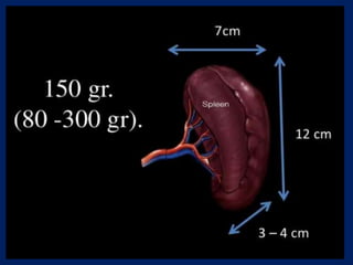
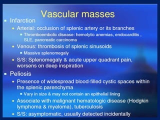
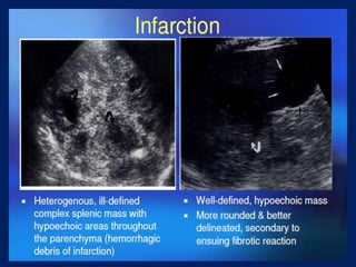
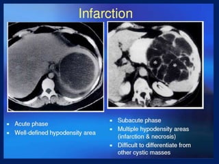
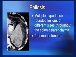
Downloaded 1,731 times
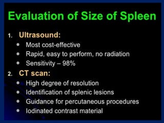


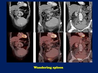
![Axial reconstructions derived from the CT portion of the PET/CT demonstrate the
‘whorled-appearance’ of the long vascular pedicle extending to the ectopic spleen.
The alternating bands of hypodensity and hyperdensity represent the splenic
vessels and surrounding fat of the twisted splenic pedicle. [red arrow = splenic
artery; yellow arrow = pancreas; white arrow = splenic vein; asterisk = spleen]
Wandering spleen](https://image.slidesharecdn.com/presentation1-140322130213-phpapp01/85/Presentation1-pptx-spleen-21-320.jpg)
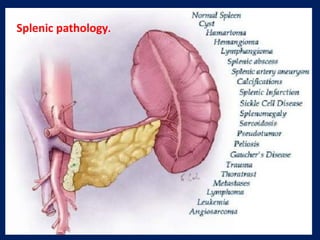


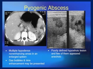
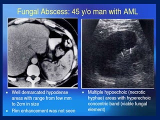
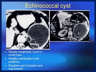
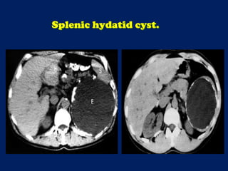
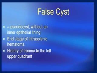
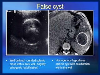
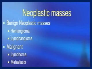

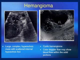
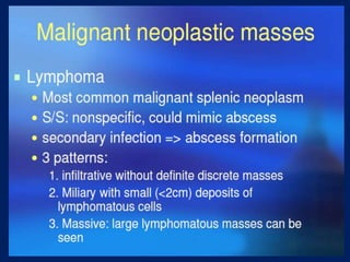
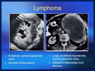

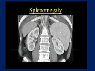
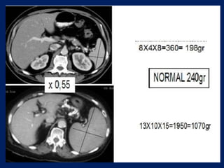
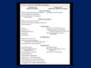
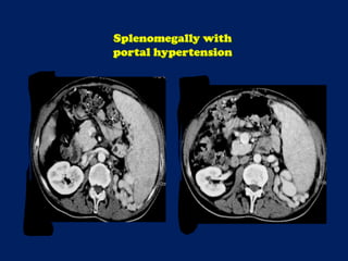
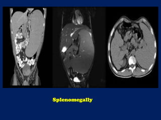
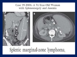


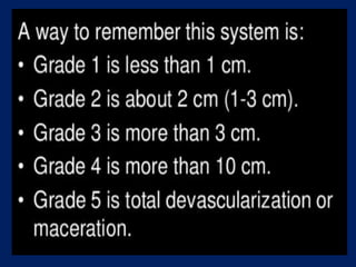
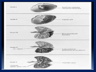
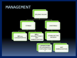
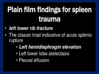
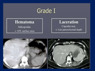
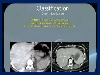
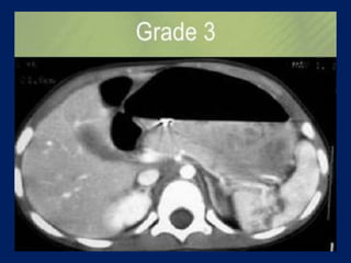
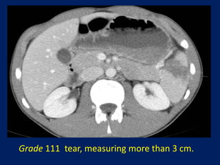
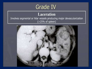
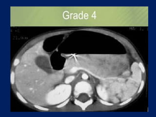
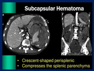
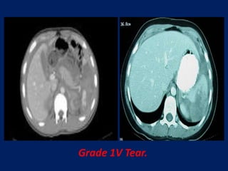
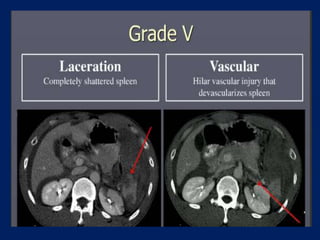
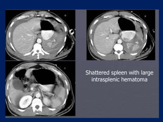


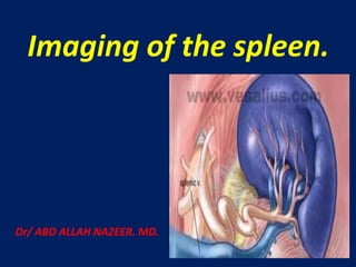
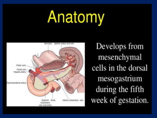
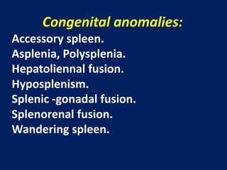
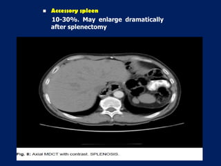
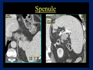
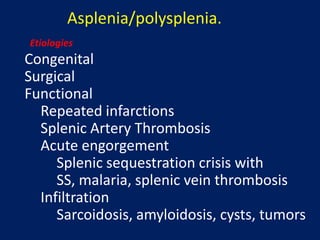
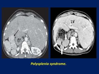
This document discusses imaging of the spleen and summarizes various congenital anomalies and pathologies that can affect the spleen. Some common congenital anomalies mentioned include accessory spleens, asplenia, polysplenia, and splenic fusions. Acquired conditions like repeated infarctions, infiltration, tumors, and cysts can also cause splenomegaly or functional asplenia. Wandering spleen is discussed as a rare congenital anomaly where the spleen lacks attachments and is mobile within the abdomen. Various grades of splenic lacerations and examples of splenic imaging findings are also briefly summarized.

![Spleen[1]](https://cdn.slidesharecdn.com/ss_thumbnails/spleen1-171112094140-thumbnail.jpg?width=640&height=640&fit=bounds)


![Imaging ofsplenic diseases [Autosaved].pptx](https://cdn.slidesharecdn.com/ss_thumbnails/imagingodsplenicdiseasesautosaved-220822020559-3118a410-thumbnail.jpg?width=640&height=640&fit=bounds)


