Downloaded 774 times
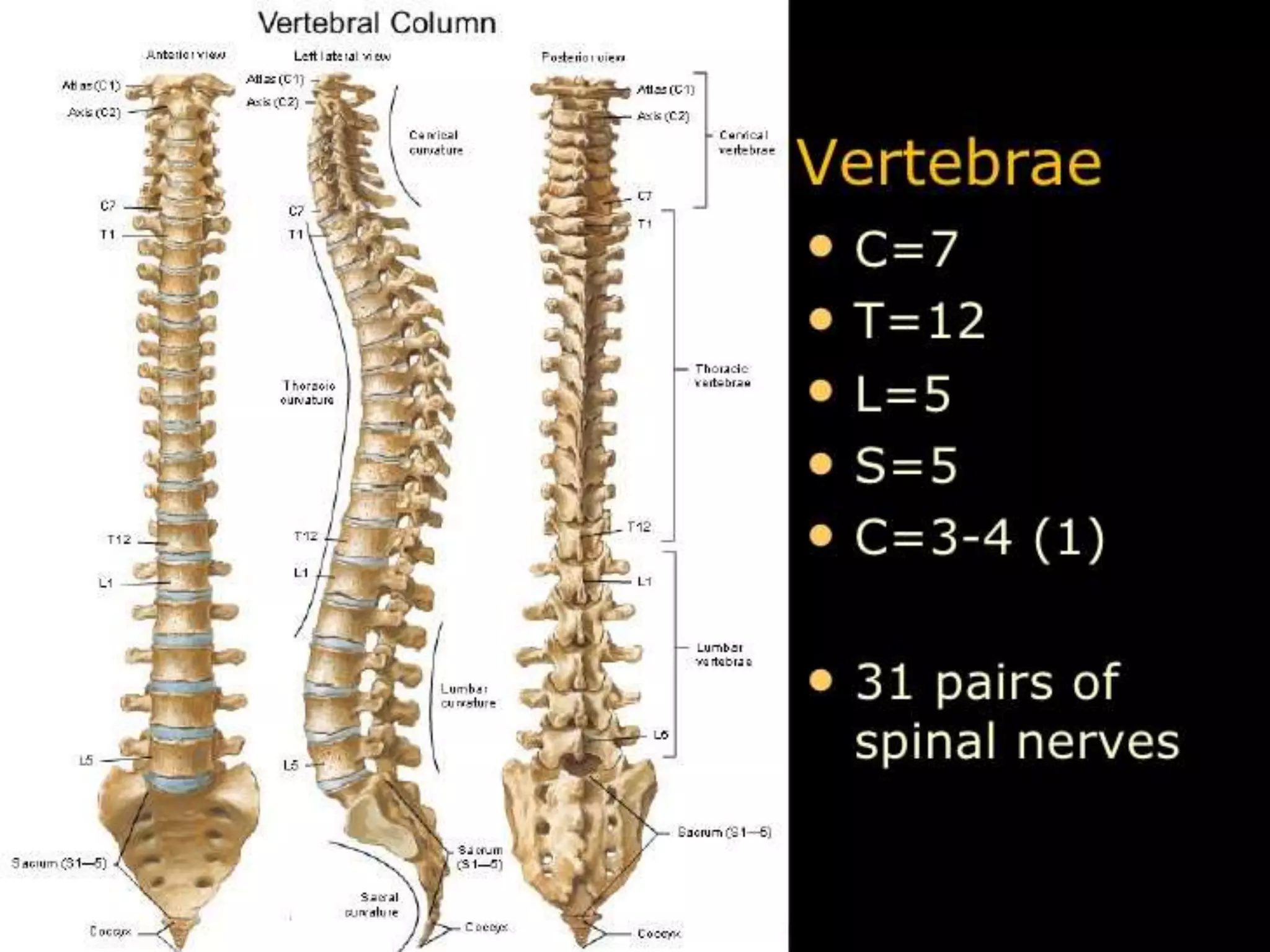
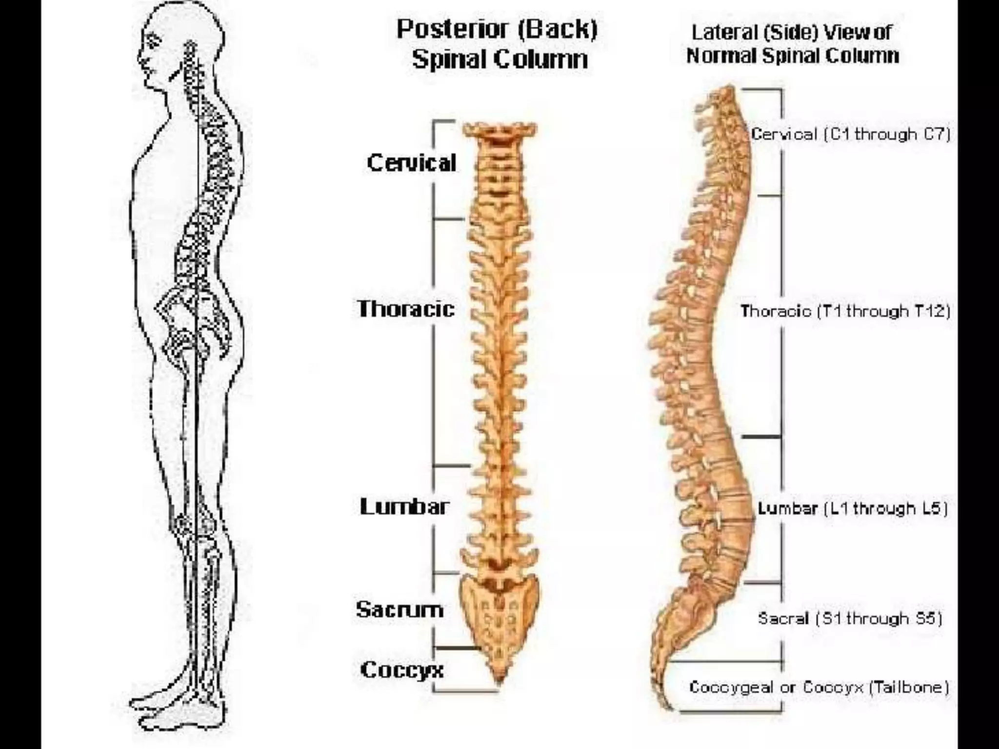
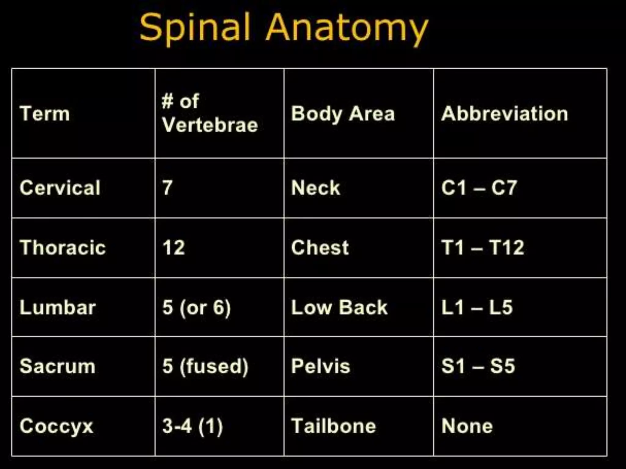
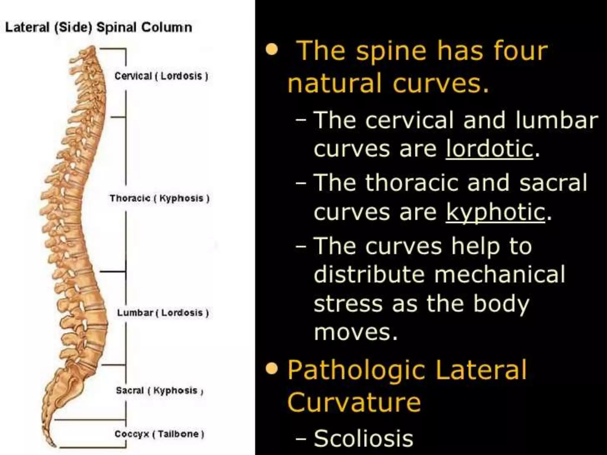
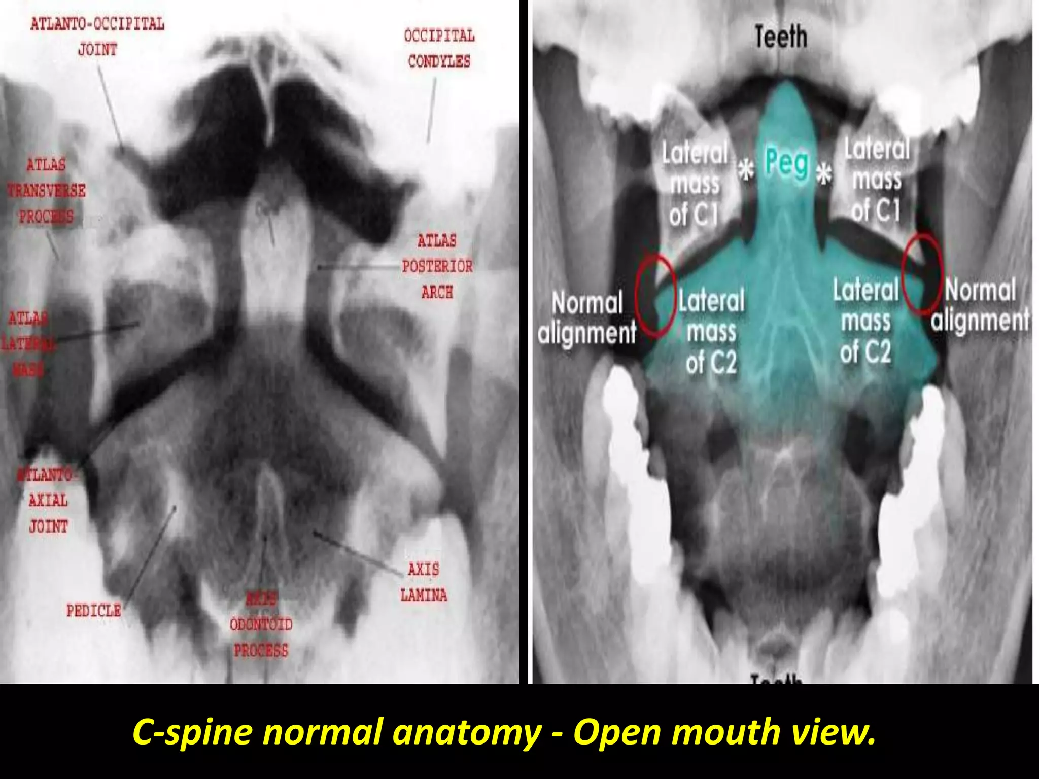
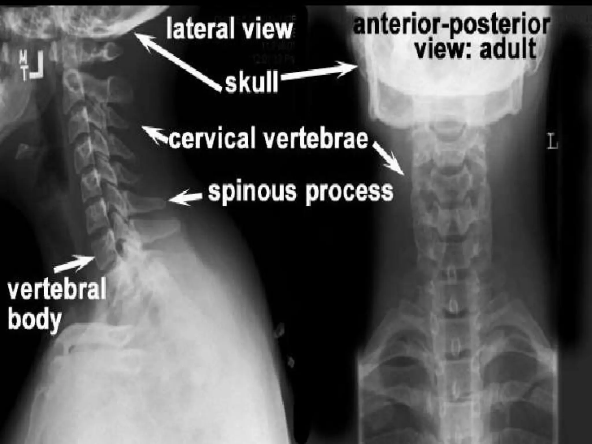
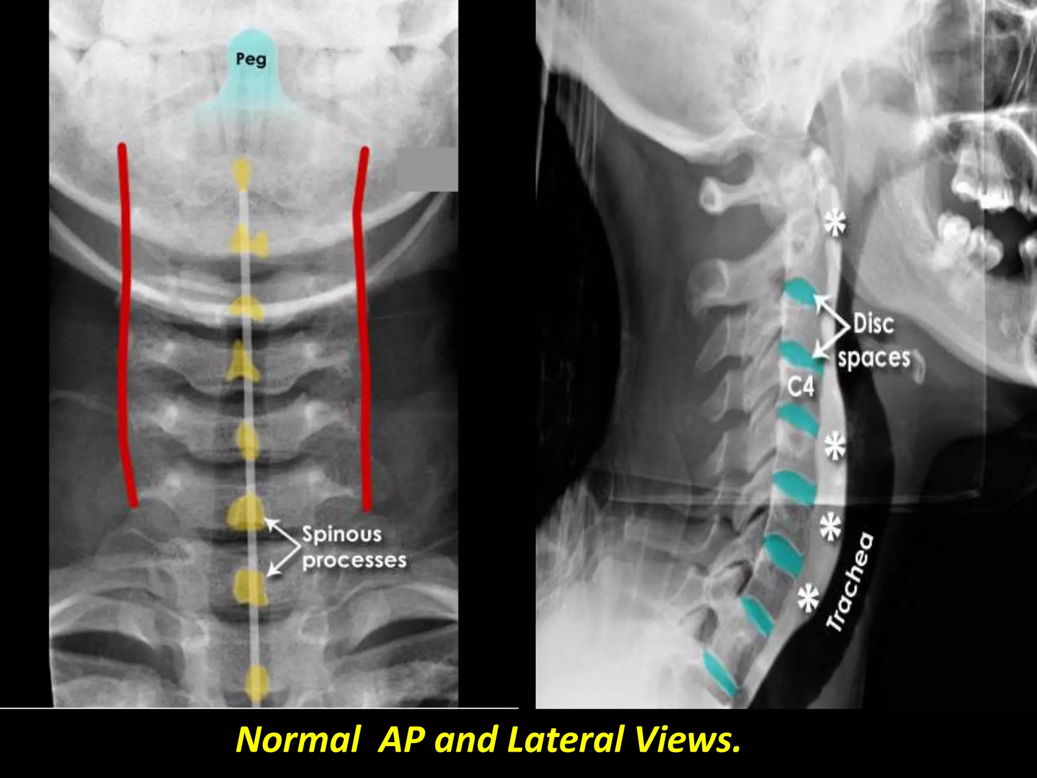
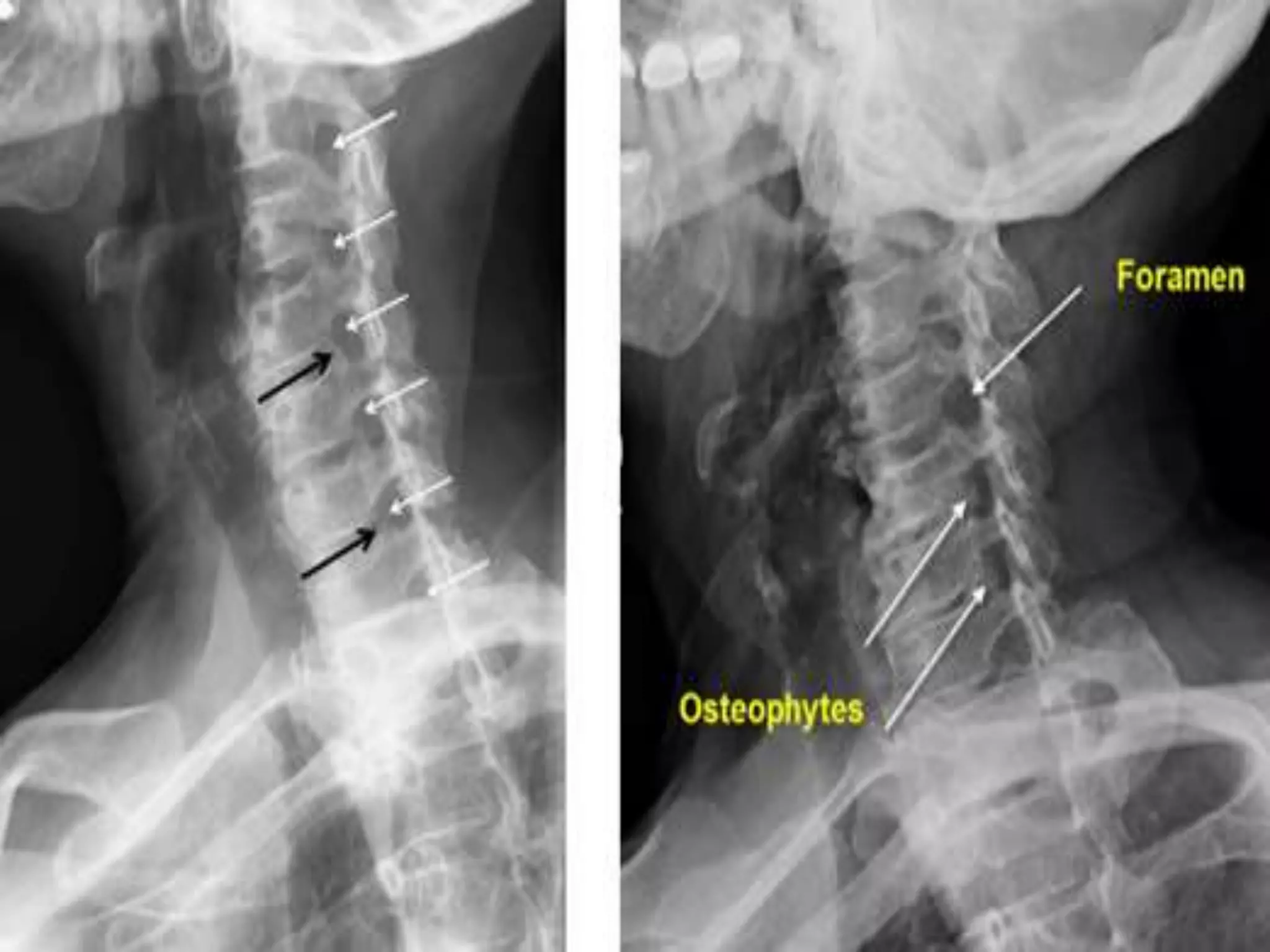
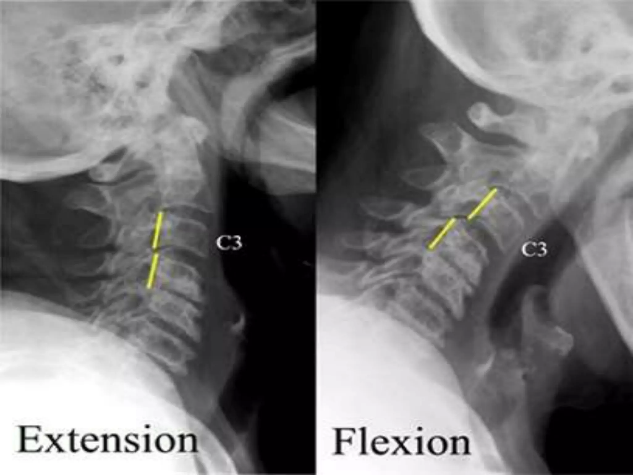
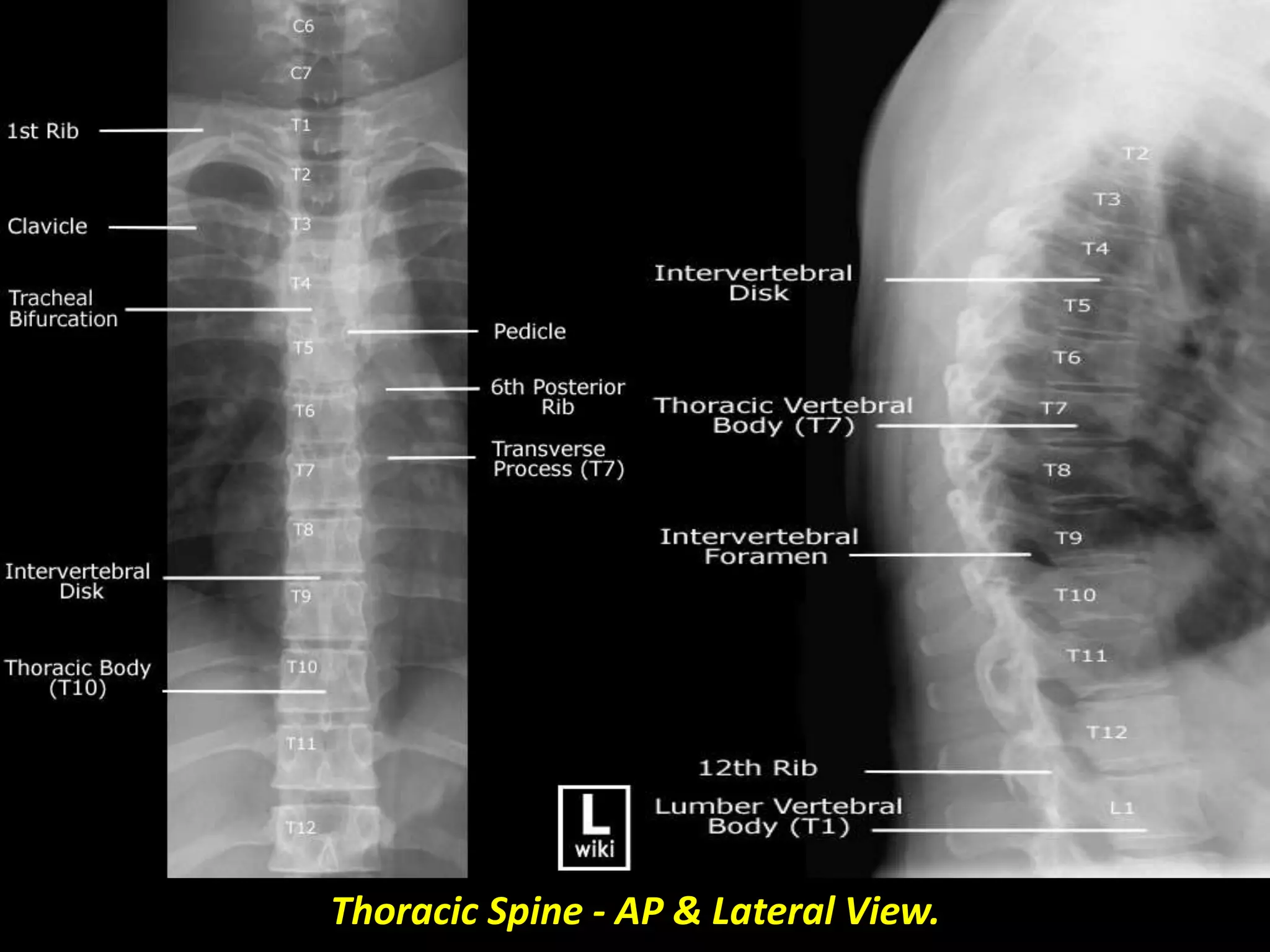
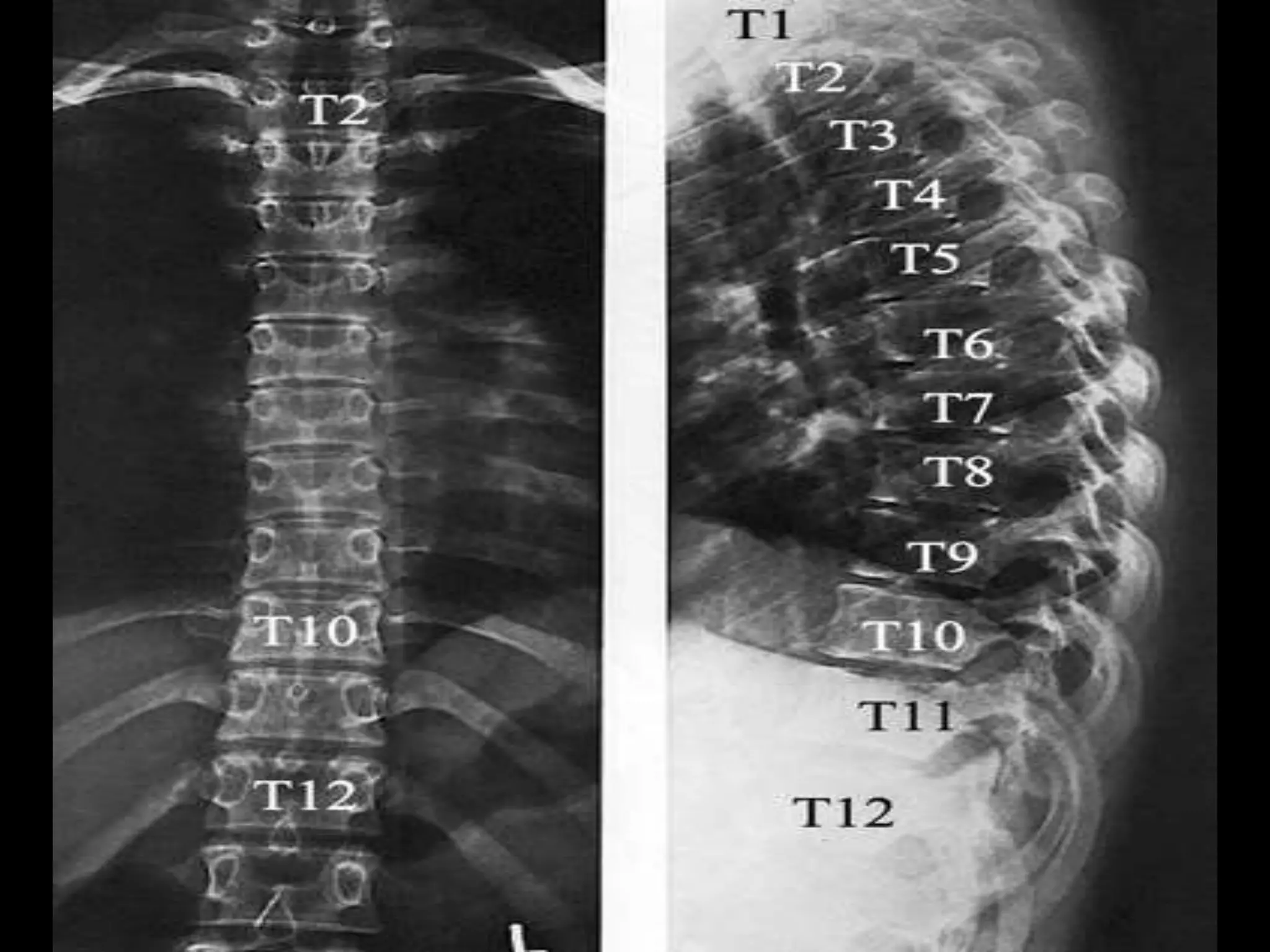
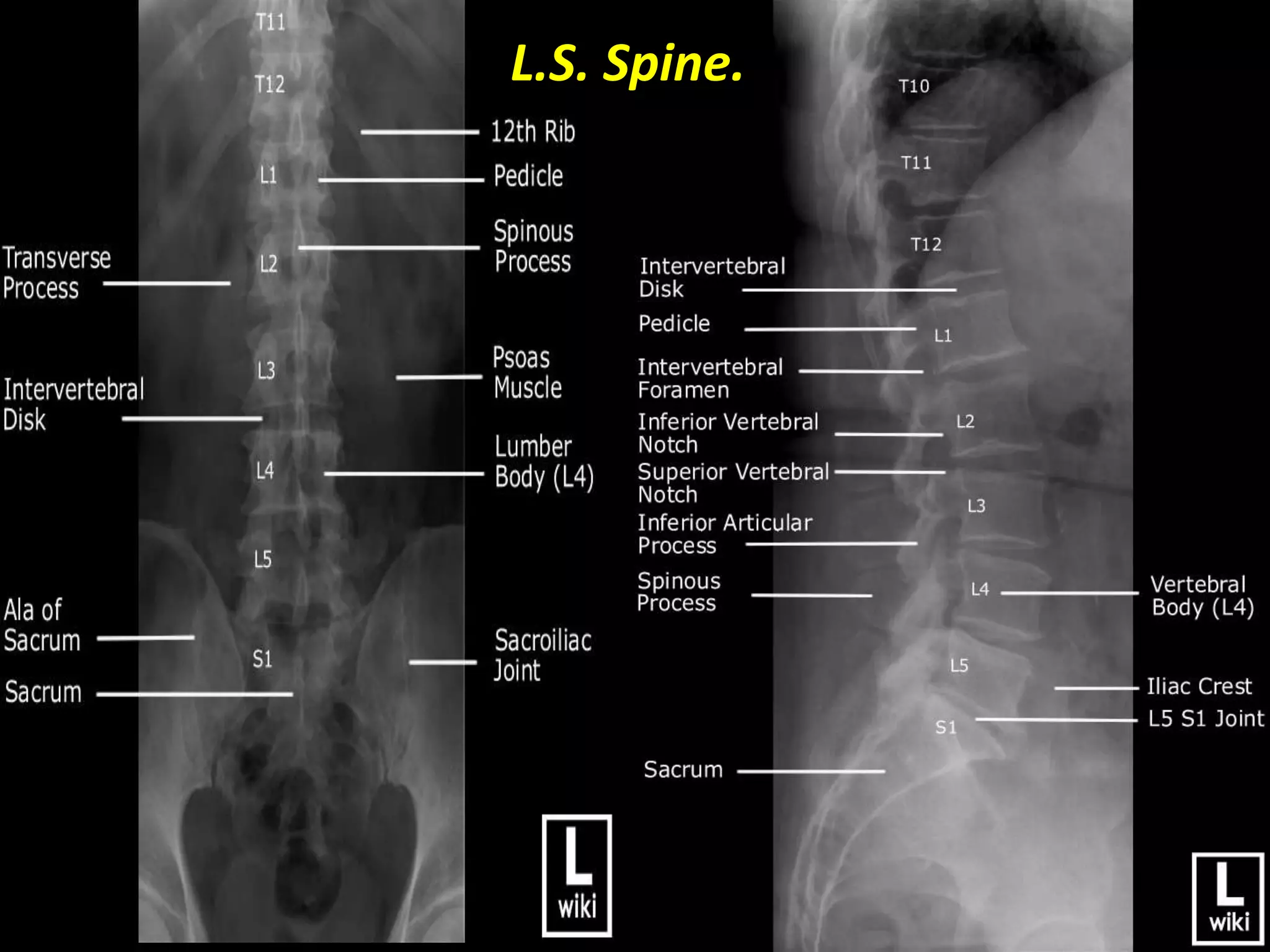
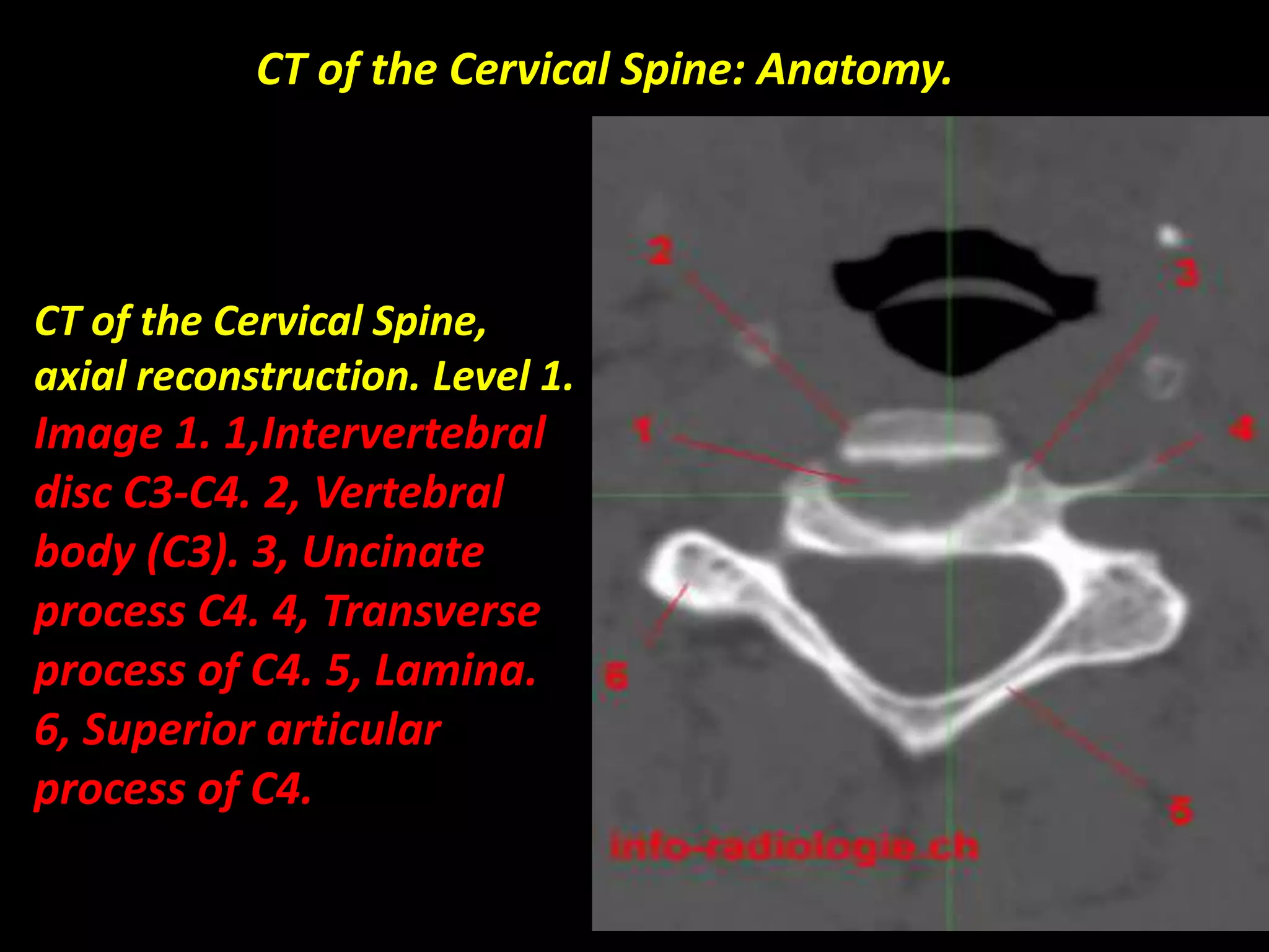
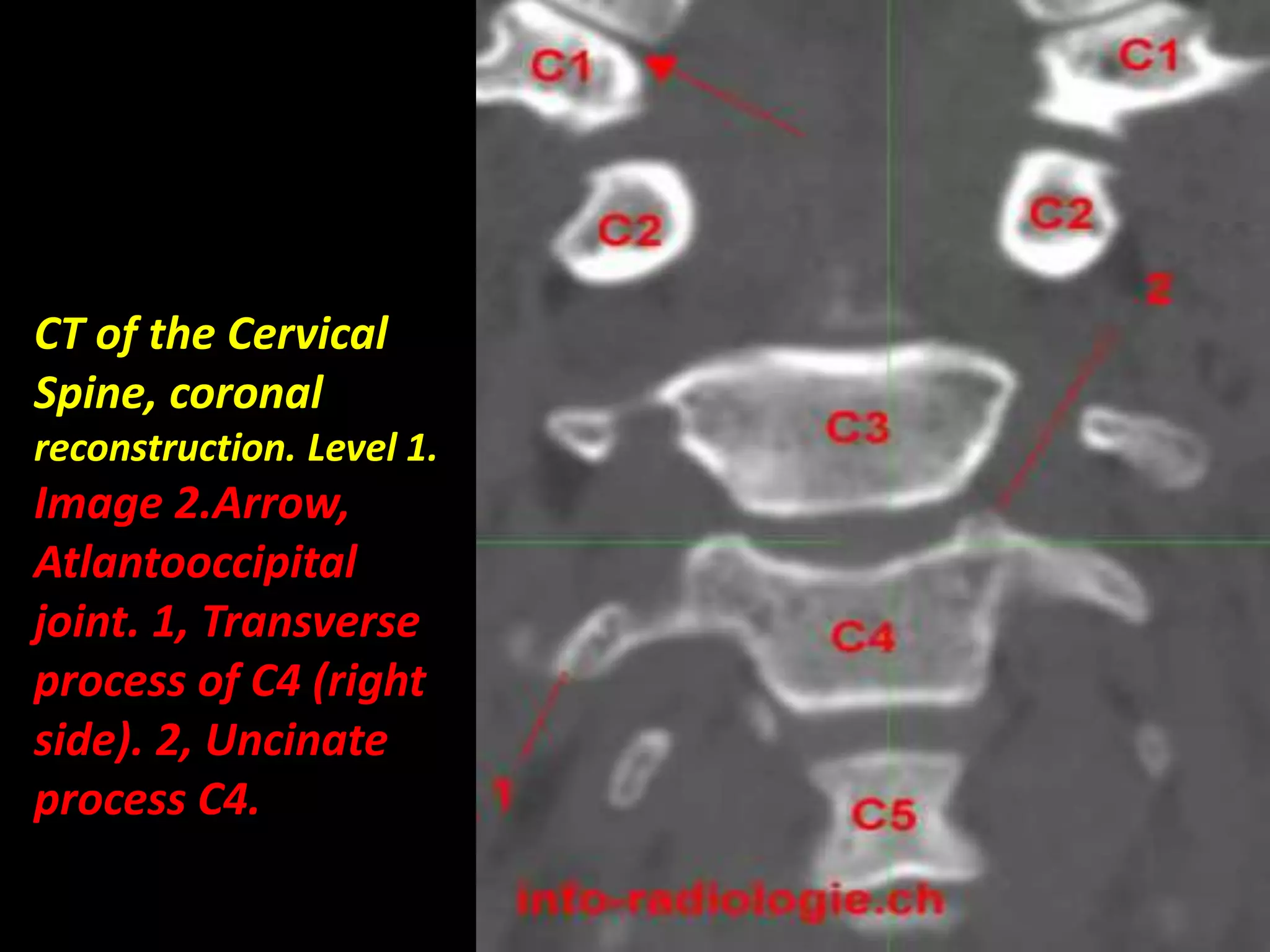
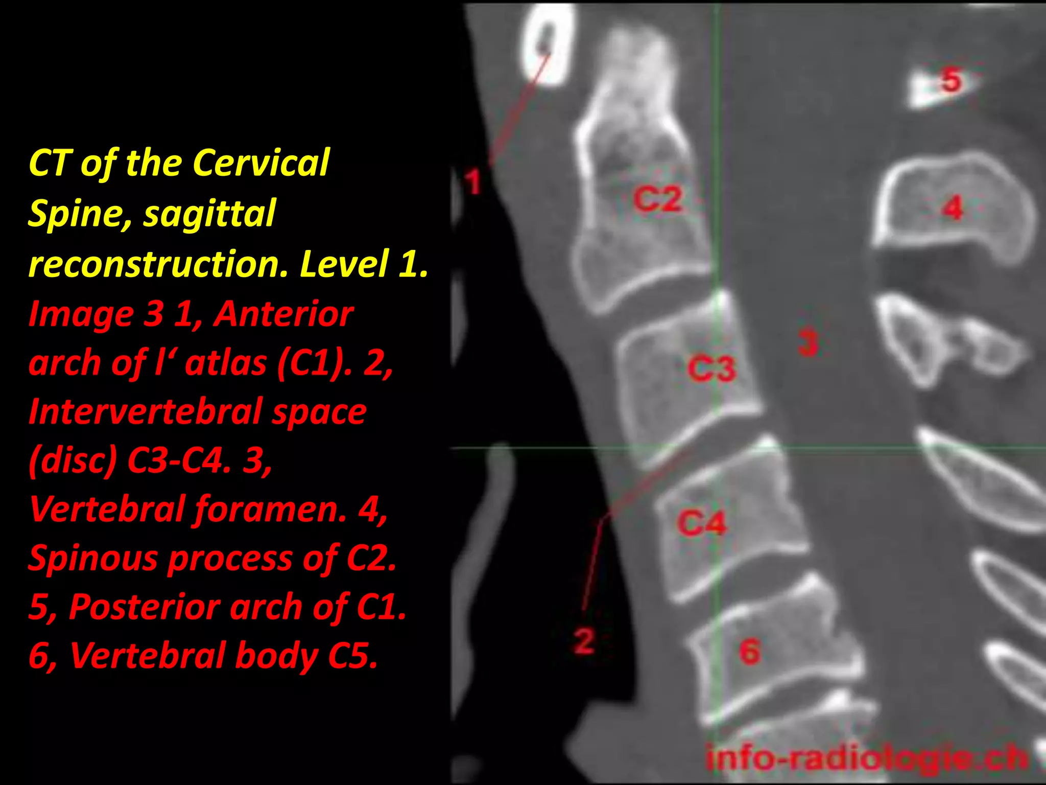
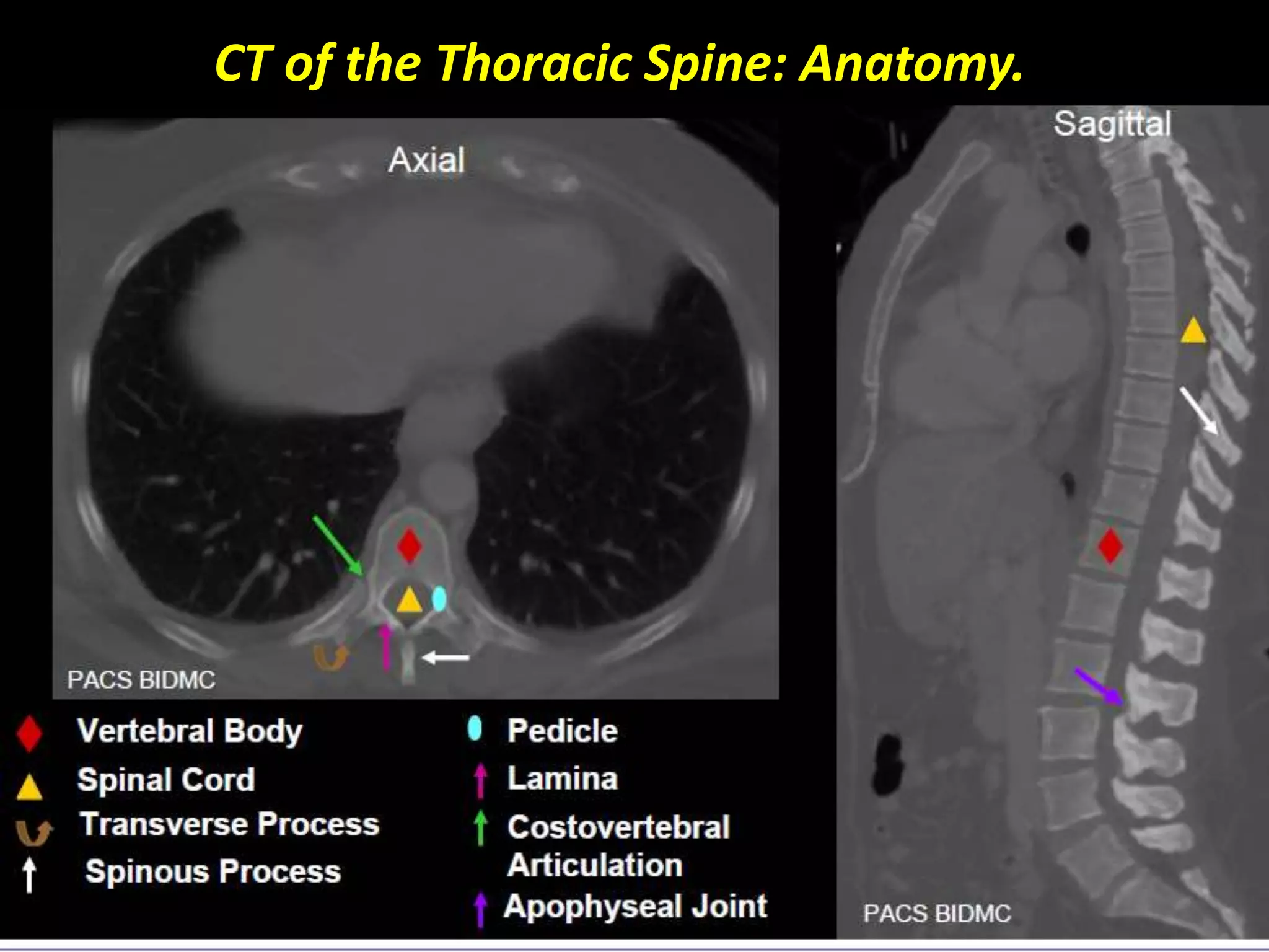
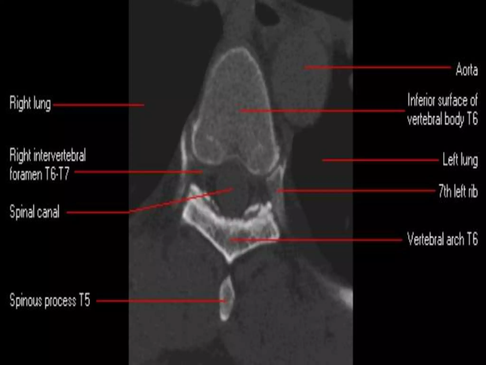
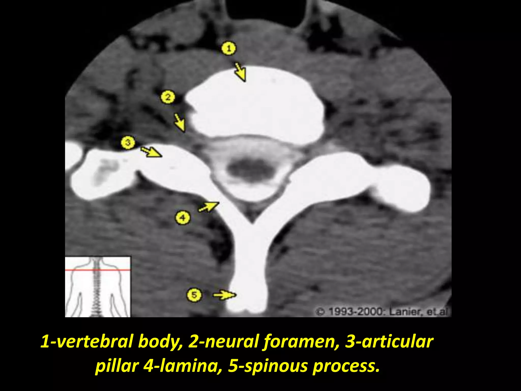
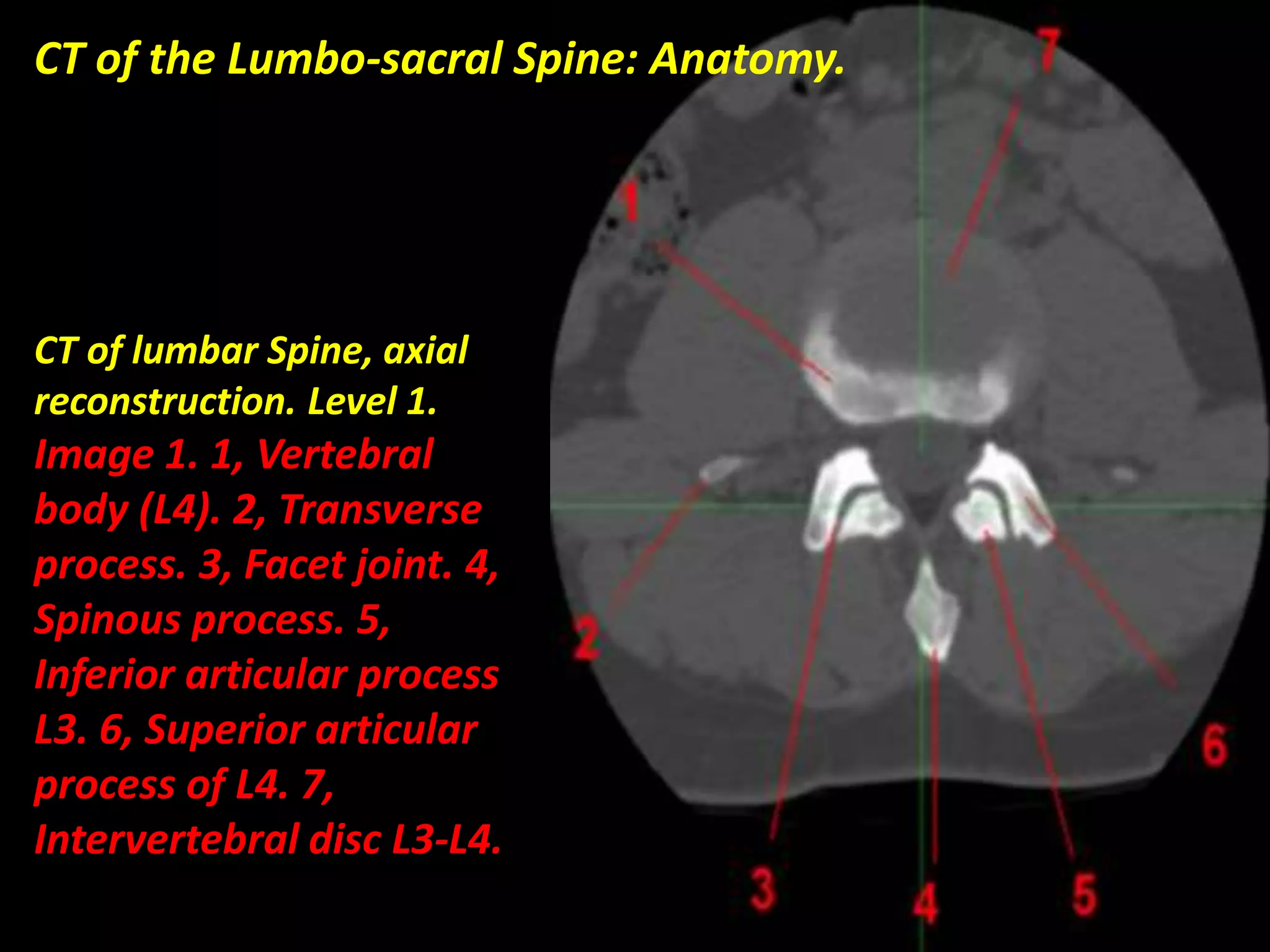
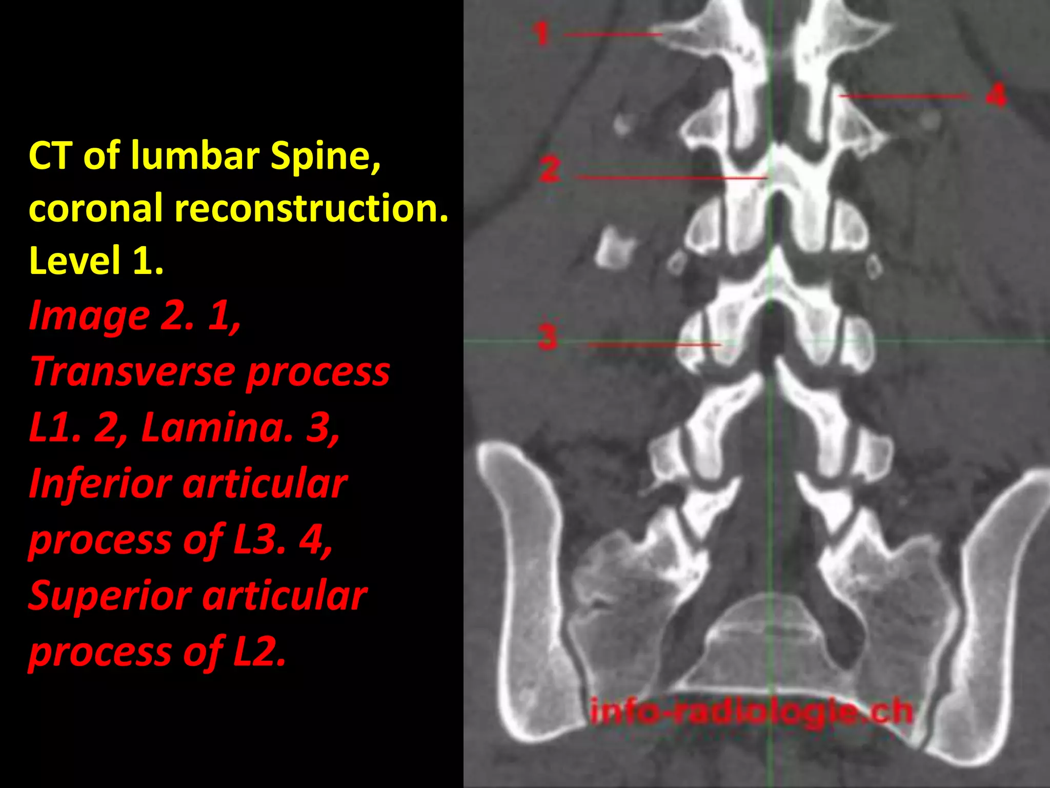
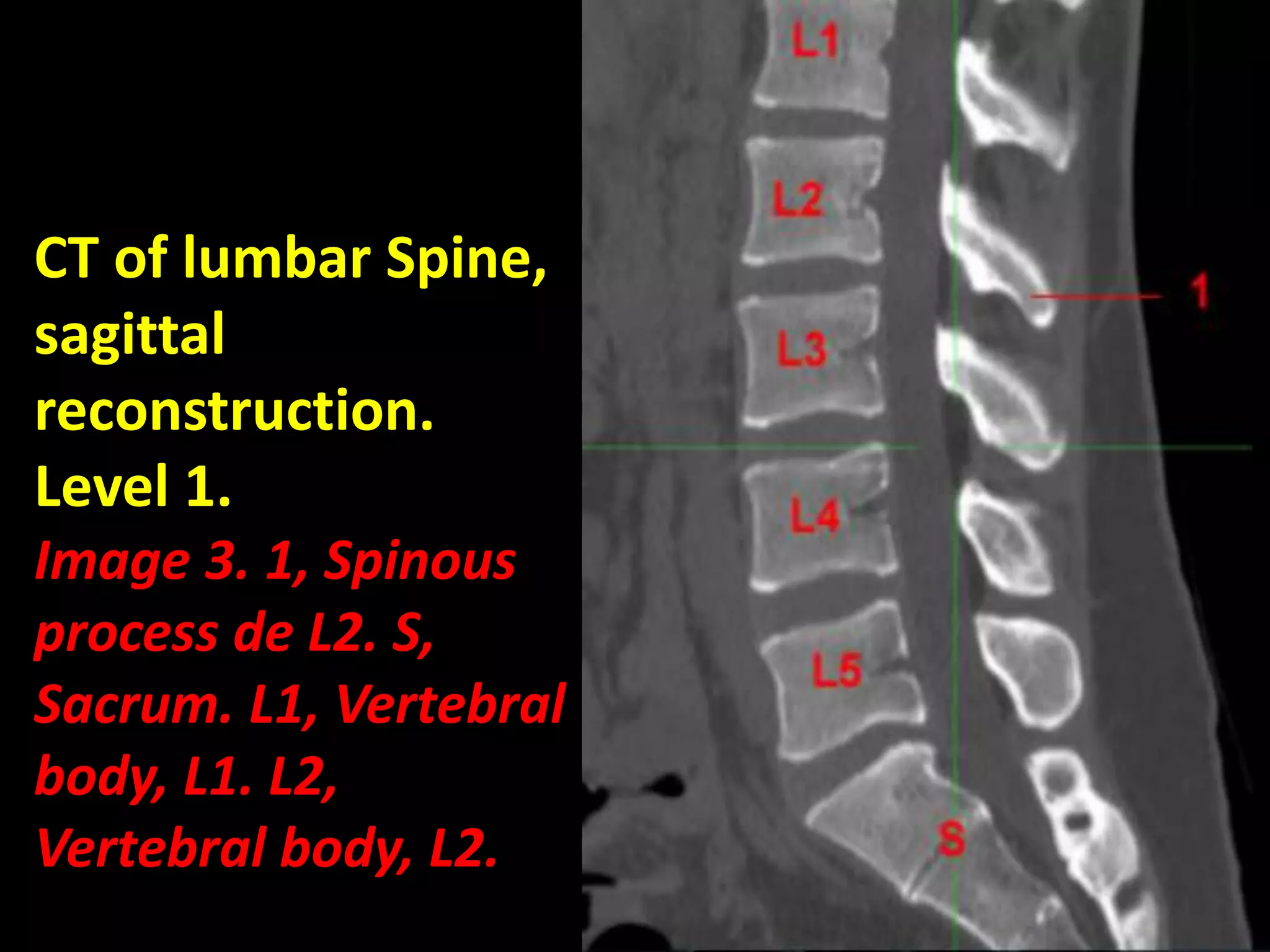
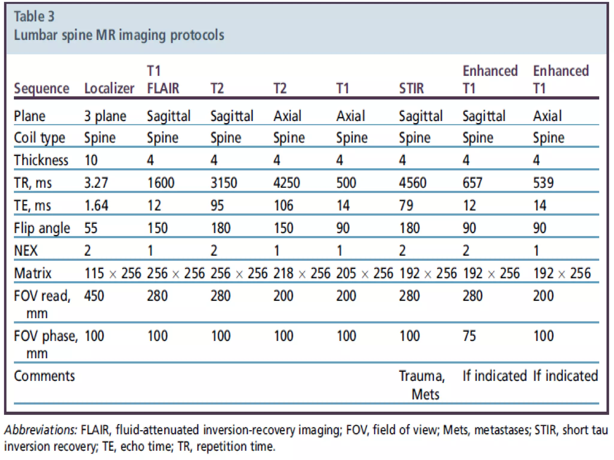
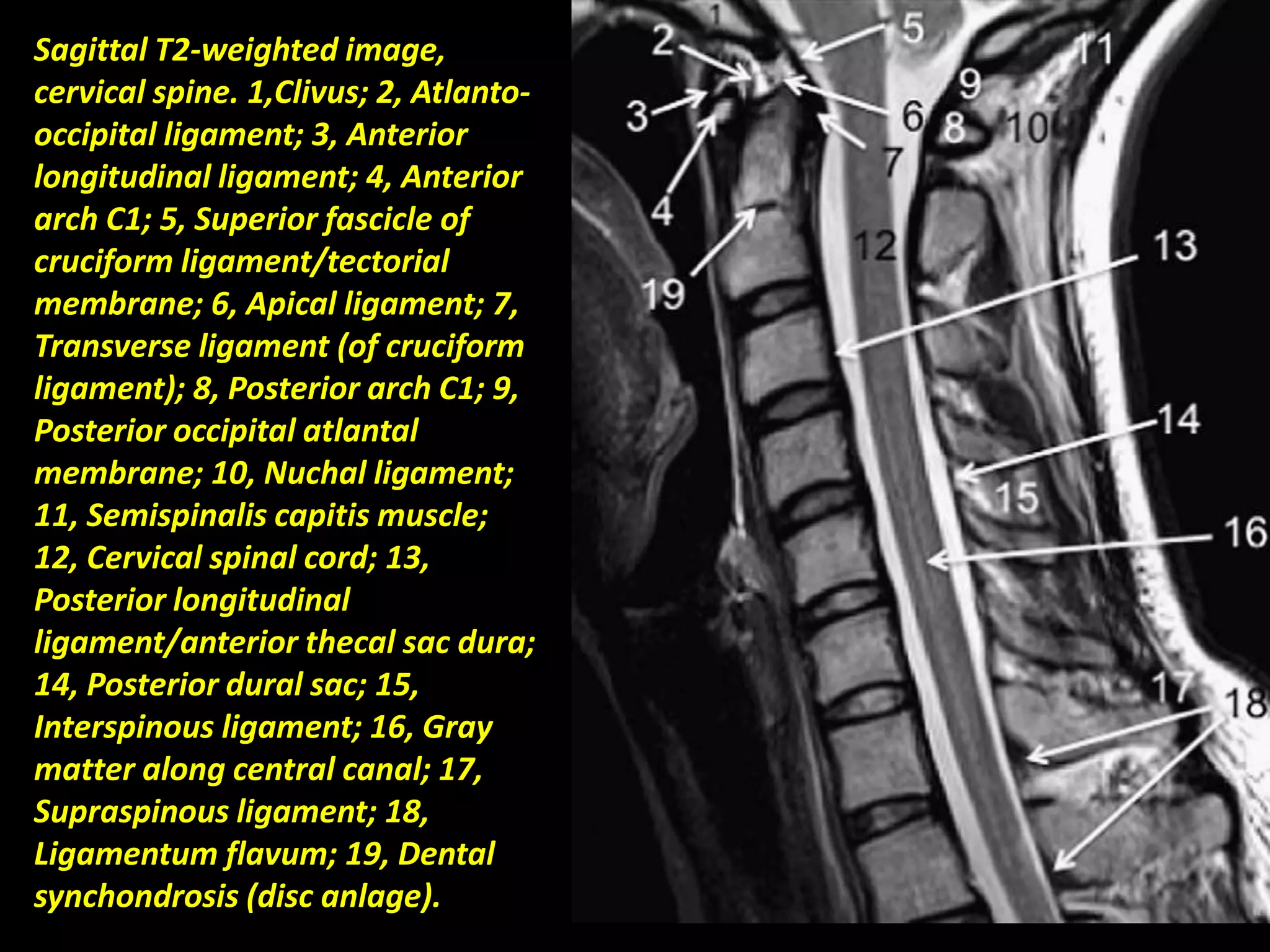
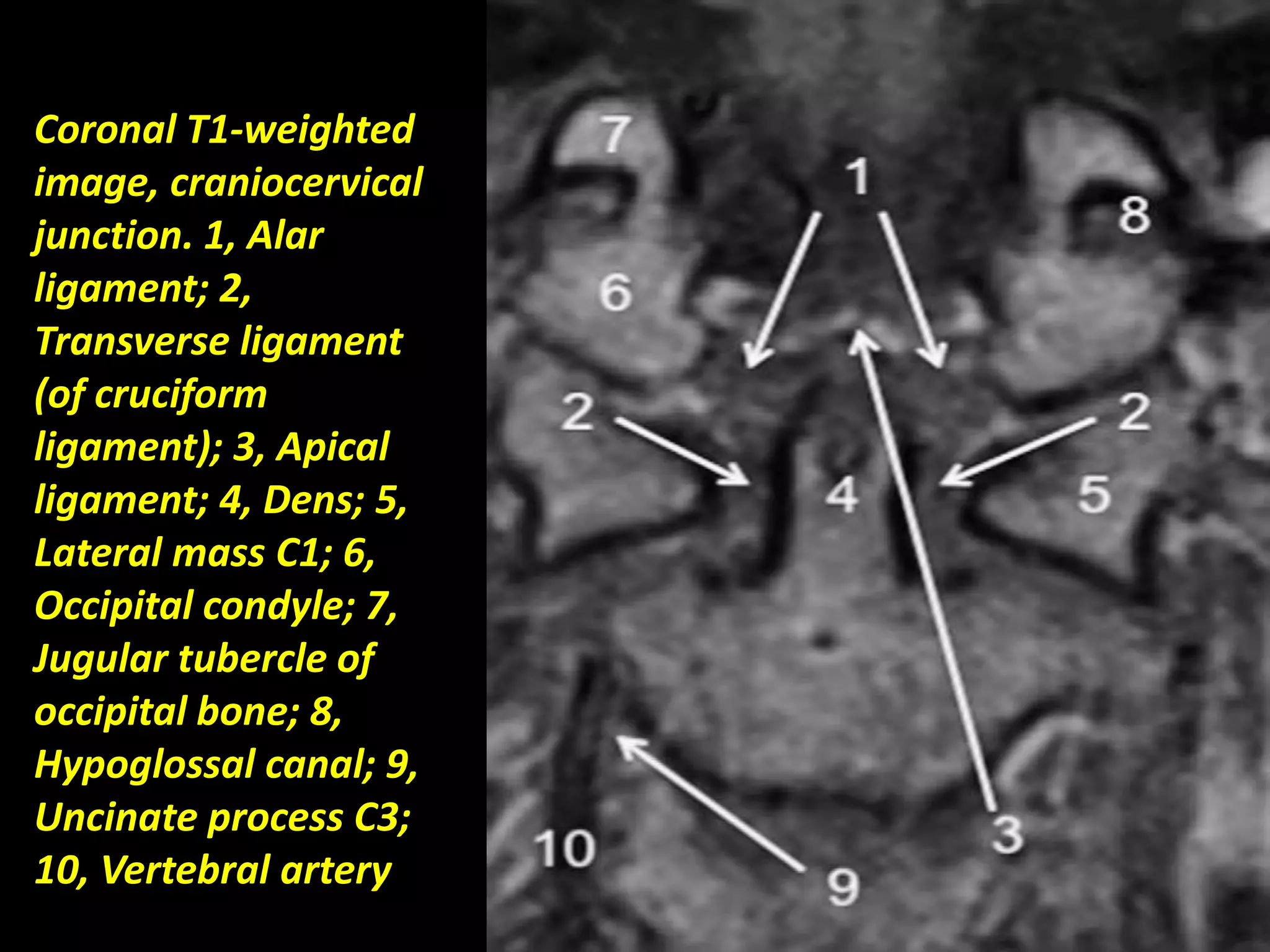
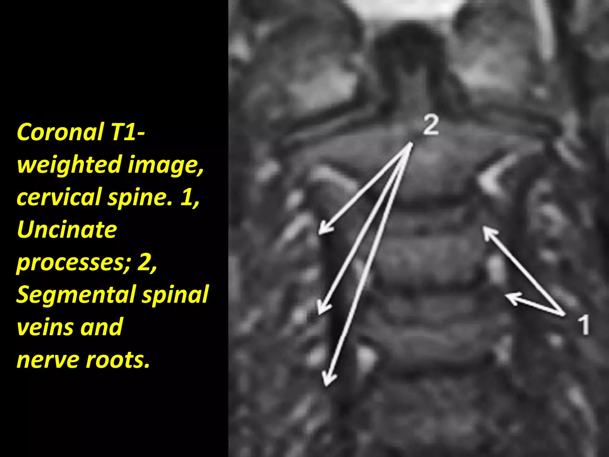
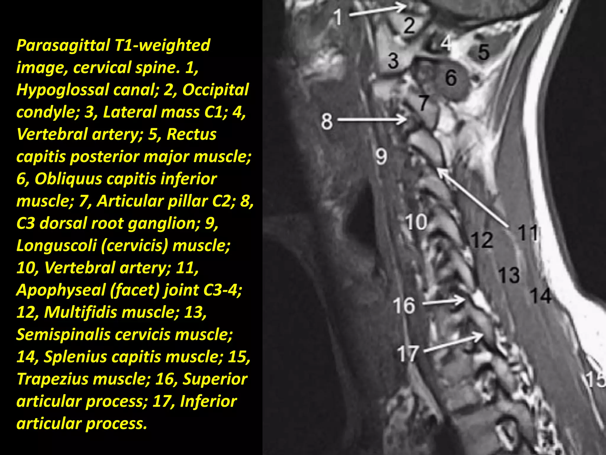
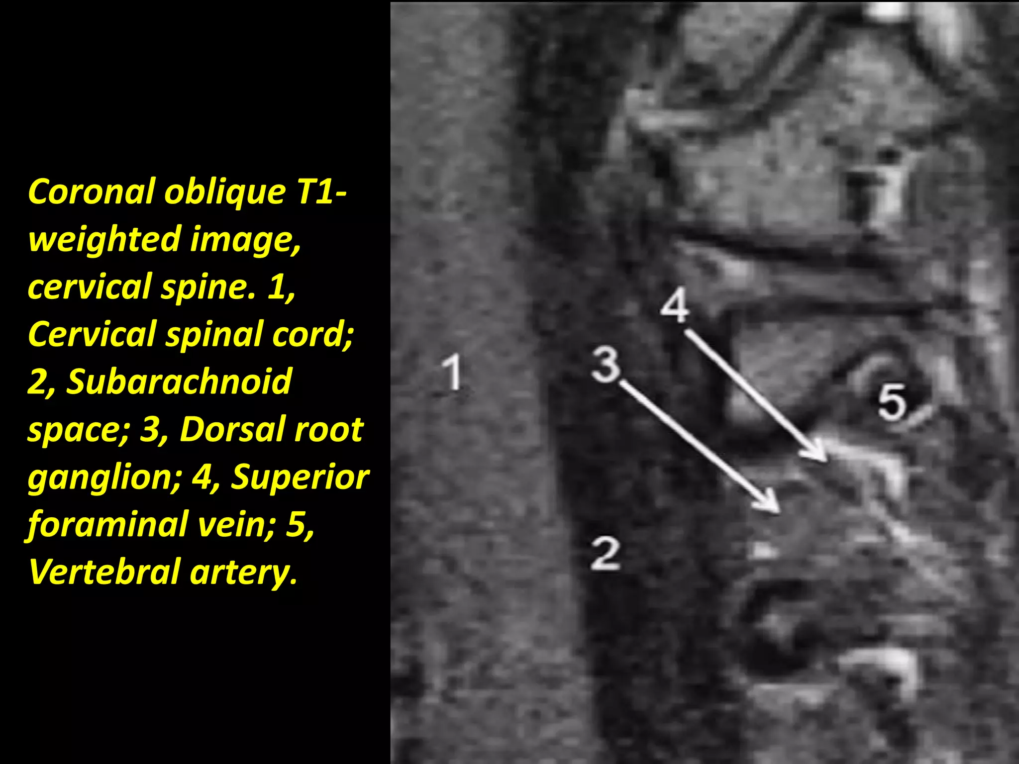

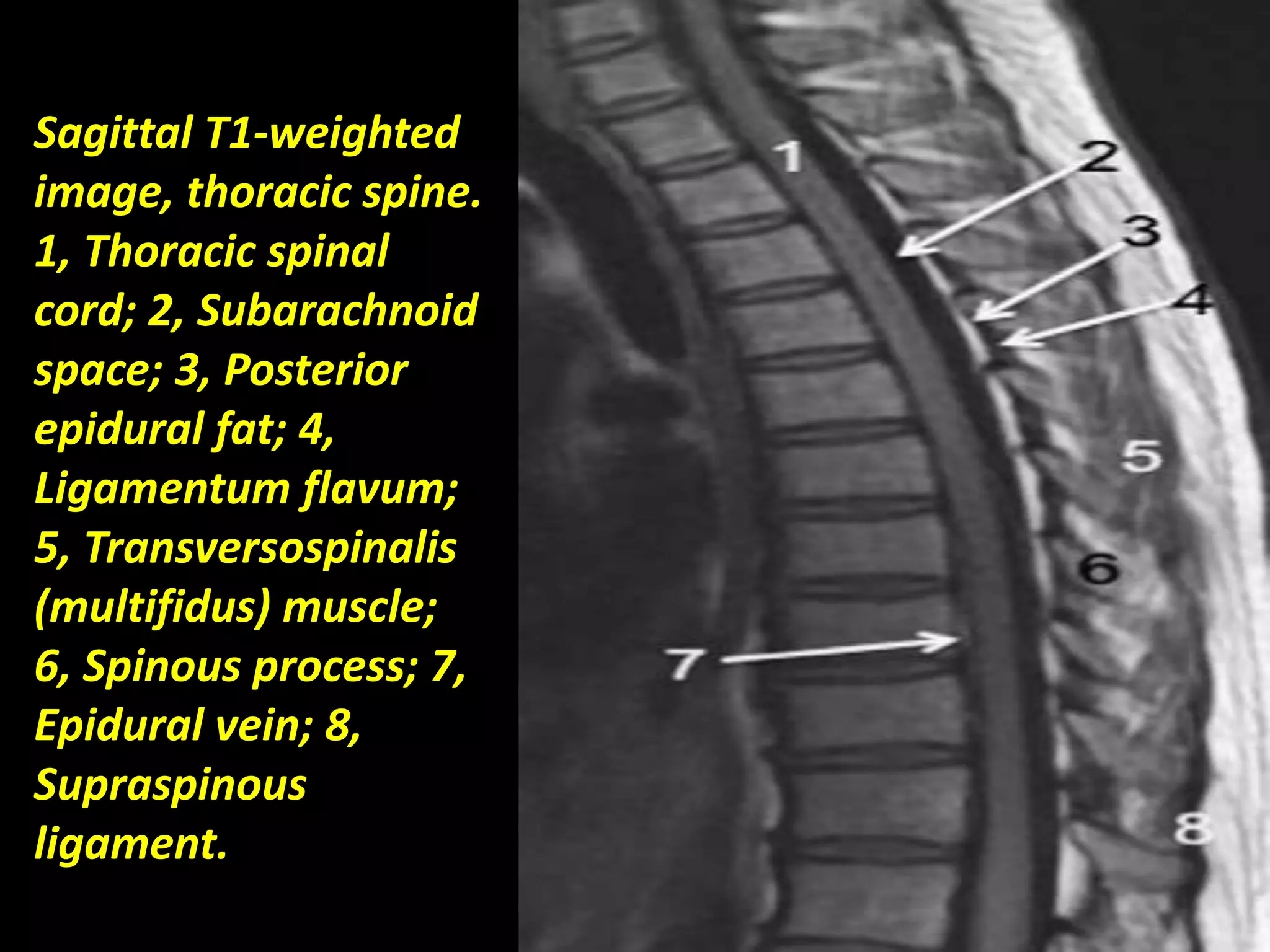
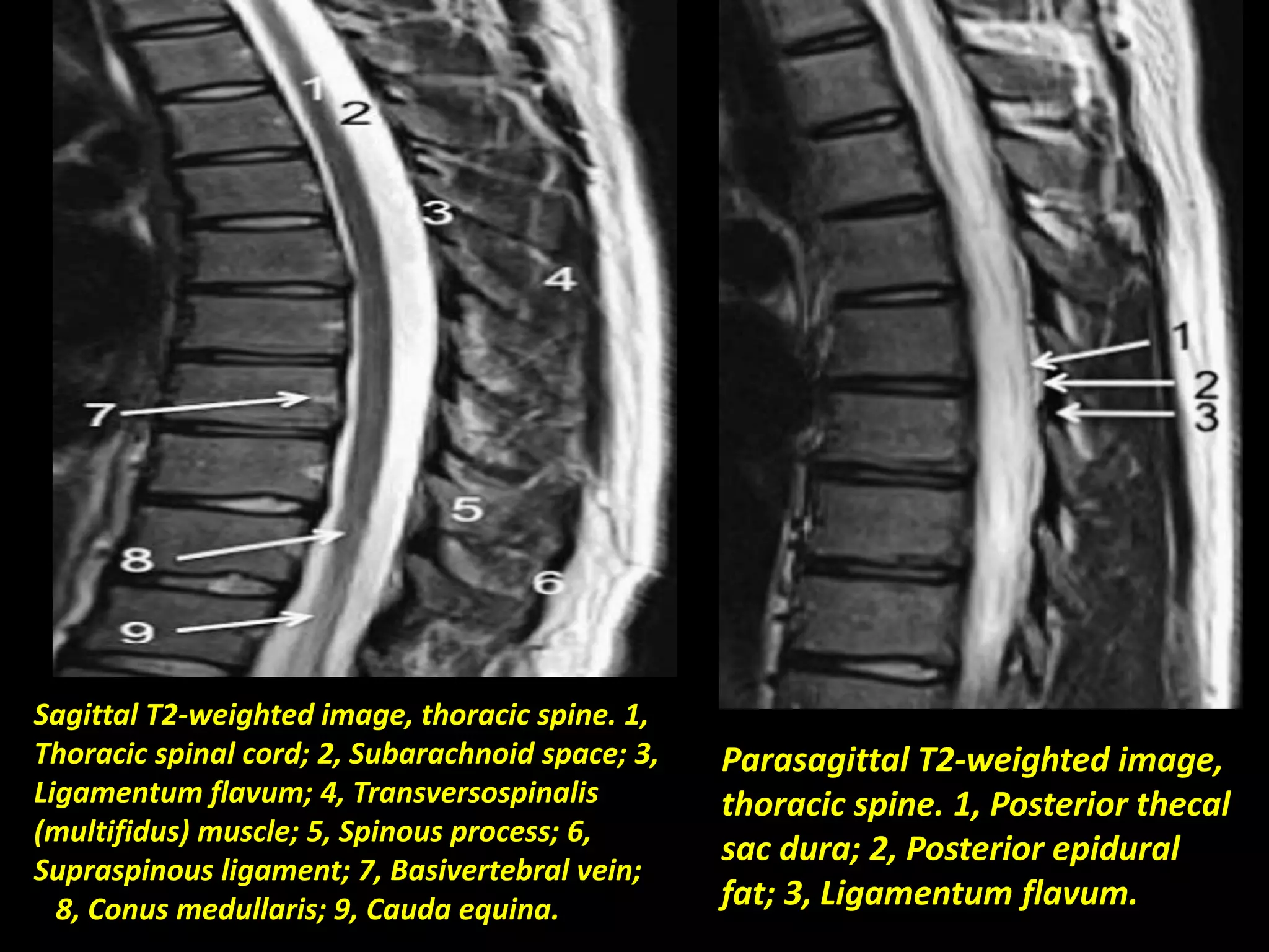
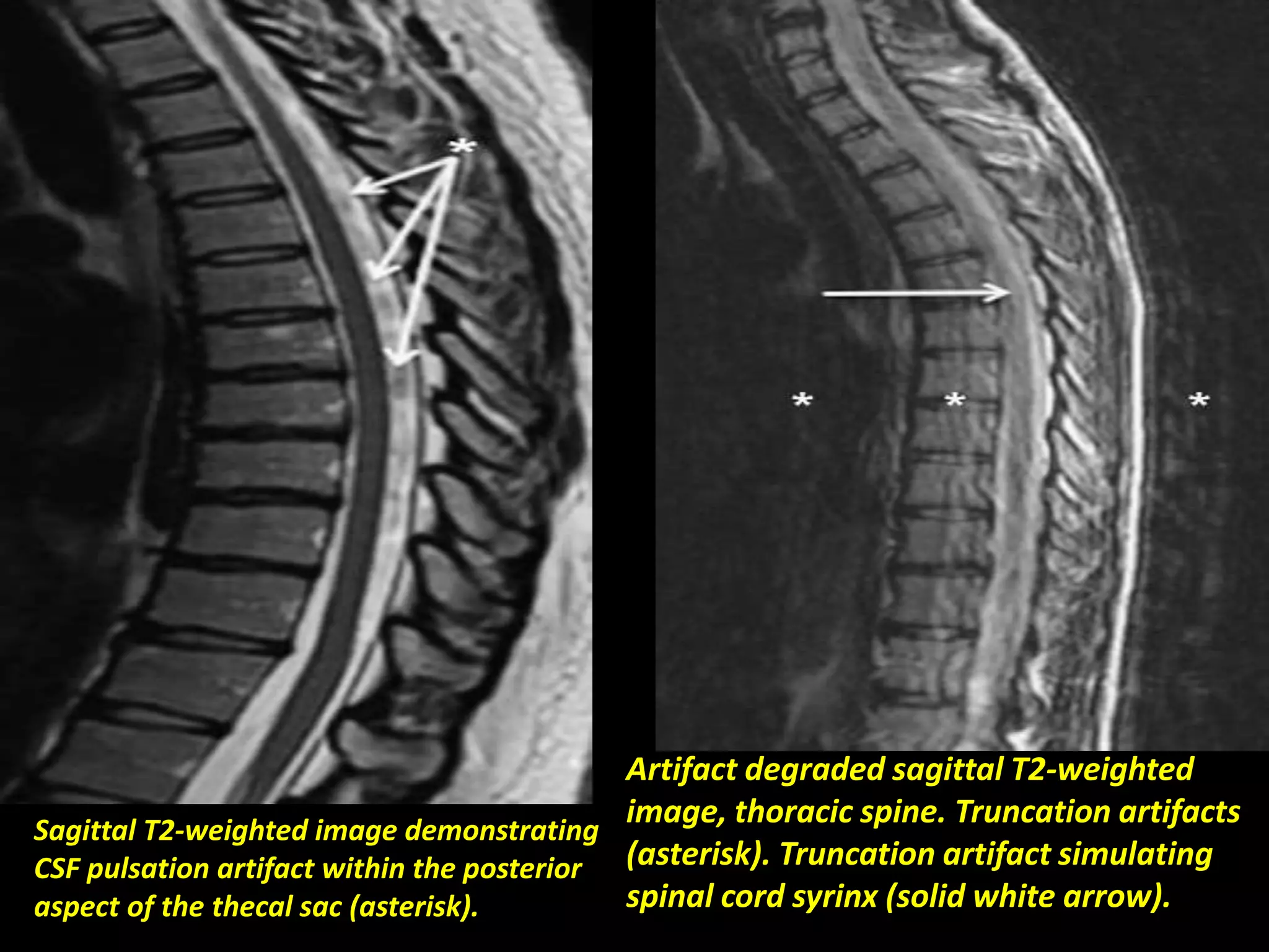
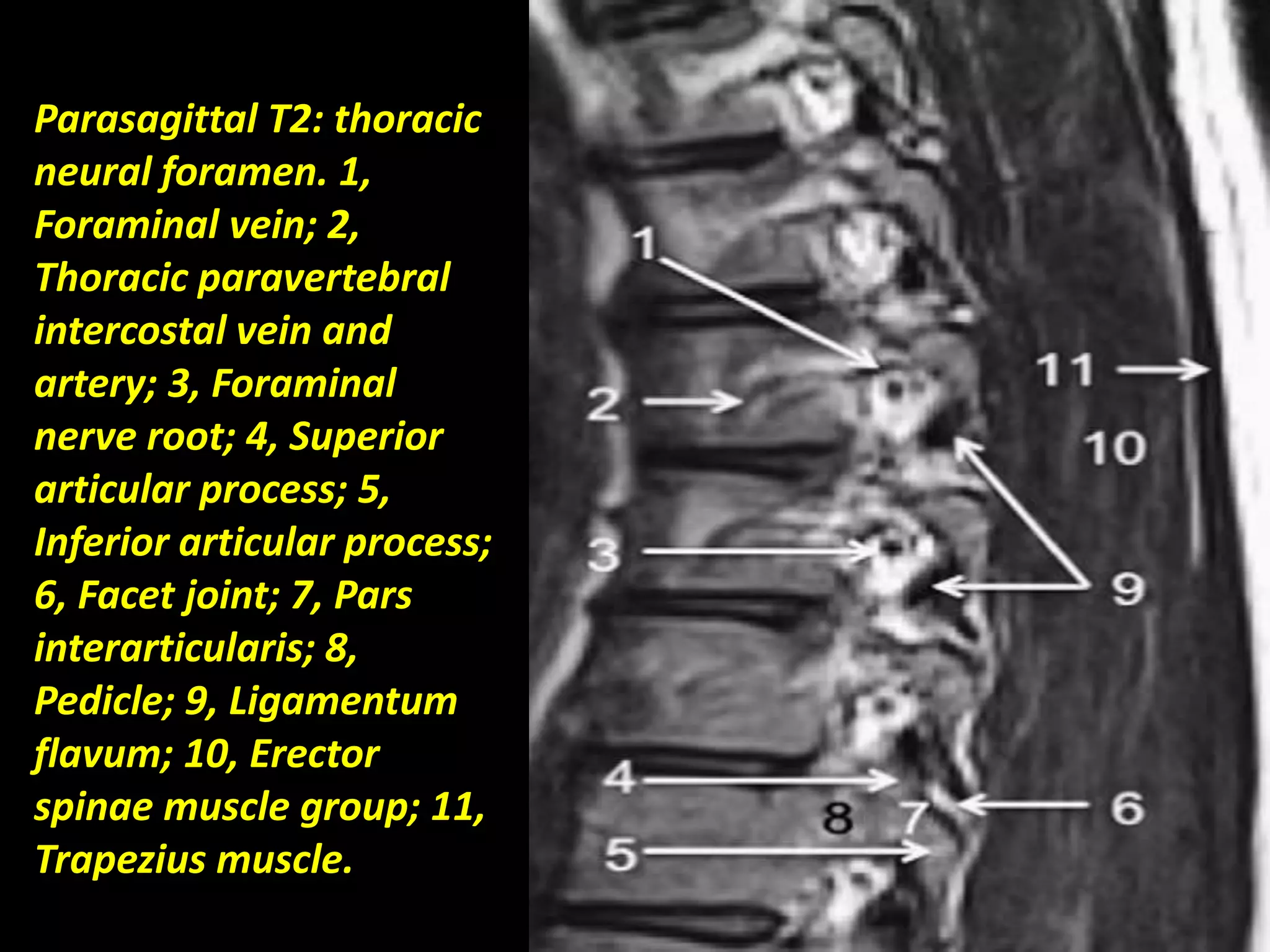
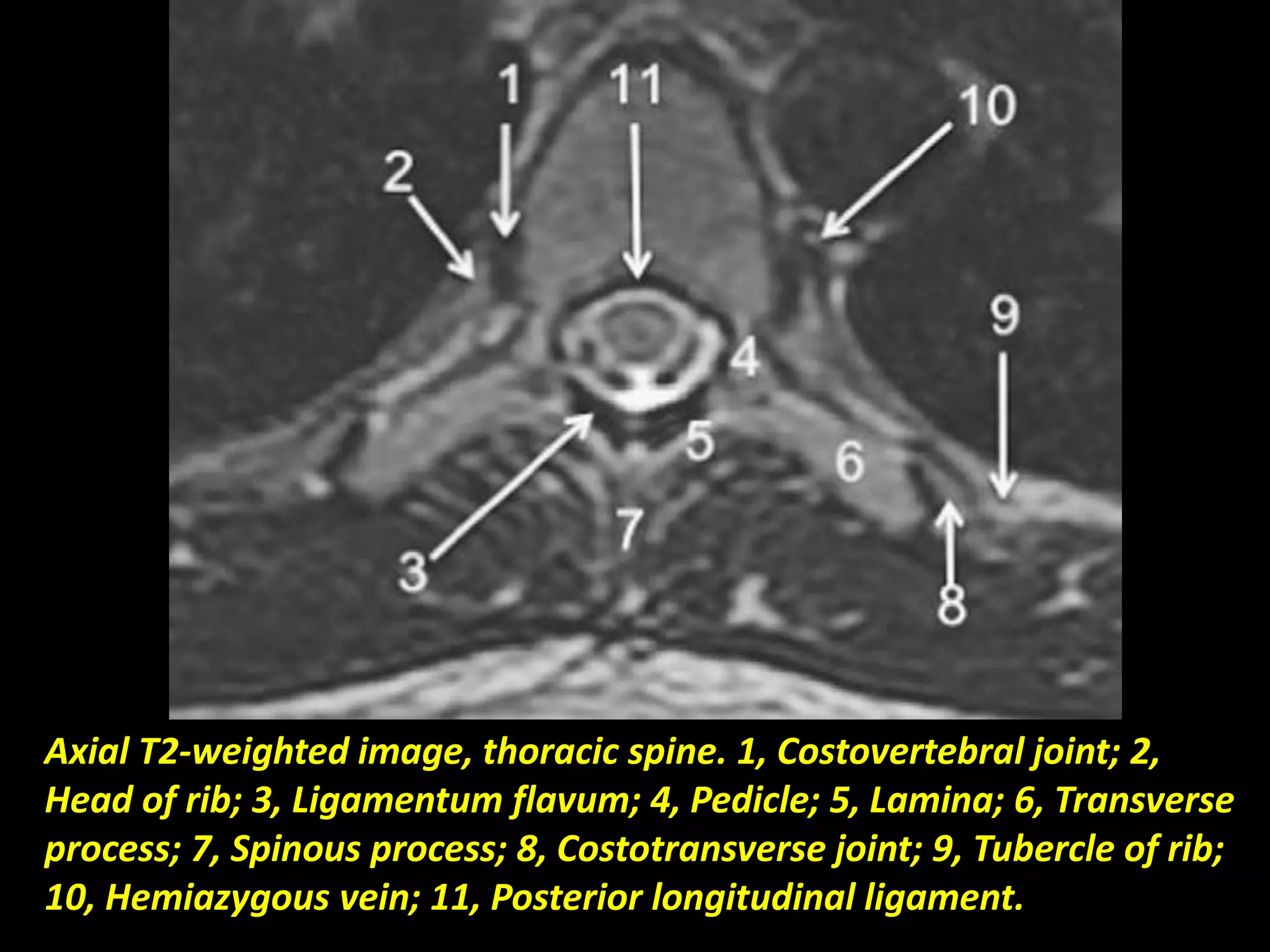
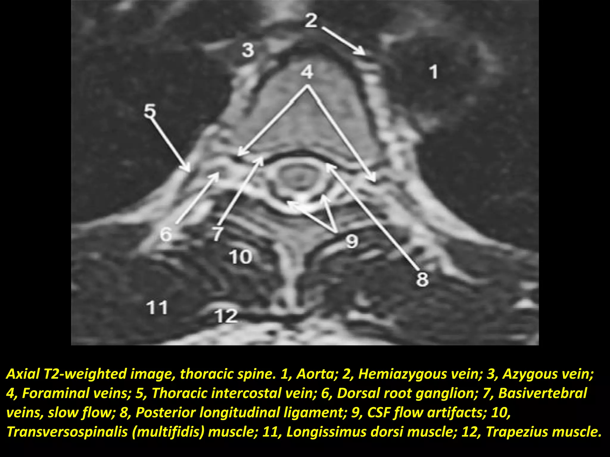
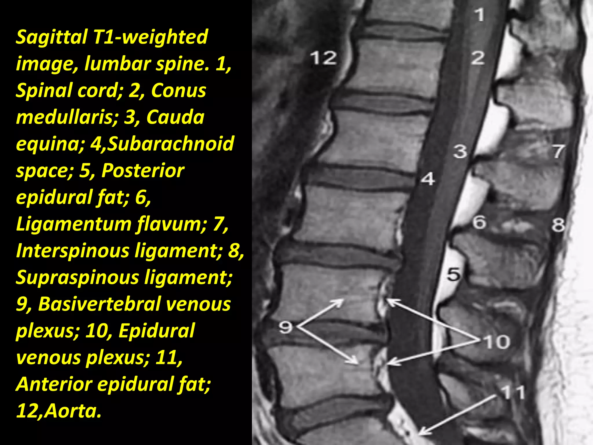
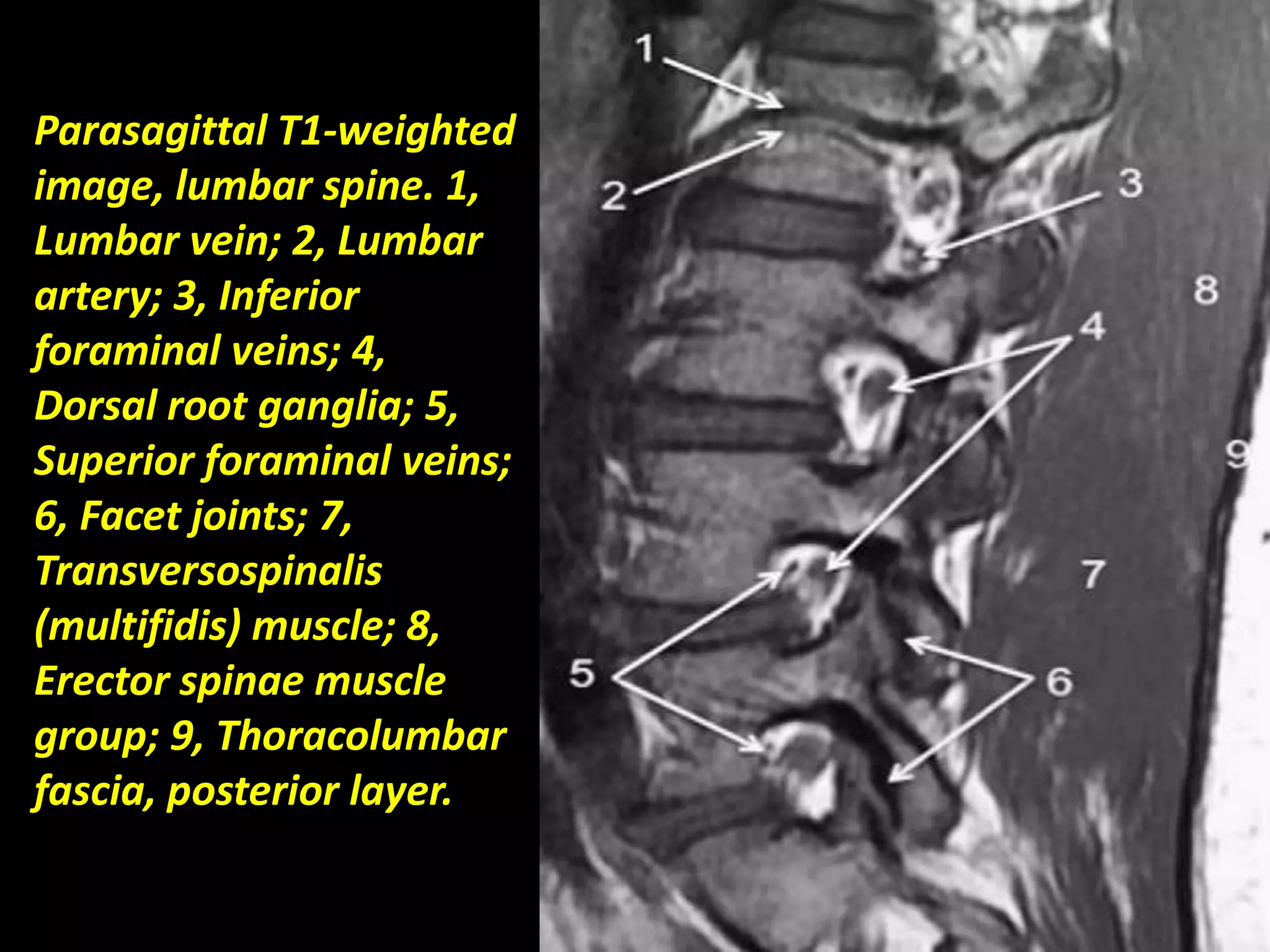
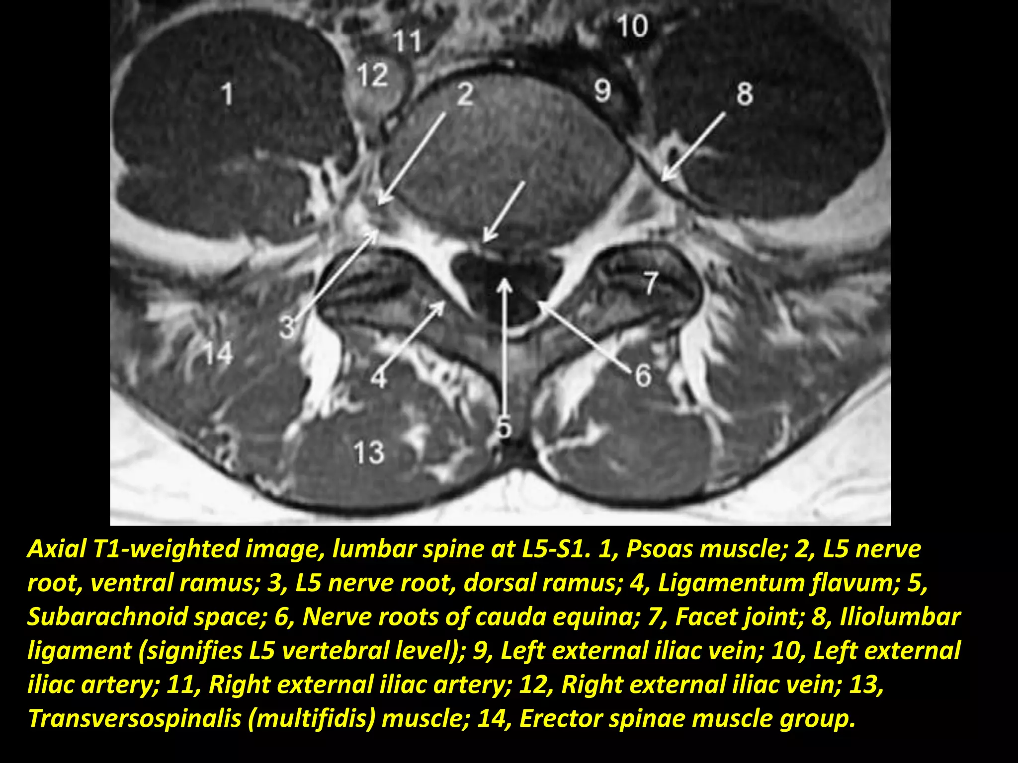


This document provides an overview of radiological anatomy of the spine as seen on different imaging modalities including radiographs, CT, and MRI. It describes normal anatomy of the cervical, thoracic, and lumbar spine in axial, sagittal, and coronal views. Key anatomical structures like vertebrae, discs, ligaments, muscles, and vasculature are labeled on various images. Imaging techniques for MRI of the spine including slice thickness and plane orientations are also discussed.


