SlideShare a Scribd company logo
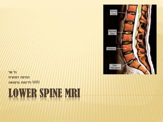
LOWER SPINE MRI
‫שי‬ ‫גל‬
‫רפואית‬ ‫הנדסה‬
MRI‫ברפואה‬ ‫לדימות‬
THINGS WE’LL TALK ABOUT
 Spine Anatomy
 Spine Injuries/Diseases
 MRI Indications
 Lumbar Spine MRI Principles
 No More Time…
SPINE ANATOMY
 8 Cervical
 12 Thoracic
 5 Lumbar
 Sacrum + Coccyx
SPINE ANATOMY
The aorta and vena cava
Separating around the level of
the L3/L4 disc space
Aorta
Vena cava
Iliac arteries
Iliac veins
Midsacral vessels
SPINE ANATOMY
 Lumbar Spine Anatomy
SPINE INJURIES/DISEASES
MRI INDICATIONS
MRI INDICATIONS
MRI‫פחות‬
‫בדימות‬ ‫יעיל‬
‫פגיעות‬ ‫של‬
‫אנטומיות‬
RI PRINCIPLESMPINESUMBARL
 T1- when contrast is dependent on differences
in longitudinal magnetic relaxation times
values between various tissues the image is
called “T1-weighted”.
 T2- when contrast is mostly determined by
differences in transverse magnetic relaxation
values the image is called “T2-weighted”.
RI PRINCIPLESMPINESUMBARL
 T1: T1-weighting is mainly used to produce a
“fat image”.
RI PRINCIPLESMPINESUMBARL
 T2- bright signal intensity of water (CSF,
nucleus pulposus) is mainly seen in images
with T2-weighting, therefor known as “water
images”.
RI PRINCIPLESMPINESUMBARL
 FastSpinEcho vs. ConventionalSpinEcho in T2-
• in a CSE sequence epidural fat & bone marrow fat have low
signal intensity, while FSE produces a much higher fat
signal.
• In FSE T2-weighted images epidural and foraminal fat may
be almost in the same intense as CSF.
• FSE T2-W images provide “water contrast” as well as “fat
contrast”.
RI PRINCIPLESMPINESUMBARL
Cont.
• Difficult to see degenerative changes/
metastases/ osteomyelitis (bone infection)
symptoms.
• Alternative option for “water image” is to use a
T2* -weighted gradient-echo (GRE) sequence
(but mostly for cervical area).
RI PRINCIPLESMPINESUMBARL
 How to reduce fat from our picture-
• Short TI inversion recovery (STIR).
• Spectral fat suppression by pre-saturation (SPIR).
RI PRINCIPLESMPINESUMBARL
 Imaging Planes-
generally, in sectional imaging, anatomic surfaces/
structures are best imaged in a plane which lies
perpendicular to the surface of interest.
• Sagittal: Images in this plane are best for demonstrating
disc herniations, mid-sagittal diameter of the spinal canal,
increase or decrease in sagittal diameter of the spinal
canal.
axial
RI PRINCIPLESMPINESUMBARL
• Axial: extent of a disc abnormality, lateral recesses of the spinal
canal are best studied in the axial plane, The foramen and its
contents can be studied in axial images (less well than in the
sagittal plane).
• Coronal: This imaging plane is used only rarely in diagnosis of
degenerative disease. Some spinal deformities such as
scoliosis or hemivertebra are imaged best in the coronal plane.
RI PRINCIPLESMPINESUMBARL
Fat areas
Fat, vascular,
infected areas
Liquid areas
Summary
Bye Bye

More Related Content

What's hot

Med 4 ortho spinal cases
Med 4 ortho spinal casesMed 4 ortho spinal cases
Med 4 ortho spinal cases
Luke Oakden-Rayner
 
X-ray spine
X-ray spineX-ray spine
MRI OF SHOULDER INJURY
MRI OF SHOULDER INJURYMRI OF SHOULDER INJURY
MRI OF SHOULDER INJURY
Krishna Kiran Karanth
 
Seminar new
Seminar newSeminar new
Seminar new
Ganesh Gadag
 
Presentation1, radiological imaging of shoulder dislocation.
Presentation1, radiological imaging of shoulder dislocation.Presentation1, radiological imaging of shoulder dislocation.
Presentation1, radiological imaging of shoulder dislocation.
Abdellah Nazeer
 
MRI-Spine/Pelvis_Biancz_Noveno
MRI-Spine/Pelvis_Biancz_NovenoMRI-Spine/Pelvis_Biancz_Noveno
MRI-Spine/Pelvis_Biancz_NovenoBiancz Noveno
 
Radiology of the spine and musc final 2012
Radiology of the spine and musc final 2012Radiology of the spine and musc final 2012
Radiology of the spine and musc final 2012
Reina Ramesh
 
Mri shoulder joint with common pathologies
Mri shoulder joint with common pathologiesMri shoulder joint with common pathologies
Mri shoulder joint with common pathologies
Gobardhan Thapa
 
MRI SPINE ANATOMY
MRI SPINE ANATOMYMRI SPINE ANATOMY
MRI SPINE ANATOMY
NeurologyKota
 
Spine radiography
Spine radiographySpine radiography
Spine radiography
Dr. Muhammad Bin Zulfiqar
 
Presentation1.pptx, normal spinal anatomy.
Presentation1.pptx, normal spinal anatomy.Presentation1.pptx, normal spinal anatomy.
Presentation1.pptx, normal spinal anatomy.Abdellah Nazeer
 
MRI LSS
MRI LSSMRI LSS
CT Cervical Spine
CT Cervical SpineCT Cervical Spine
CT Cervical Spine
Dr. Yash Kumar Achantani
 
Spine mri bir 2 copy
Spine mri bir 2 copySpine mri bir 2 copy
Spine mri bir 2 copy
Winston Rennie
 
MRI slides, second yr students 2008
MRI slides, second yr students 2008MRI slides, second yr students 2008
MRI slides, second yr students 2008
Harp
 
Mri knee
Mri kneeMri knee
PATHOLOGICAL INVESTIGATIONS AND IMAGING TECHNIQUES IN NEUROMUSCULAR DISORDERS...
PATHOLOGICAL INVESTIGATIONS AND IMAGING TECHNIQUES IN NEUROMUSCULAR DISORDERS...PATHOLOGICAL INVESTIGATIONS AND IMAGING TECHNIQUES IN NEUROMUSCULAR DISORDERS...
PATHOLOGICAL INVESTIGATIONS AND IMAGING TECHNIQUES IN NEUROMUSCULAR DISORDERS...
shuchij10
 
RADIOGRAPHY OF CHEST AND SPINE
RADIOGRAPHY OF CHEST AND SPINERADIOGRAPHY OF CHEST AND SPINE
RADIOGRAPHY OF CHEST AND SPINE
kunalj000
 
Us and mri shoulder us 20140611 webversie
Us and mri shoulder us 20140611 webversieUs and mri shoulder us 20140611 webversie
Us and mri shoulder us 20140611 webversieartejee
 

What's hot (20)

Med 4 ortho spinal cases
Med 4 ortho spinal casesMed 4 ortho spinal cases
Med 4 ortho spinal cases
 
X-ray spine
X-ray spineX-ray spine
X-ray spine
 
MRI OF SHOULDER INJURY
MRI OF SHOULDER INJURYMRI OF SHOULDER INJURY
MRI OF SHOULDER INJURY
 
Seminar new
Seminar newSeminar new
Seminar new
 
Presentation1, radiological imaging of shoulder dislocation.
Presentation1, radiological imaging of shoulder dislocation.Presentation1, radiological imaging of shoulder dislocation.
Presentation1, radiological imaging of shoulder dislocation.
 
MRI-Spine/Pelvis_Biancz_Noveno
MRI-Spine/Pelvis_Biancz_NovenoMRI-Spine/Pelvis_Biancz_Noveno
MRI-Spine/Pelvis_Biancz_Noveno
 
Radiology of the spine and musc final 2012
Radiology of the spine and musc final 2012Radiology of the spine and musc final 2012
Radiology of the spine and musc final 2012
 
Mri shoulder joint with common pathologies
Mri shoulder joint with common pathologiesMri shoulder joint with common pathologies
Mri shoulder joint with common pathologies
 
MRI SPINE ANATOMY
MRI SPINE ANATOMYMRI SPINE ANATOMY
MRI SPINE ANATOMY
 
MR maging In Orthopaedics
MR maging In OrthopaedicsMR maging In Orthopaedics
MR maging In Orthopaedics
 
Spine radiography
Spine radiographySpine radiography
Spine radiography
 
Presentation1.pptx, normal spinal anatomy.
Presentation1.pptx, normal spinal anatomy.Presentation1.pptx, normal spinal anatomy.
Presentation1.pptx, normal spinal anatomy.
 
MRI LSS
MRI LSSMRI LSS
MRI LSS
 
CT Cervical Spine
CT Cervical SpineCT Cervical Spine
CT Cervical Spine
 
Spine mri bir 2 copy
Spine mri bir 2 copySpine mri bir 2 copy
Spine mri bir 2 copy
 
MRI slides, second yr students 2008
MRI slides, second yr students 2008MRI slides, second yr students 2008
MRI slides, second yr students 2008
 
Mri knee
Mri kneeMri knee
Mri knee
 
PATHOLOGICAL INVESTIGATIONS AND IMAGING TECHNIQUES IN NEUROMUSCULAR DISORDERS...
PATHOLOGICAL INVESTIGATIONS AND IMAGING TECHNIQUES IN NEUROMUSCULAR DISORDERS...PATHOLOGICAL INVESTIGATIONS AND IMAGING TECHNIQUES IN NEUROMUSCULAR DISORDERS...
PATHOLOGICAL INVESTIGATIONS AND IMAGING TECHNIQUES IN NEUROMUSCULAR DISORDERS...
 
RADIOGRAPHY OF CHEST AND SPINE
RADIOGRAPHY OF CHEST AND SPINERADIOGRAPHY OF CHEST AND SPINE
RADIOGRAPHY OF CHEST AND SPINE
 
Us and mri shoulder us 20140611 webversie
Us and mri shoulder us 20140611 webversieUs and mri shoulder us 20140611 webversie
Us and mri shoulder us 20140611 webversie
 

Viewers also liked

Mri – spine
Mri – spineMri – spine
Mri – spine
vinod naneria
 
Nomenclature in spine mri
Nomenclature in spine mriNomenclature in spine mri
Nomenclature in spine mriHieder Al-Shami
 
BASICS OF MRI
BASICS OF MRIBASICS OF MRI
BASICS OF MRI
Krishna Kiran Karanth
 
Biomechanics of lumbar spine
Biomechanics of lumbar spineBiomechanics of lumbar spine
Biomechanics of lumbar spineVenus Pagare
 
Mri cervical & dorsal spine
Mri cervical & dorsal spineMri cervical & dorsal spine
Mri cervical & dorsal spine
Mohamed Shaaban
 
Anatomy of Vertebral Column
Anatomy of Vertebral ColumnAnatomy of Vertebral Column
Anatomy of Vertebral Column
Sharmin Susiwala
 
Anatomy of spinal cord
Anatomy of spinal cordAnatomy of spinal cord
Anatomy of spinal cordMBBS IMS MSU
 
Spinal anatomy and biomechanics
Spinal anatomy and biomechanicsSpinal anatomy and biomechanics
Spinal anatomy and biomechanicsAmbrish Verma
 
X ray c-spine
X ray c-spine X ray c-spine
X ray c-spine
Rajaoct
 
Low back pain...2
Low back pain...2Low back pain...2
Low back pain...2
Pranav Khawale
 
Appropriate imaging for low back pain
Appropriate imaging for low back painAppropriate imaging for low back pain
Appropriate imaging for low back pain
SpinePlus
 
Imaging of spinal trauma
Imaging of spinal traumaImaging of spinal trauma
Imaging of spinal traumaVishal Sankpal
 
Biomechanics of spine
Biomechanics of  spineBiomechanics of  spine
Biomechanics of spine
Muhammad Mustaqeem
 
Development Of Vertebral Column
Development Of Vertebral ColumnDevelopment Of Vertebral Column
Development Of Vertebral ColumnAnan
 
Lec 1 biomechanics of the spine
Lec 1 biomechanics of the spineLec 1 biomechanics of the spine
Lec 1 biomechanics of the spine
madiha123anees
 
MRI of Knee joint-- hossam massoud
MRI of Knee joint-- hossam massoudMRI of Knee joint-- hossam massoud
MRI of Knee joint-- hossam massoud
Hossam Massoud
 
Assessment of cervical spine
Assessment of cervical spineAssessment of cervical spine
Assessment of cervical spine
khushali52
 

Viewers also liked (19)

Mri – spine
Mri – spineMri – spine
Mri – spine
 
Nomenclature in spine mri
Nomenclature in spine mriNomenclature in spine mri
Nomenclature in spine mri
 
Anatomy of spine
Anatomy of spineAnatomy of spine
Anatomy of spine
 
BASICS OF MRI
BASICS OF MRIBASICS OF MRI
BASICS OF MRI
 
Biomechanics of lumbar spine
Biomechanics of lumbar spineBiomechanics of lumbar spine
Biomechanics of lumbar spine
 
Mri cervical & dorsal spine
Mri cervical & dorsal spineMri cervical & dorsal spine
Mri cervical & dorsal spine
 
Anatomy of Vertebral Column
Anatomy of Vertebral ColumnAnatomy of Vertebral Column
Anatomy of Vertebral Column
 
Anatomy of spinal cord
Anatomy of spinal cordAnatomy of spinal cord
Anatomy of spinal cord
 
Spinal anatomy and biomechanics
Spinal anatomy and biomechanicsSpinal anatomy and biomechanics
Spinal anatomy and biomechanics
 
X ray c-spine
X ray c-spine X ray c-spine
X ray c-spine
 
Low back pain...2
Low back pain...2Low back pain...2
Low back pain...2
 
Appropriate imaging for low back pain
Appropriate imaging for low back painAppropriate imaging for low back pain
Appropriate imaging for low back pain
 
Imaging of spinal trauma
Imaging of spinal traumaImaging of spinal trauma
Imaging of spinal trauma
 
Biomechanics of spine
Biomechanics of  spineBiomechanics of  spine
Biomechanics of spine
 
Development Of Vertebral Column
Development Of Vertebral ColumnDevelopment Of Vertebral Column
Development Of Vertebral Column
 
Lec 1 biomechanics of the spine
Lec 1 biomechanics of the spineLec 1 biomechanics of the spine
Lec 1 biomechanics of the spine
 
MRI of Knee joint-- hossam massoud
MRI of Knee joint-- hossam massoudMRI of Knee joint-- hossam massoud
MRI of Knee joint-- hossam massoud
 
Spine
SpineSpine
Spine
 
Assessment of cervical spine
Assessment of cervical spineAssessment of cervical spine
Assessment of cervical spine
 

Similar to Lower Spine MRI (1) - עותק

Image interpretation (for radiology residents)
Image interpretation (for radiology residents)Image interpretation (for radiology residents)
Image interpretation (for radiology residents)
Riham Dessouky
 
MRI sholdure
MRI sholdureMRI sholdure
MRI sholdure
عبدالله فهد
 
mri in orthopaedics its importance and understanding
mri in orthopaedics its importance and understandingmri in orthopaedics its importance and understanding
mri in orthopaedics its importance and understanding
AamirMalik429799
 
Imaging of shoulder - Dr. Vishal Sankpal
Imaging of shoulder - Dr. Vishal SankpalImaging of shoulder - Dr. Vishal Sankpal
Imaging of shoulder - Dr. Vishal Sankpal
Vishal Sankpal
 
Spine biomechanics2
Spine biomechanics2Spine biomechanics2
Spine biomechanics2
Jayant Sharma
 
Rotator cuff
Rotator cuffRotator cuff
Rotator cuff
Joy Saha
 
Foundations of Diagnostic Imaging for Physical Therapist
Foundations of Diagnostic Imaging for Physical TherapistFoundations of Diagnostic Imaging for Physical Therapist
Foundations of Diagnostic Imaging for Physical TherapistDana Tew
 
Cervical spine injuries
Cervical spine injuries Cervical spine injuries
Cervical spine injuries
Sj Karthik
 
MRI in orthopaedics
MRI in orthopaedicsMRI in orthopaedics
MRI in orthopaedics
KARNA VENKATESWARA REDDY
 
Rotatory cuff syndrome & Scapular Dyskinesia
Rotatory cuff syndrome & Scapular DyskinesiaRotatory cuff syndrome & Scapular Dyskinesia
Rotatory cuff syndrome & Scapular Dyskinesia
Dr. Manoj Parida
 
Radiological Examination of Shoulder and Elbow
Radiological Examination of Shoulder and ElbowRadiological Examination of Shoulder and Elbow
Radiological Examination of Shoulder and Elbow
Hein Htet Zaw
 
CME SPINAL INJURY.pptx
CME SPINAL INJURY.pptxCME SPINAL INJURY.pptx
CME SPINAL INJURY.pptx
mieyoi
 
MRI-in Spine PPT.pptx
MRI-in Spine PPT.pptxMRI-in Spine PPT.pptx
MRI-in Spine PPT.pptx
ssusere26312
 
Mri in corellation to surgery
Mri in corellation to surgeryMri in corellation to surgery
Mri in corellation to surgery
Shoulder Library
 
diagnosticimaginginoromaxillofacialsurgery-141214161540-conversion-gate02.pptx
diagnosticimaginginoromaxillofacialsurgery-141214161540-conversion-gate02.pptxdiagnosticimaginginoromaxillofacialsurgery-141214161540-conversion-gate02.pptx
diagnosticimaginginoromaxillofacialsurgery-141214161540-conversion-gate02.pptx
SamarElgendi
 
how to read spine mri
 how to read spine mri how to read spine mri
how to read spine mri
FK UNCEN
 
MRI KNEE OF ORTHOPEDIC IMPORTANCE
MRI KNEE OF ORTHOPEDIC IMPORTANCEMRI KNEE OF ORTHOPEDIC IMPORTANCE
MRI KNEE OF ORTHOPEDIC IMPORTANCE
Benthungo Tungoe
 
Cervical spine trauma
Cervical spine traumaCervical spine trauma
Cervical spine trauma
Dr. Soe Moe Htoo
 
Spine_Radiography.pptx
Spine_Radiography.pptxSpine_Radiography.pptx
Spine_Radiography.pptx
shyam sunder
 

Similar to Lower Spine MRI (1) - עותק (20)

Image interpretation (for radiology residents)
Image interpretation (for radiology residents)Image interpretation (for radiology residents)
Image interpretation (for radiology residents)
 
MRI sholdure
MRI sholdureMRI sholdure
MRI sholdure
 
mri in orthopaedics its importance and understanding
mri in orthopaedics its importance and understandingmri in orthopaedics its importance and understanding
mri in orthopaedics its importance and understanding
 
Imaging of shoulder - Dr. Vishal Sankpal
Imaging of shoulder - Dr. Vishal SankpalImaging of shoulder - Dr. Vishal Sankpal
Imaging of shoulder - Dr. Vishal Sankpal
 
How to Read MRI.pptx
How to Read MRI.pptxHow to Read MRI.pptx
How to Read MRI.pptx
 
Spine biomechanics2
Spine biomechanics2Spine biomechanics2
Spine biomechanics2
 
Rotator cuff
Rotator cuffRotator cuff
Rotator cuff
 
Foundations of Diagnostic Imaging for Physical Therapist
Foundations of Diagnostic Imaging for Physical TherapistFoundations of Diagnostic Imaging for Physical Therapist
Foundations of Diagnostic Imaging for Physical Therapist
 
Cervical spine injuries
Cervical spine injuries Cervical spine injuries
Cervical spine injuries
 
MRI in orthopaedics
MRI in orthopaedicsMRI in orthopaedics
MRI in orthopaedics
 
Rotatory cuff syndrome & Scapular Dyskinesia
Rotatory cuff syndrome & Scapular DyskinesiaRotatory cuff syndrome & Scapular Dyskinesia
Rotatory cuff syndrome & Scapular Dyskinesia
 
Radiological Examination of Shoulder and Elbow
Radiological Examination of Shoulder and ElbowRadiological Examination of Shoulder and Elbow
Radiological Examination of Shoulder and Elbow
 
CME SPINAL INJURY.pptx
CME SPINAL INJURY.pptxCME SPINAL INJURY.pptx
CME SPINAL INJURY.pptx
 
MRI-in Spine PPT.pptx
MRI-in Spine PPT.pptxMRI-in Spine PPT.pptx
MRI-in Spine PPT.pptx
 
Mri in corellation to surgery
Mri in corellation to surgeryMri in corellation to surgery
Mri in corellation to surgery
 
diagnosticimaginginoromaxillofacialsurgery-141214161540-conversion-gate02.pptx
diagnosticimaginginoromaxillofacialsurgery-141214161540-conversion-gate02.pptxdiagnosticimaginginoromaxillofacialsurgery-141214161540-conversion-gate02.pptx
diagnosticimaginginoromaxillofacialsurgery-141214161540-conversion-gate02.pptx
 
how to read spine mri
 how to read spine mri how to read spine mri
how to read spine mri
 
MRI KNEE OF ORTHOPEDIC IMPORTANCE
MRI KNEE OF ORTHOPEDIC IMPORTANCEMRI KNEE OF ORTHOPEDIC IMPORTANCE
MRI KNEE OF ORTHOPEDIC IMPORTANCE
 
Cervical spine trauma
Cervical spine traumaCervical spine trauma
Cervical spine trauma
 
Spine_Radiography.pptx
Spine_Radiography.pptxSpine_Radiography.pptx
Spine_Radiography.pptx
 

Lower Spine MRI (1) - עותק

  • 1. LOWER SPINE MRI ‫שי‬ ‫גל‬ ‫רפואית‬ ‫הנדסה‬ MRI‫ברפואה‬ ‫לדימות‬
  • 2. THINGS WE’LL TALK ABOUT  Spine Anatomy  Spine Injuries/Diseases  MRI Indications  Lumbar Spine MRI Principles  No More Time…
  • 3. SPINE ANATOMY  8 Cervical  12 Thoracic  5 Lumbar  Sacrum + Coccyx
  • 4. SPINE ANATOMY The aorta and vena cava Separating around the level of the L3/L4 disc space Aorta Vena cava Iliac arteries Iliac veins Midsacral vessels
  • 5. SPINE ANATOMY  Lumbar Spine Anatomy
  • 9. RI PRINCIPLESMPINESUMBARL  T1- when contrast is dependent on differences in longitudinal magnetic relaxation times values between various tissues the image is called “T1-weighted”.  T2- when contrast is mostly determined by differences in transverse magnetic relaxation values the image is called “T2-weighted”.
  • 10. RI PRINCIPLESMPINESUMBARL  T1: T1-weighting is mainly used to produce a “fat image”.
  • 11. RI PRINCIPLESMPINESUMBARL  T2- bright signal intensity of water (CSF, nucleus pulposus) is mainly seen in images with T2-weighting, therefor known as “water images”.
  • 12. RI PRINCIPLESMPINESUMBARL  FastSpinEcho vs. ConventionalSpinEcho in T2- • in a CSE sequence epidural fat & bone marrow fat have low signal intensity, while FSE produces a much higher fat signal. • In FSE T2-weighted images epidural and foraminal fat may be almost in the same intense as CSF. • FSE T2-W images provide “water contrast” as well as “fat contrast”.
  • 13. RI PRINCIPLESMPINESUMBARL Cont. • Difficult to see degenerative changes/ metastases/ osteomyelitis (bone infection) symptoms. • Alternative option for “water image” is to use a T2* -weighted gradient-echo (GRE) sequence (but mostly for cervical area).
  • 14. RI PRINCIPLESMPINESUMBARL  How to reduce fat from our picture- • Short TI inversion recovery (STIR). • Spectral fat suppression by pre-saturation (SPIR).
  • 15. RI PRINCIPLESMPINESUMBARL  Imaging Planes- generally, in sectional imaging, anatomic surfaces/ structures are best imaged in a plane which lies perpendicular to the surface of interest. • Sagittal: Images in this plane are best for demonstrating disc herniations, mid-sagittal diameter of the spinal canal, increase or decrease in sagittal diameter of the spinal canal. axial
  • 16. RI PRINCIPLESMPINESUMBARL • Axial: extent of a disc abnormality, lateral recesses of the spinal canal are best studied in the axial plane, The foramen and its contents can be studied in axial images (less well than in the sagittal plane). • Coronal: This imaging plane is used only rarely in diagnosis of degenerative disease. Some spinal deformities such as scoliosis or hemivertebra are imaged best in the coronal plane.
  • 17. RI PRINCIPLESMPINESUMBARL Fat areas Fat, vascular, infected areas Liquid areas Summary