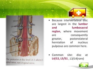
SPINEProlapseintervertebral disc  Spinal StenosisSpondylosisSpondylolysthesisspondylolysis
PROLAPSED INTERVERTEBRAL DISC (PID)
In PID, gelatinous nucleus pulposus squeezes through the fibres of the annulus fibrosusand bulges posteriorly or postrolaterallybeneath the posterior longitudinal ligamentCauses: Herniation of intervertebral disc, senile degeneration of disc, obesity, sudden jerk, sprain, trauma to spine. History of: Over-straining of lumber spine, lifting weight, violent coughing, sudden stooping or twisting.
Because intervertebral disc are largest in the lumbar and lumbosacral region, where movement are consequently greater, posterolateralherniation of nucleus pulposus are common here.Common site: disc at L4/L5, L5/S1 , L3/L4(rare)
Types of herniation (Anatomy)posterolateral disc herniation – protrusion is usually posterolateral into vertebral canal, compress the roots of a spinal nerveprotruded disc usually compresses next lower nerve as that nerve crosses level of disc in its path to its foramen (eg.protrusion of fifth lumbar disc usually affects S1 instead of L5)         central (posterior) herniation:in the lower lumbar segments, central herniation may result in S1 radiculopathyless frequently, a protruded disc above second lumbar vertebra may compress spinal cord itself or or may result in caudaequinasyndromelateral disc herniation:may compress the nerve root above the level of the herniationL4 nerve root is most often involved & patient typically have intense radicular pain
CLINICAL FEATURESYoung adultBack pain – Location: in lower back, radiates to gluteal region, back of thigh, calf, foot ; worse by: flexion (bending forwards) movement, coughing, stooping, turning, walking ; better by: rest, extension.Compensatory scoliosisSymptoms depend on the structure involved and degree of compression:  pressure on ligament – backache
 pressure on dural envelope of the nerve root – severe pain referred to the buttock and lower limb (sciatica)
pressure to the nerve itself – numbness, parasthesia,and muscle weakness
Compression of caudaequina – urinary retentionSignsMidline tenderness of the low backParavertebral muscle spasm – compensatory scoliosisStraight leg raising test (SLR) +veSciatic strecth test +veCross SLR  maybe +veFemoral strecth test maybe +ve (indicate prolapse at L3/L4)
InvestigationX-Ray :  lumbo-sacral spine	• Narrowed disc spaces.• Loss of lumber lordosis.• Compensatory scoliosis.CT scan lumber spine	• Outline of soft tissues.• Bulging out disc.MRI lumber spine	• Intervertebral disc protrusion.• Compression of nerve root.
Management Rest, Reduction, Removal & RehabilitationConservativeHeat therapy, NSAIDsBed rests – During Acute attackIn severe cases- traction is applied to leg or pelvis, provided there is no cord compression.Reginmobility gradually.Advice on spinal posturalRestrict jerky movements, avoid forward bending, lifting weight, reduce weight if obese.OperativeIndication : Caudaequina syndrome does not clear up within 6hours of starting bed rest and traction ( emergency!)Failed of conservative treatmentNeurological deteriorationFrequently recurring attackNerve decompression- Laminotomy+Diskectomy(through post approach between adjacent vertebral  laminae, dural sac is retracted to one side and bulging disc exposed. The friable partially shredded  material is removed. )
SPINAL STENOSIs
Definition :	 Narrowing of spinal canal results in cord/root compression.Causes:Congenital stenosis - Idiopathic, osteopetrosis, achondroplasiaSpine degenerative - OA- narrowing spinal canal d/t hypertrophy of facet and ligflavum associated with osteophyte.Spine instability - supporting lig torn/ stretched from severe back injury- bone move forwardDisc herniationDegenerative spondylolisthesis- decreases its AP diameter Trauma
CLINICAL FEATURESElderly – late 5th / 6th decade
Back pain -  worse by extension, relieved by sitting/ forward leaning
Numbness and paraesthesia in thighs, legs or feet
Spinal (neurological) claudication
Neurological symptom exercebrated by walking / standingDifferentiating claudication
InvestigationLateral view XRAYs- Look for degenerative changes like spondylolisthesis, disc degeneration and disc height lost with osteophytes formationCT scans (with myelogram): canal narrowingMRI: evaluate cord/ root compression, extent of spinal cord narrowing
ManagementConservative: Control the symptomsactivity modification
Physiotherapy - Instruction in spinal posture, flexion exercise
Analgesia - NSAIDs, epidural injection
Protect neurological function – Vitamin B complexOperative:Endoscopic spine  decompression (laminectomy +/- facetectomy)Degenerative Disc Disease
Degenerative disc disease involves the degeneration of intervertebraldiscs. Disc properties change lead to decrease mechanical propertiesWith increasing age, the discs can lose flexibility, elasticity, and shock absorbing characteristics. They also become thinner as they dehydrate. When all that happens, the discs change from a supple state that allows fluid movement to a stiff and rigid state that restricts your movement and causes pain.
Tiny tears or cracks in the annulus fibrosus may forced out the nucleus pulposus through the tears or cracks, which causes the disc to bulge, break or rupture.It can take place throughout the spine, but it most often occurs in the discs in the lower back (lumbar region) and the neck (cervical region).
CF - chronic back or neckpainw/out radiculopathyHigh risk : smoke cigarettes ,heavy physical work (repeated heavy lifting), obeseA sudden (acute) injury leading to a herniated disc (such as a fall) may also begin the degeneration process.As the space between the vertebrae gets smaller, there is less padding between them, and the spine becomes less stable. The body reacts to this by constructing bony growths called bone spurs (osteophytes). Bone spurs can put pressure on the spinal nerve roots or spinal cord, resulting in pain and affecting nerve function.
Osteophyte on intervertebral foramina compress spinal nerve Hypertrophic changes at vertebral margins with spur formationDegeneration of lumbar IV disc
Disc Problems
ManagementConservative:  Rest, activity modification, NSAIDs, +/- muscle relaxants Physical therapy: stretching,    strengthening, weight control Lumbar bracingOperative:  Lumbar fusion, disc replacement
Spondylosis
Spondylosis (spinal OA) - degenerative disorder that may cause loss of normal spinal structure and function.Degenerative changes in discs, facets, and uncovertebral jointmay affect the cervical (neck), thoracic (mid-back), or lumbar (low back) regions of the spine.CF: Cervical (Neck) : axial, neck pain, UL pain (spread into the shoulder or down the arm), paresthesia +/- weakness. Site: disc at C5/C6, C6/C7
Thoracic (Mid-Back) : pain triggered by forward flexion and hyperextension
Lumbar (Low Back) : >40, Pain and morning stiffness , worse by movementCan result in cord or root compression : myelopathy/radiculopathy

Spine

  • 1.
    SPINEProlapseintervertebral disc Spinal StenosisSpondylosisSpondylolysthesisspondylolysis
  • 2.
  • 3.
    In PID, gelatinousnucleus pulposus squeezes through the fibres of the annulus fibrosusand bulges posteriorly or postrolaterallybeneath the posterior longitudinal ligamentCauses: Herniation of intervertebral disc, senile degeneration of disc, obesity, sudden jerk, sprain, trauma to spine. History of: Over-straining of lumber spine, lifting weight, violent coughing, sudden stooping or twisting.
  • 4.
    Because intervertebral discare largest in the lumbar and lumbosacral region, where movement are consequently greater, posterolateralherniation of nucleus pulposus are common here.Common site: disc at L4/L5, L5/S1 , L3/L4(rare)
  • 5.
    Types of herniation(Anatomy)posterolateral disc herniation – protrusion is usually posterolateral into vertebral canal, compress the roots of a spinal nerveprotruded disc usually compresses next lower nerve as that nerve crosses level of disc in its path to its foramen (eg.protrusion of fifth lumbar disc usually affects S1 instead of L5)         central (posterior) herniation:in the lower lumbar segments, central herniation may result in S1 radiculopathyless frequently, a protruded disc above second lumbar vertebra may compress spinal cord itself or or may result in caudaequinasyndromelateral disc herniation:may compress the nerve root above the level of the herniationL4 nerve root is most often involved & patient typically have intense radicular pain
  • 7.
    CLINICAL FEATURESYoung adultBackpain – Location: in lower back, radiates to gluteal region, back of thigh, calf, foot ; worse by: flexion (bending forwards) movement, coughing, stooping, turning, walking ; better by: rest, extension.Compensatory scoliosisSymptoms depend on the structure involved and degree of compression: pressure on ligament – backache
  • 8.
    pressure ondural envelope of the nerve root – severe pain referred to the buttock and lower limb (sciatica)
  • 9.
    pressure to thenerve itself – numbness, parasthesia,and muscle weakness
  • 10.
    Compression of caudaequina– urinary retentionSignsMidline tenderness of the low backParavertebral muscle spasm – compensatory scoliosisStraight leg raising test (SLR) +veSciatic strecth test +veCross SLR maybe +veFemoral strecth test maybe +ve (indicate prolapse at L3/L4)
  • 11.
    InvestigationX-Ray : lumbo-sacral spine • Narrowed disc spaces.• Loss of lumber lordosis.• Compensatory scoliosis.CT scan lumber spine • Outline of soft tissues.• Bulging out disc.MRI lumber spine • Intervertebral disc protrusion.• Compression of nerve root.
  • 12.
    Management Rest, Reduction,Removal & RehabilitationConservativeHeat therapy, NSAIDsBed rests – During Acute attackIn severe cases- traction is applied to leg or pelvis, provided there is no cord compression.Reginmobility gradually.Advice on spinal posturalRestrict jerky movements, avoid forward bending, lifting weight, reduce weight if obese.OperativeIndication : Caudaequina syndrome does not clear up within 6hours of starting bed rest and traction ( emergency!)Failed of conservative treatmentNeurological deteriorationFrequently recurring attackNerve decompression- Laminotomy+Diskectomy(through post approach between adjacent vertebral laminae, dural sac is retracted to one side and bulging disc exposed. The friable partially shredded material is removed. )
  • 13.
  • 14.
    Definition : Narrowingof spinal canal results in cord/root compression.Causes:Congenital stenosis - Idiopathic, osteopetrosis, achondroplasiaSpine degenerative - OA- narrowing spinal canal d/t hypertrophy of facet and ligflavum associated with osteophyte.Spine instability - supporting lig torn/ stretched from severe back injury- bone move forwardDisc herniationDegenerative spondylolisthesis- decreases its AP diameter Trauma
  • 15.
    CLINICAL FEATURESElderly –late 5th / 6th decade
  • 16.
    Back pain - worse by extension, relieved by sitting/ forward leaning
  • 17.
    Numbness and paraesthesiain thighs, legs or feet
  • 18.
  • 19.
    Neurological symptom exercebratedby walking / standingDifferentiating claudication
  • 20.
    InvestigationLateral view XRAYs-Look for degenerative changes like spondylolisthesis, disc degeneration and disc height lost with osteophytes formationCT scans (with myelogram): canal narrowingMRI: evaluate cord/ root compression, extent of spinal cord narrowing
  • 22.
    ManagementConservative: Control thesymptomsactivity modification
  • 23.
    Physiotherapy - Instructionin spinal posture, flexion exercise
  • 24.
    Analgesia - NSAIDs,epidural injection
  • 25.
    Protect neurological function– Vitamin B complexOperative:Endoscopic spine decompression (laminectomy +/- facetectomy)Degenerative Disc Disease
  • 26.
    Degenerative disc diseaseinvolves the degeneration of intervertebraldiscs. Disc properties change lead to decrease mechanical propertiesWith increasing age, the discs can lose flexibility, elasticity, and shock absorbing characteristics. They also become thinner as they dehydrate. When all that happens, the discs change from a supple state that allows fluid movement to a stiff and rigid state that restricts your movement and causes pain.
  • 27.
    Tiny tears orcracks in the annulus fibrosus may forced out the nucleus pulposus through the tears or cracks, which causes the disc to bulge, break or rupture.It can take place throughout the spine, but it most often occurs in the discs in the lower back (lumbar region) and the neck (cervical region).
  • 28.
    CF - chronicback or neckpainw/out radiculopathyHigh risk : smoke cigarettes ,heavy physical work (repeated heavy lifting), obeseA sudden (acute) injury leading to a herniated disc (such as a fall) may also begin the degeneration process.As the space between the vertebrae gets smaller, there is less padding between them, and the spine becomes less stable. The body reacts to this by constructing bony growths called bone spurs (osteophytes). Bone spurs can put pressure on the spinal nerve roots or spinal cord, resulting in pain and affecting nerve function.
  • 29.
    Osteophyte on intervertebralforamina compress spinal nerve Hypertrophic changes at vertebral margins with spur formationDegeneration of lumbar IV disc
  • 30.
  • 31.
    ManagementConservative: Rest,activity modification, NSAIDs, +/- muscle relaxants Physical therapy: stretching, strengthening, weight control Lumbar bracingOperative: Lumbar fusion, disc replacement
  • 32.
  • 33.
    Spondylosis (spinal OA) - degenerativedisorder that may cause loss of normal spinal structure and function.Degenerative changes in discs, facets, and uncovertebral jointmay affect the cervical (neck), thoracic (mid-back), or lumbar (low back) regions of the spine.CF: Cervical (Neck) : axial, neck pain, UL pain (spread into the shoulder or down the arm), paresthesia +/- weakness. Site: disc at C5/C6, C6/C7
  • 34.
    Thoracic (Mid-Back) : paintriggered by forward flexion and hyperextension
  • 35.
    Lumbar (Low Back): >40, Pain and morning stiffness , worse by movementCan result in cord or root compression : myelopathy/radiculopathy