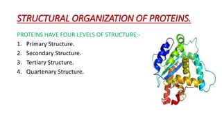
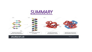
The document discusses the four levels of protein structure:
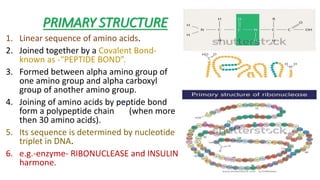
1) Primary structure is the linear sequence of amino acids joined by peptide bonds.
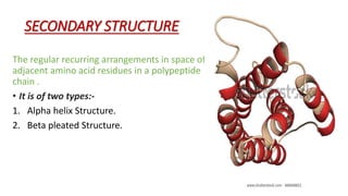
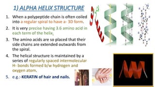
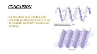
2) Secondary structure involves regular patterns of amino acids, such as alpha helices and beta pleated sheets formed by hydrogen bonds.
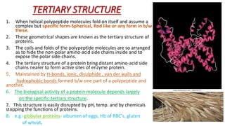
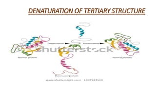
3) Tertiary structure is the specific 3D shape proteins fold into, formed by interactions between different parts of the polypeptide chain.
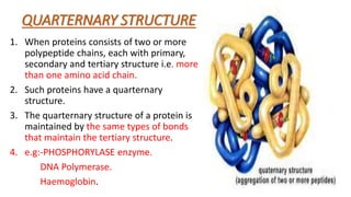
4) Quaternary structure refers to proteins made of multiple polypeptide chains that interact to form a complex.


![Photoisomerisation of aromaric compounds [recovered]](https://cdn.slidesharecdn.com/ss_thumbnails/photoisomerisationofaromariccompoundsrecovered-210612142707-thumbnail.jpg?width=640&height=640&fit=bounds)


![Catalysis by solid bases [recovered] [autosaved]](https://cdn.slidesharecdn.com/ss_thumbnails/catalysisbysolidbasesrecoveredautosaved-210612140910-thumbnail.jpg?width=640&height=640&fit=bounds)


