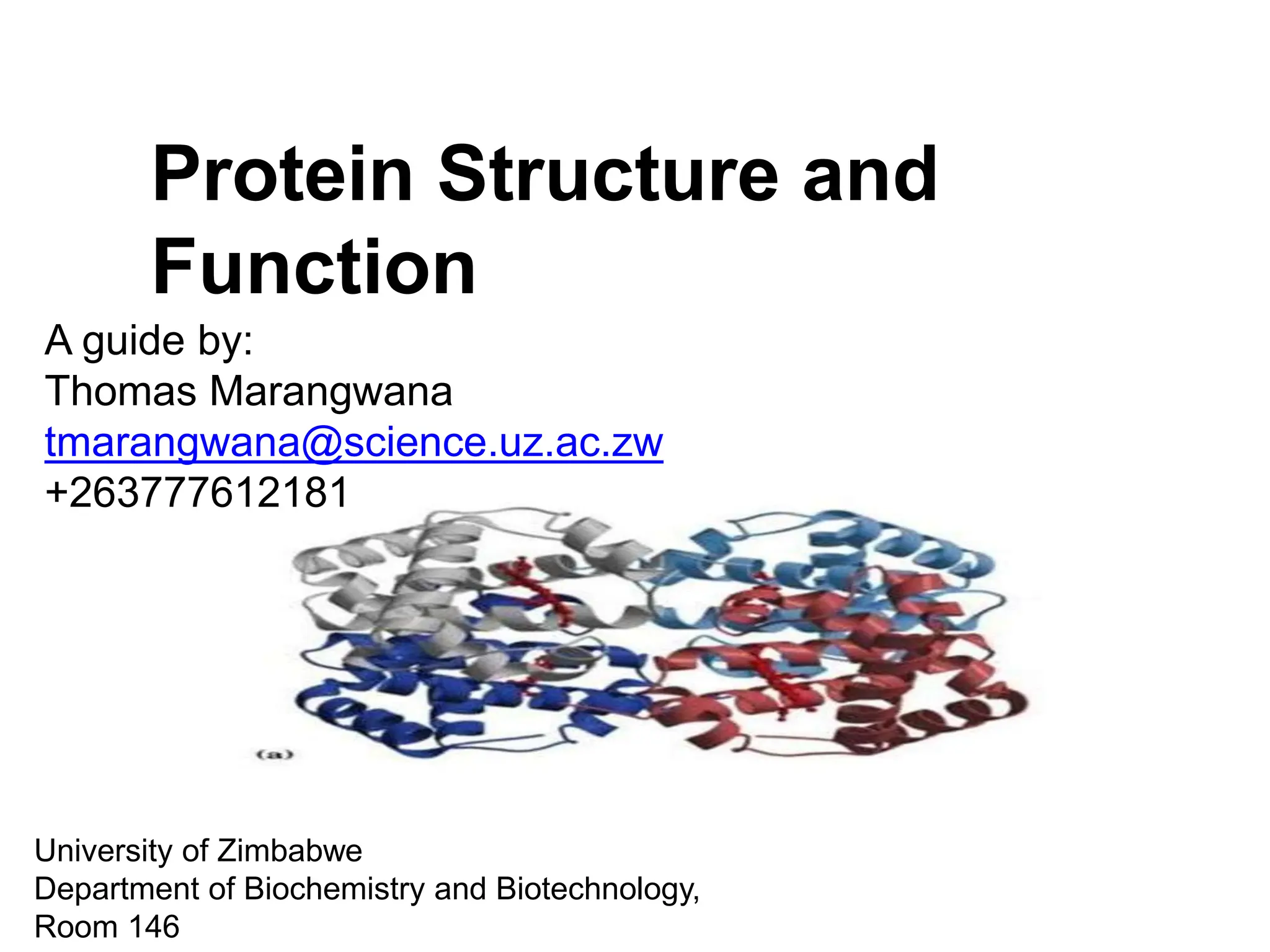
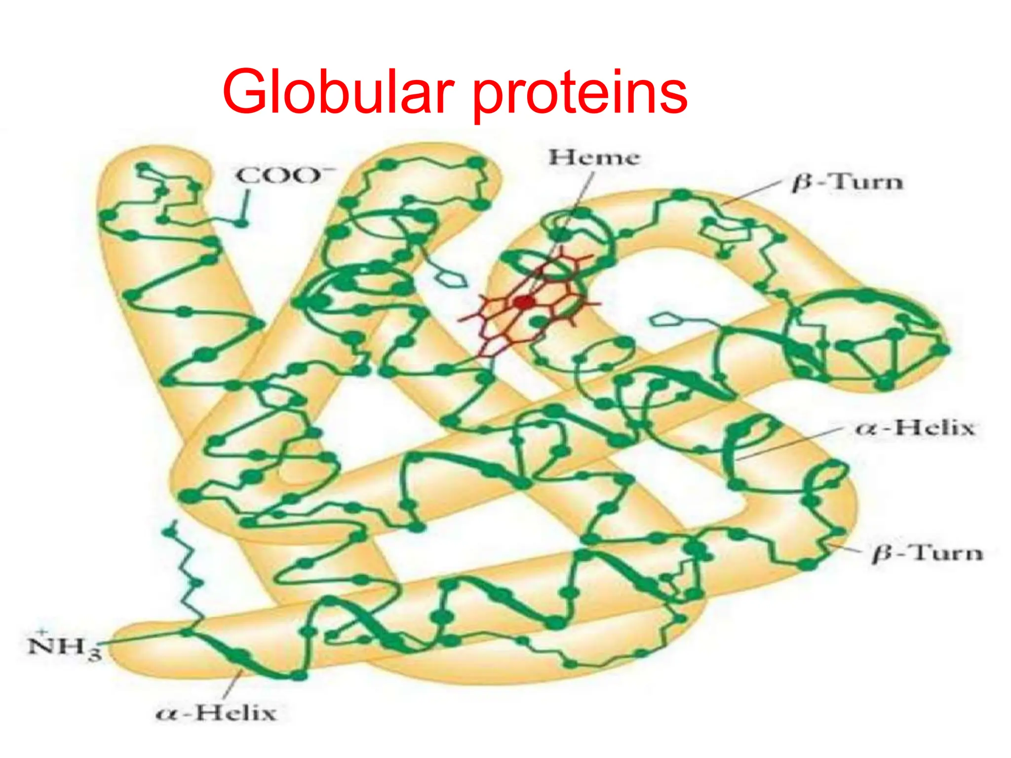
Protein Structure and
Function
A guide by:
Thomas Marangwana
tmarangwana@science.uz.ac.zw
+263777612181
University of Zimbabwe
Department of Biochemistry and Biotechnology,
Room 146
Proteins
• Proteins are the most abundant organic molecules
of the living system.
• They constitute about 50% of the cellular dry weight.
• They constitute the fundamental basis of structure
and function of life.
• In 1839, Dutch chemist G.J. Mulder was first
to describe about proteins.
• The term protein is derived from a Greek
word proteios, meaning first place.
• The proteins are nitrogenous macromolecules that
are composed of many amino acids.
Peptide formation
• Carboxyl group of one amino acid (with side
chain R1)forms acovalent peptide bond with α-
amino group of another amino acid (with side
chain R2) by removal of a water molecule.
• The result is : Dipeptide.
• The dipeptide can then form a second peptide
bond with a third amino acid (with side chain R3)
to give tripeptide.
• Repetition of this process generates a
polypeptide or protein of specific aminoacid
sequence.
Peptide formation
Structure of proteins
• Proteins have different levels of
organisation
i. Primary Structure
ii. Secondary Structure
iii. Tertiary Structure
iv. Quaternary Structure
Structure of proteins
Forces that determine the structure
• Primary structure: determined by
covalent bonds
• Secondary structure: determined by
weak forces
Protein structures
1. Primary Structure
• The primary structure of protein refers to
the sequence of amino acids present in
the polypeptide chain, it contains all the
information necessary to make a protein.
• Amino acids are covalently linked by
peptide bonds or covalent bonds.
• Eachcomponent amino acid in a polypeptide
is called a "residue” or “moiety”.
1. Primary Structure
• By convention the primary structure of
protein starts from the amino terminal
(N) end and ends in the carboxyl terminal
(C) end.
2. Secondary structure
• It is a local, regularly occurring structure in
proteins and is mainly formed through hydrogen
bonds between backbone atoms.
• Pauling & Corey studied the secondary
structures and proposed 2 conformations i.e.,
i. α helix
ii. β sheets
2. Secondary structure
α-Helices
• The a-helix is a rod like structure. It is
one peptide chain which is coiled so
that adjacent residues are 1.5A apart.
• There are H-bonds between the N-H and
the C=O groups of the backbone. These
link the peptide bonds between every
fourth amino acids.
• The side chains all extend outwards from
the helix.
α-Helices
• Although the helix itself is quite a rigid
structure, it can be curved or kinked,
which lends some flexibility to the protein
structure.
• Two or more polypeptides can entwine to
form very long, very stable, structures
called α-helical coiled coils.
• These are often found in keratin (hair),
myosin (muscle), epidermin (skin), and
fibrin (blood clots).
α-Helices
• Right handed spiral structure.
• Side chain extend outwards.
• Stabilized by H bonding that are arranged
such that the peptide Carbonyl oxygen (nth
residue) bonds with amide hydrogen (n+4 th
residue).
α-Helices
• Amino acids per turn – 3.6
• Pitch is 5.4 A°
• Alpha helical segments, are found in many
globular proteins like myoglobin
• Length ~12 residues and ~3 helical turns.
phi = -60 degrees, psi = -45 degrees
α-Helices
β-Sheets
• A polypeptide in a β-sheet is called a β-
strand, and is almost fully extended (not
coiled) so there is 3.5A between adjacent
residues.
• Formed when 2 or more polypeptides line up
side by side.
• The β-strands in a sheet can either run in
the same (parallel) or different (anti-
parallel) direction.
• They are stabilized by hydrogen bond
between NH and carbonyl groups of
adjacent chains.
β-Sheets
β-Sheets
• β sheets come in two varieties.
i. Antiparallel β sheet – neighboring
hydrogen bonded polypeptide chains
run in opposite direction.
ii. Parallel β sheet - hydrogen bonded
chains extend in the same direction.
β-Sheets
Protein folding
• The peptide bond allows for rotation around
it and therefore the protein can fold and
orient the R groups in favorable positions.
• Weak non-covalent interactions will hold
the protein in its functional shape – these
are weak and will take many to hold the
shape.
• Protein folding occurs in the cytosol.
Protein folding
Protein folding patterns
• 2 regular folding patterns have been
identified
– formed between the bonds of the
peptide backbone.
i. α-helix – protein turns like a spiral –
fibrous proteins (hair, nails, horns).
ii. β-sheet – protein folds back on itself as
in a ribbon – globular protein.
Protein folding patterns
3. Tertiary structure
• The tertiary structure defines the specific overall 3-D
shape of the protein, how the peptide chain folds so
that sidechains are packed and organized.
• Tertiary structure is stabilized by various types of
interactions between the side-chains of the peptide
chain.
• These interactions are:
i. Disulfide bonds
ii. Hydrophobic interactions
iii. Hydrogen bonds
iv. Ionic interactions
v. Vander Waals force
3. Tertiary structure
1. Disulphide Bonds
• If two cysteine side chains are close to one
another and the local environment is
conducive to oxidation, then they can form
a disulphide bond/bridge.
• The residues can either be in the same
peptide chain (forming a loop) or in
different chains.
• They mainly occur in secreted proteins and in
the parts of membrane proteins which face
the outside.
• NB, disuphide bridges only form after the
protein has folded into its tertiary structure.
1. Disulphide Bonds
2. Hydrophobic Interactions
• Close attraction of non-polar R groups
through dispersion forces.
• They are non attractive interactions,
but results from the inability of water
to form hydrogen bonds with certain
side chains.
• Very weak but collective interactions
over large area stabilize structure.
• Repel polar and charged
molecules/particles.
2. Hydrophobic Interactions
3. Hydrogen Bonds
• When unfolded, all polar/hydrophillic
sidechains can interact via H-bonds with
water. When the protein folds, they must H-
bond to each other and exclude much of the
water. All groups capable of forming a
hydrogen bond MUST, hence H-bonding in
the backbone (C=O to N-H) by way of
helices and sheets is an efficient way of
ensuring stability
3. Hydrogen Bonds
• The capacity of proteins to form hydrogen
bonds is an important determinant of
protein stability.
• Hydrogen bonds can be between backbone
groups, as in helices and sheets; between
side chains, such as serine or threonine O-H
groups and carbonyl carbons of side chains
(-C=O); and between backbone groups and
side chain groups.
3. Hydrogen Bonds
4. Ion Pairs
• When amino acid side chains of opposite
charge are in close proximity, they can
form an ion pair (also called a salt bridge).
• Since charge is affected by pH, so is the
formation and the breakage of these ion
pairs.
• Ions on R groups form salt bridges
through ionic bonds.
• NH3 +and COO- areas of the protein
attract and form ionic bonds.
4. Ion Pairs
Non-Covalent Bonds
• Hydrophobic interactions, hydrogen bonds
and salt bridges are all non-covalent
interactions.
• These are all relatively weak interactions but
the large number in a protein combine to
give the overall stability of the structure.
• If these interactions are maintained the
protein keeps its tertiary ("native")
structure.
• However once the bonds maintaining the
structure begin to be disrupted the tertiary
structure is destroyed and the protein is
said to be "denatured
Interactions
Globular proteins
• Globular proteins fold up into compact,
spherical shapes.
• Their functions include biosynthesis,
transport and metabolism, eg, myoglobin
is a globular protein that stores oxygen in
the muscles.
• Myoglobin is a single peptide chain that is
mostly α-helix –
• The O2 binding pocket is formed by a heme
group and specific amino acid side-chains
that are brought into position by the tertiary
structure
Globular proteins
Fibrous proteins
• Fibrous proteins consist of long fibers and are
mainly structural proteins, eg α-keratins are
fibrous proteins that make hair, fur, nails and
skin.
-hair is made of twined fibrils, which are
braids of three α –helices (similar to the
triple helix structure of collagen)
- the α -helices are held together by
disulfide bonds.
• β -keratins are fibrous proteins found in
feathers and scales that are made up mostly of
β -pleated sheets
Fibrous proteins
Subunits and Quaternary
Structure
• Proteins containing more than one
polypeptide chain exhibit an additional
level of structural organisation.
• In this case, each polypeptide chain is a
"subunit". Quaternary (4D) structure
describes the way the subunits are arranged
together and the nature of their contacts.
• Subunits associate by non-covalent
interactions similar to those involved in
tertiary structure.
.
Subunits and Quaternary
Structure
• There are two kinds of quaternary
structures: both are multisubunit proteins.
i. Homodimer : association between identical
polypeptide chains.
ii. Heterodimer : interactions between
subunits of very different structures.
Subunits and Quaternary
Structure
Subunits and Quaternary
Structure
• Quaternary structure adds stability by
decreasing the surface/volume ratio of
smaller subunit
• Simplifies the construction of large
complexes
– viral capsids and proteosomes
THANK YOU

5. Protein structure and function and amino.pptx

  • 1.
    Protein Structure and Function Aguide by: Thomas Marangwana tmarangwana@science.uz.ac.zw +263777612181 University of Zimbabwe Department of Biochemistry and Biotechnology, Room 146
  • 2.
    Proteins • Proteins arethe most abundant organic molecules of the living system. • They constitute about 50% of the cellular dry weight. • They constitute the fundamental basis of structure and function of life. • In 1839, Dutch chemist G.J. Mulder was first to describe about proteins. • The term protein is derived from a Greek word proteios, meaning first place. • The proteins are nitrogenous macromolecules that are composed of many amino acids.
  • 3.
    Peptide formation • Carboxylgroup of one amino acid (with side chain R1)forms acovalent peptide bond with α- amino group of another amino acid (with side chain R2) by removal of a water molecule. • The result is : Dipeptide. • The dipeptide can then form a second peptide bond with a third amino acid (with side chain R3) to give tripeptide. • Repetition of this process generates a polypeptide or protein of specific aminoacid sequence.
  • 4.
  • 5.
    Structure of proteins •Proteins have different levels of organisation i. Primary Structure ii. Secondary Structure iii. Tertiary Structure iv. Quaternary Structure
  • 6.
  • 7.
    Forces that determinethe structure • Primary structure: determined by covalent bonds • Secondary structure: determined by weak forces
  • 8.
  • 9.
    1. Primary Structure •The primary structure of protein refers to the sequence of amino acids present in the polypeptide chain, it contains all the information necessary to make a protein. • Amino acids are covalently linked by peptide bonds or covalent bonds. • Eachcomponent amino acid in a polypeptide is called a "residue” or “moiety”.
  • 10.
    1. Primary Structure •By convention the primary structure of protein starts from the amino terminal (N) end and ends in the carboxyl terminal (C) end.
  • 11.
    2. Secondary structure •It is a local, regularly occurring structure in proteins and is mainly formed through hydrogen bonds between backbone atoms. • Pauling & Corey studied the secondary structures and proposed 2 conformations i.e., i. α helix ii. β sheets
  • 12.
  • 13.
    α-Helices • The a-helixis a rod like structure. It is one peptide chain which is coiled so that adjacent residues are 1.5A apart. • There are H-bonds between the N-H and the C=O groups of the backbone. These link the peptide bonds between every fourth amino acids. • The side chains all extend outwards from the helix.
  • 14.
    α-Helices • Although thehelix itself is quite a rigid structure, it can be curved or kinked, which lends some flexibility to the protein structure. • Two or more polypeptides can entwine to form very long, very stable, structures called α-helical coiled coils. • These are often found in keratin (hair), myosin (muscle), epidermin (skin), and fibrin (blood clots).
  • 15.
    α-Helices • Right handedspiral structure. • Side chain extend outwards. • Stabilized by H bonding that are arranged such that the peptide Carbonyl oxygen (nth residue) bonds with amide hydrogen (n+4 th residue).
  • 16.
    α-Helices • Amino acidsper turn – 3.6 • Pitch is 5.4 A° • Alpha helical segments, are found in many globular proteins like myoglobin • Length ~12 residues and ~3 helical turns. phi = -60 degrees, psi = -45 degrees
  • 17.
  • 18.
    β-Sheets • A polypeptidein a β-sheet is called a β- strand, and is almost fully extended (not coiled) so there is 3.5A between adjacent residues. • Formed when 2 or more polypeptides line up side by side. • The β-strands in a sheet can either run in the same (parallel) or different (anti- parallel) direction. • They are stabilized by hydrogen bond between NH and carbonyl groups of adjacent chains.
  • 19.
  • 20.
    β-Sheets • β sheetscome in two varieties. i. Antiparallel β sheet – neighboring hydrogen bonded polypeptide chains run in opposite direction. ii. Parallel β sheet - hydrogen bonded chains extend in the same direction.
  • 21.
  • 22.
    Protein folding • Thepeptide bond allows for rotation around it and therefore the protein can fold and orient the R groups in favorable positions. • Weak non-covalent interactions will hold the protein in its functional shape – these are weak and will take many to hold the shape. • Protein folding occurs in the cytosol.
  • 23.
  • 24.
    Protein folding patterns •2 regular folding patterns have been identified – formed between the bonds of the peptide backbone. i. α-helix – protein turns like a spiral – fibrous proteins (hair, nails, horns). ii. β-sheet – protein folds back on itself as in a ribbon – globular protein.
  • 25.
  • 26.
    3. Tertiary structure •The tertiary structure defines the specific overall 3-D shape of the protein, how the peptide chain folds so that sidechains are packed and organized. • Tertiary structure is stabilized by various types of interactions between the side-chains of the peptide chain. • These interactions are: i. Disulfide bonds ii. Hydrophobic interactions iii. Hydrogen bonds iv. Ionic interactions v. Vander Waals force
  • 27.
  • 28.
    1. Disulphide Bonds •If two cysteine side chains are close to one another and the local environment is conducive to oxidation, then they can form a disulphide bond/bridge. • The residues can either be in the same peptide chain (forming a loop) or in different chains. • They mainly occur in secreted proteins and in the parts of membrane proteins which face the outside. • NB, disuphide bridges only form after the protein has folded into its tertiary structure.
  • 29.
  • 30.
    2. Hydrophobic Interactions •Close attraction of non-polar R groups through dispersion forces. • They are non attractive interactions, but results from the inability of water to form hydrogen bonds with certain side chains. • Very weak but collective interactions over large area stabilize structure. • Repel polar and charged molecules/particles.
  • 31.
  • 32.
    3. Hydrogen Bonds •When unfolded, all polar/hydrophillic sidechains can interact via H-bonds with water. When the protein folds, they must H- bond to each other and exclude much of the water. All groups capable of forming a hydrogen bond MUST, hence H-bonding in the backbone (C=O to N-H) by way of helices and sheets is an efficient way of ensuring stability
  • 33.
    3. Hydrogen Bonds •The capacity of proteins to form hydrogen bonds is an important determinant of protein stability. • Hydrogen bonds can be between backbone groups, as in helices and sheets; between side chains, such as serine or threonine O-H groups and carbonyl carbons of side chains (-C=O); and between backbone groups and side chain groups.
  • 34.
  • 35.
    4. Ion Pairs •When amino acid side chains of opposite charge are in close proximity, they can form an ion pair (also called a salt bridge). • Since charge is affected by pH, so is the formation and the breakage of these ion pairs. • Ions on R groups form salt bridges through ionic bonds. • NH3 +and COO- areas of the protein attract and form ionic bonds.
  • 36.
  • 37.
    Non-Covalent Bonds • Hydrophobicinteractions, hydrogen bonds and salt bridges are all non-covalent interactions. • These are all relatively weak interactions but the large number in a protein combine to give the overall stability of the structure. • If these interactions are maintained the protein keeps its tertiary ("native") structure. • However once the bonds maintaining the structure begin to be disrupted the tertiary structure is destroyed and the protein is said to be "denatured
  • 38.
  • 39.
    Globular proteins • Globularproteins fold up into compact, spherical shapes. • Their functions include biosynthesis, transport and metabolism, eg, myoglobin is a globular protein that stores oxygen in the muscles. • Myoglobin is a single peptide chain that is mostly α-helix – • The O2 binding pocket is formed by a heme group and specific amino acid side-chains that are brought into position by the tertiary structure
  • 40.
  • 41.
    Fibrous proteins • Fibrousproteins consist of long fibers and are mainly structural proteins, eg α-keratins are fibrous proteins that make hair, fur, nails and skin. -hair is made of twined fibrils, which are braids of three α –helices (similar to the triple helix structure of collagen) - the α -helices are held together by disulfide bonds. • β -keratins are fibrous proteins found in feathers and scales that are made up mostly of β -pleated sheets
  • 42.
  • 43.
    Subunits and Quaternary Structure •Proteins containing more than one polypeptide chain exhibit an additional level of structural organisation. • In this case, each polypeptide chain is a "subunit". Quaternary (4D) structure describes the way the subunits are arranged together and the nature of their contacts. • Subunits associate by non-covalent interactions similar to those involved in tertiary structure. .
  • 44.
    Subunits and Quaternary Structure •There are two kinds of quaternary structures: both are multisubunit proteins. i. Homodimer : association between identical polypeptide chains. ii. Heterodimer : interactions between subunits of very different structures.
  • 45.
  • 46.
    Subunits and Quaternary Structure •Quaternary structure adds stability by decreasing the surface/volume ratio of smaller subunit • Simplifies the construction of large complexes – viral capsids and proteosomes
  • 48.