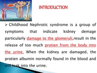
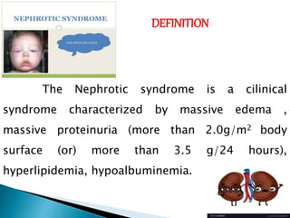
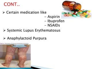
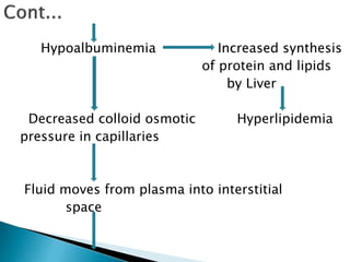
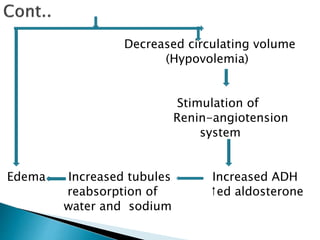
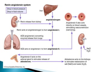
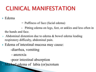
INTRODUCTION
 Childhood Nephrotic syndrome is a group of
symptoms that indicate kidney demage
particularly damage to the glomeruli,result in the
release of too much protien from the body into
the urine. When the kidney are damaged, the
protien albumin normally found in the blood and
will leak into the urine.
DEFINITION
Nephrotic syndrome is a manifestation
of glomerular disease characterized by
protienuria , Hypoprotienaemia,
hyprerlipidemia and generalized edema.
- Aruchamy
DEFINITION
The Nephrotic syndrome is a cilinical
syndrome characterized by massive edema ,
massive proteinuria (more than 2.0g/m2 body
surface (or) more than 3.5 g/24 hours),
hyperlipidemia, hypoalbuminemia.
INCIDENCE
 Incidence of the condition is to 7 per 1000
children.
 It occurs commonly between the ages of 2-5
years
 Predominantly males to female ratio 2:1.
CAUSES
1.Primary renal causes:-
 Minimal change nephropathy
 Focal glomerulosclerosis
 Immune complex glomerulo nephritis
 Membrano proliferative glomerulonephritis
 Acute post streptococcal glomerulonephritis
 Membranous nephropathy
 Congenital Nephrosis
CONT..
2.Systemic causes
 Infection
 Toxins: mercurial, bismuth , gold etc.
 Allergies: poison oak, bee sting , serum sickness, inhaled
pollens, food allergy.
 cardiovascular disorders: sickle cell disease
Passive congestive heart failure
Renal vein thrombosis
 Certain medication like
- Aspirin
- Ibuprofen
- NSAIDs
CONT..
 Systemic Lupus Erythematosus
 Anaphylactoid Purpura
Nephrotic syndrome can be classified as
1. According to age
a. Congenital Nephrotic syndrome
b. Infantile Nephrotic Syndrome
c. Childhood Nephrotic Syndrome
2. According to the Etiological Factor
a. Primary Nephrotic syndrome
b. Secondary nephrotic syndrome
1.According to age
a. Congenital Nephrotic syndrome
 A hereditary form of nephrotic syndrome, present at
birth or before 3 months of life.
 It may be primary or secondary. The main causes is
congenital infections such as syphillis, cytomegalovirus,
rubella & toxoplasmosis etc.
 Proteinuria & edema are significant early findings, often
immediately after birth.
 The disease doesn’t respond well to therapy. Death
usually occurs before the age of 2 years.
B.Infantile Nephrotic syndrome:
 Onset between 3 months and 1 year of age.
 It can be caused by infections, drugs, Malignancies
and most of the case idiopathic.
C. Childhood Nephrotic Syndrome:
 Onset between 2 and 8 years.
2. According to Etiological Factors
 The classification of Nephrotic Syndrome
according to etiological factors are as follows;
A. Primary Nephrotic Syndrome:
 It is the most common type of childhood Nephrotic
syndrome which begins in the kidneys and affects
only the kidney.
 90% of case are Idiopathic in childhood.
 It responds to immunosuppressive therapy.
B.Secondary Nephrotic syndrome:
 When the nephrotic syndrome occurs as a part of a
recognized systemic disease (or) results from some
evident cause. It is termed secondary Nephrotic
syndrome.
These may include
 Systemic collagen vascular disorders.
 Infections
 Other causes includes certain medications such as –
Aspirin , Ibuprofen, Non-steriod anti-inflammatory
drugs, exposure to chemicals like mercury and lithium.
 Systemic Lupus Erythematosus.
 Anaphylactoid Purpura.
Due to any causes
Glomerular damage
Increased permeability of glomerular base
membrane
Plasma protein escape into the urine
Hypoalbuminemia Increased synthesis
of protein and lipids
by Liver
Decreased colloid osmotic Hyperlipidemia
pressure in capillaries
Fluid moves from plasma into interstitial
space
Decreased circulating volume
(Hypovolemia)
Stimulation of
Renin-angiotension
system
Edema Increased tubules Increased ADH
reabsorption of ↑ed aldosterone
water and sodium
 Edema
- Puffiness of face (facial edema)
- Pitting edema on legs, feet, or ankles and less often in
the hands and face.
 Abdominal distention due to edema & bowel edema leading
respiratory difficulty, abdominal pain.
 Edema of intestinal mucosa may cause:
-diarrhea, vomiting
- anorexia
-poor intestinal absorption
 Marked edema of labia (or)scrotum
 Hyperlipidemia
 Hypoalbuminemia
 Proteinuria
 Weight gain
 Risk of Clotting
 Urine alterations:
- decreased urine out put with dark
- Frothy urine
- Dark urine( Heamaturia
In addition, the child will show
-pale & irritability
-easily fatigue& activity intolerance
- lethargic.
 Blood pressure normal (or) slightly increase
 Susceptibility to infection
EDEMA
DARK URINE
FROTHY URINE
RESPIRATORY DIFFICULTY
ABDOMINAL PAIN
FEVER
WIEGHT
GAIN
HYPOALBUMINEMIA
High blood
pressure
CLINICAL
MANIFESTATION
LOSS OF APPETITE
LETHARGY
DIARRHEA
DIAGNOSTIC EVALUATION
Diagnostic Evaluation:
 History Collection
Physical Examination
Complete blood test
Kidney Function test
Liver Function test
BUN
 Urine analysis:
Proteinuria is reflected
by urinary excretion of
patient that reaches
levels in excess of 2
mg/ 24 hours.
Dipstick test for
albumin in urine.
Urine specific gravity:
Increased specific
gravity due to
proteinuria.
 Ultrasound of the kidney
Cont…
 Kidney biopsy
Management:
1.Bed rest & high protein diet: With restriction of
fluid intake are important aspects of management.
 Protein replacement dietary (or) intravenous
albumin.
Conti………
2. Steroid therapy:
With oral prednisolone is the most significant aspect
of Management of Nephrotic syndrome.
 Initially a daily dose of 2mg/ kg/ day in 2 to 3 divided
doses for at least 4 to 6 weeks & then gradually tapered off
(or) abruptly stopped, after another 4 to 6 wks.
 Antacid is given along with prednisolone to prevent
gastric complication.
Are prescribed in the presence of severe edema &
massive ascites.
 Frusemide 1-3 mg/ kg/ day/ in 2 divided doses
alone (or ) with Spironolactone 2-3 mg/ kg/ day/
in 2 divided doses is given.
 Rapid fluid loss shouldn’t be attempted in 8 to
12 hrs.
 Potassium supplementation to be given along
with diuretics.
3. Diuretics:
4. Albumin infusion
 1gm/kg/day) may be given in case of massive edema &
ascites.
 It helps to shift the fluid from interstitial space into he
vascular system.
 Diuretic therapy is given in combination of albumin
infusion.
 Plasma (or ) blood transfusion may be given in some
cases to treat hypoalbuminemia.
5. Immunosuppressive drugs
 (methrotrexate, cyclophosphamide, cyclosporine,
chlorambucil ) may be administered along with
prednisolone in case of frequent relapses & in
steroid dependent cases.
 maintenance (or)for children who fail to respond
of electrolyte imbalance to steroids.
6.Renal transplantation
 Is indicated in end stage renal failure due to
steroid resistant glomerulosclerosis.
NURSING MANAGEMENT:
ASSESSMENT:
 Assess for signs and symptoms of fluid volume
excess.
 Assess for signs of electrolyte
imbalance(hypokalemia, hyponetremia,
hypernatremia).
 Assess protein loss & nutritional status.
 Assess for side effects from medication
administration.
 Assess for signs of decrease a functioning..
 Assess for signs of ineffective breathing pattern
&pulmonary infection.
 Assess for signs of infection (fever, increase WBC
count, positive culture results)
 Assess for skin breakdown from severe edema.
 Assess child’s comfort level &ability to tolerate
activity.
 Assess child’s &family coping response to illness.
INTREVENTIONS:
1.Monitor & maintain fluid balance:
 Assess hydration status frequently
 Monitor ascites by monitoring abdominal
girth.
 Closely monitor edematous areas report
changes as indicated.
 Measure & record weight daily, report
changes as indicated.
2.Perform measure to correct electrolyte
imbalance as indicated.
 Continually monitor
appetite & nutritional
intake.
3. Encourage & nutritional
intake &proper nutritional
status.
 Provide diet high in calories & proteins
Cont…
 Offer food in small quantities in attractive
manner.
 Decrease sodium intake
(avoidance of high sodium
foods, diet with no salt).
Cont…
4.Frequently reassess cardiovascular status &
institute supportive measures as indicated.
5.Monitor for pain &provide relief measures as
needed.
 Assist child to find comfortable position .
 Administer analgesics as needed.
 Use pharmacologic pain relief method as
appropriate.
 Provide emotional support to child &family.
COMPLICATION
Renal Failure
Pericardial effusion
Hydrothorax
Atheroscelerosis
NEPHROTIC SYNDROME- Condition in pediatric

NEPHROTIC SYNDROME- Condition in pediatric

  • 3.
    INTRODUCTION  Childhood Nephroticsyndrome is a group of symptoms that indicate kidney demage particularly damage to the glomeruli,result in the release of too much protien from the body into the urine. When the kidney are damaged, the protien albumin normally found in the blood and will leak into the urine.
  • 4.
    DEFINITION Nephrotic syndrome isa manifestation of glomerular disease characterized by protienuria , Hypoprotienaemia, hyprerlipidemia and generalized edema. - Aruchamy
  • 5.
    DEFINITION The Nephrotic syndromeis a cilinical syndrome characterized by massive edema , massive proteinuria (more than 2.0g/m2 body surface (or) more than 3.5 g/24 hours), hyperlipidemia, hypoalbuminemia.
  • 6.
    INCIDENCE  Incidence ofthe condition is to 7 per 1000 children.  It occurs commonly between the ages of 2-5 years  Predominantly males to female ratio 2:1.
  • 7.
    CAUSES 1.Primary renal causes:- Minimal change nephropathy  Focal glomerulosclerosis  Immune complex glomerulo nephritis  Membrano proliferative glomerulonephritis  Acute post streptococcal glomerulonephritis  Membranous nephropathy  Congenital Nephrosis
  • 8.
    CONT.. 2.Systemic causes  Infection Toxins: mercurial, bismuth , gold etc.  Allergies: poison oak, bee sting , serum sickness, inhaled pollens, food allergy.  cardiovascular disorders: sickle cell disease Passive congestive heart failure Renal vein thrombosis
  • 9.
     Certain medicationlike - Aspirin - Ibuprofen - NSAIDs CONT..  Systemic Lupus Erythematosus  Anaphylactoid Purpura
  • 10.
    Nephrotic syndrome canbe classified as 1. According to age a. Congenital Nephrotic syndrome b. Infantile Nephrotic Syndrome c. Childhood Nephrotic Syndrome 2. According to the Etiological Factor a. Primary Nephrotic syndrome b. Secondary nephrotic syndrome
  • 11.
    1.According to age a.Congenital Nephrotic syndrome  A hereditary form of nephrotic syndrome, present at birth or before 3 months of life.  It may be primary or secondary. The main causes is congenital infections such as syphillis, cytomegalovirus, rubella & toxoplasmosis etc.  Proteinuria & edema are significant early findings, often immediately after birth.  The disease doesn’t respond well to therapy. Death usually occurs before the age of 2 years.
  • 12.
    B.Infantile Nephrotic syndrome: Onset between 3 months and 1 year of age.  It can be caused by infections, drugs, Malignancies and most of the case idiopathic. C. Childhood Nephrotic Syndrome:  Onset between 2 and 8 years.
  • 13.
    2. According toEtiological Factors  The classification of Nephrotic Syndrome according to etiological factors are as follows; A. Primary Nephrotic Syndrome:  It is the most common type of childhood Nephrotic syndrome which begins in the kidneys and affects only the kidney.  90% of case are Idiopathic in childhood.  It responds to immunosuppressive therapy.
  • 14.
    B.Secondary Nephrotic syndrome: When the nephrotic syndrome occurs as a part of a recognized systemic disease (or) results from some evident cause. It is termed secondary Nephrotic syndrome. These may include  Systemic collagen vascular disorders.  Infections  Other causes includes certain medications such as – Aspirin , Ibuprofen, Non-steriod anti-inflammatory drugs, exposure to chemicals like mercury and lithium.  Systemic Lupus Erythematosus.  Anaphylactoid Purpura.
  • 15.
    Due to anycauses Glomerular damage Increased permeability of glomerular base membrane Plasma protein escape into the urine
  • 16.
    Hypoalbuminemia Increased synthesis ofprotein and lipids by Liver Decreased colloid osmotic Hyperlipidemia pressure in capillaries Fluid moves from plasma into interstitial space
  • 17.
    Decreased circulating volume (Hypovolemia) Stimulationof Renin-angiotension system Edema Increased tubules Increased ADH reabsorption of ↑ed aldosterone water and sodium
  • 19.
     Edema - Puffinessof face (facial edema) - Pitting edema on legs, feet, or ankles and less often in the hands and face.  Abdominal distention due to edema & bowel edema leading respiratory difficulty, abdominal pain.  Edema of intestinal mucosa may cause: -diarrhea, vomiting - anorexia -poor intestinal absorption  Marked edema of labia (or)scrotum
  • 20.
     Hyperlipidemia  Hypoalbuminemia Proteinuria  Weight gain  Risk of Clotting  Urine alterations: - decreased urine out put with dark - Frothy urine - Dark urine( Heamaturia
  • 21.
    In addition, thechild will show -pale & irritability -easily fatigue& activity intolerance - lethargic.  Blood pressure normal (or) slightly increase  Susceptibility to infection
  • 22.
    EDEMA DARK URINE FROTHY URINE RESPIRATORYDIFFICULTY ABDOMINAL PAIN FEVER WIEGHT GAIN HYPOALBUMINEMIA High blood pressure CLINICAL MANIFESTATION
  • 23.
  • 24.
  • 25.
    Diagnostic Evaluation:  HistoryCollection Physical Examination Complete blood test Kidney Function test Liver Function test BUN
  • 26.
     Urine analysis: Proteinuriais reflected by urinary excretion of patient that reaches levels in excess of 2 mg/ 24 hours. Dipstick test for albumin in urine. Urine specific gravity: Increased specific gravity due to proteinuria.
  • 27.
     Ultrasound ofthe kidney Cont…  Kidney biopsy
  • 28.
    Management: 1.Bed rest &high protein diet: With restriction of fluid intake are important aspects of management.  Protein replacement dietary (or) intravenous albumin.
  • 29.
    Conti……… 2. Steroid therapy: Withoral prednisolone is the most significant aspect of Management of Nephrotic syndrome.  Initially a daily dose of 2mg/ kg/ day in 2 to 3 divided doses for at least 4 to 6 weeks & then gradually tapered off (or) abruptly stopped, after another 4 to 6 wks.  Antacid is given along with prednisolone to prevent gastric complication.
  • 30.
    Are prescribed inthe presence of severe edema & massive ascites.  Frusemide 1-3 mg/ kg/ day/ in 2 divided doses alone (or ) with Spironolactone 2-3 mg/ kg/ day/ in 2 divided doses is given.  Rapid fluid loss shouldn’t be attempted in 8 to 12 hrs.  Potassium supplementation to be given along with diuretics. 3. Diuretics:
  • 31.
    4. Albumin infusion 1gm/kg/day) may be given in case of massive edema & ascites.  It helps to shift the fluid from interstitial space into he vascular system.  Diuretic therapy is given in combination of albumin infusion.  Plasma (or ) blood transfusion may be given in some cases to treat hypoalbuminemia.
  • 32.
    5. Immunosuppressive drugs (methrotrexate, cyclophosphamide, cyclosporine, chlorambucil ) may be administered along with prednisolone in case of frequent relapses & in steroid dependent cases.  maintenance (or)for children who fail to respond of electrolyte imbalance to steroids.
  • 33.
    6.Renal transplantation  Isindicated in end stage renal failure due to steroid resistant glomerulosclerosis.
  • 34.
    NURSING MANAGEMENT: ASSESSMENT:  Assessfor signs and symptoms of fluid volume excess.  Assess for signs of electrolyte imbalance(hypokalemia, hyponetremia, hypernatremia).  Assess protein loss & nutritional status.  Assess for side effects from medication administration.
  • 35.
     Assess forsigns of decrease a functioning..  Assess for signs of ineffective breathing pattern &pulmonary infection.  Assess for signs of infection (fever, increase WBC count, positive culture results)  Assess for skin breakdown from severe edema.  Assess child’s comfort level &ability to tolerate activity.  Assess child’s &family coping response to illness.
  • 36.
    INTREVENTIONS: 1.Monitor & maintainfluid balance:  Assess hydration status frequently  Monitor ascites by monitoring abdominal girth.  Closely monitor edematous areas report changes as indicated.  Measure & record weight daily, report changes as indicated. 2.Perform measure to correct electrolyte imbalance as indicated.
  • 37.
     Continually monitor appetite& nutritional intake. 3. Encourage & nutritional intake &proper nutritional status.  Provide diet high in calories & proteins Cont…
  • 38.
     Offer foodin small quantities in attractive manner.  Decrease sodium intake (avoidance of high sodium foods, diet with no salt). Cont…
  • 39.
    4.Frequently reassess cardiovascularstatus & institute supportive measures as indicated. 5.Monitor for pain &provide relief measures as needed.  Assist child to find comfortable position .  Administer analgesics as needed.  Use pharmacologic pain relief method as appropriate.  Provide emotional support to child &family.
  • 40.