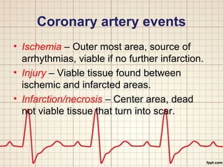
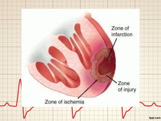
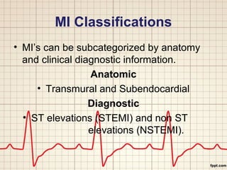
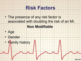
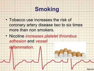
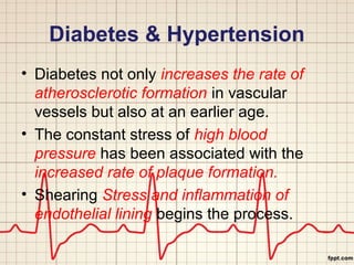
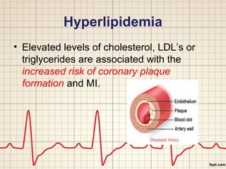
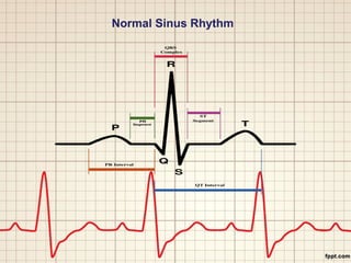
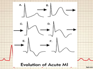
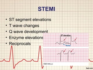
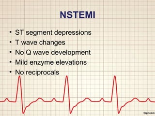
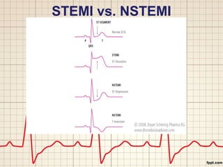
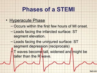
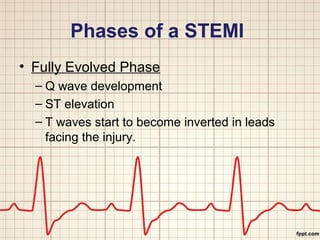
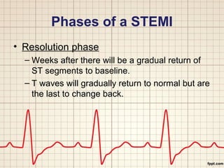
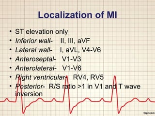
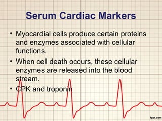
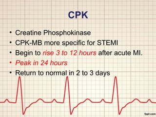
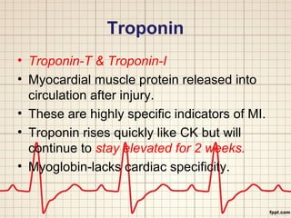
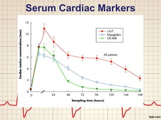
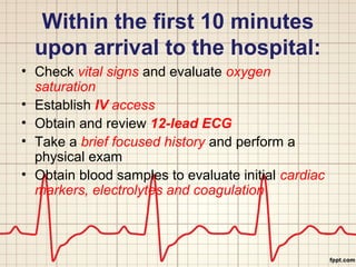
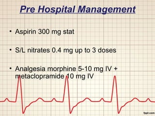
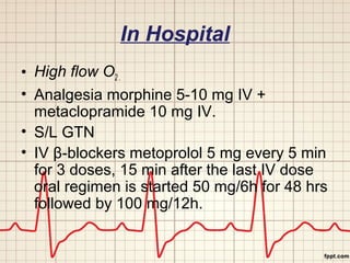
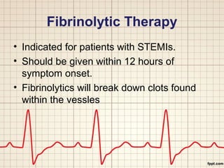
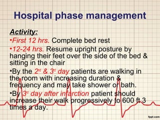
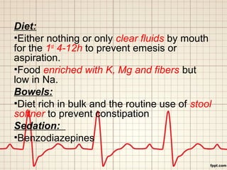
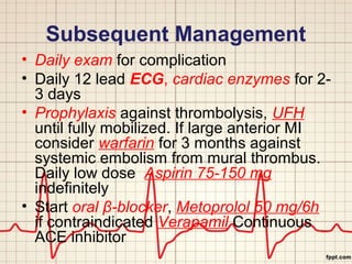
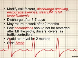
Myocardial Infarction 
Dr. Abdur Rehan 
Dr. Muhammad Aamir Iqbal 
West Medical Ward
Definition 
• Know as heart attack 
• An MI occurs when there is a diminished 
blood supply to the heart which leads to 
myocardial cell damage and ischemia. 
• Contractile function stops in the necrotic 
areas of the heart. 
• Ischemia usually occurs due to blockage 
of the coronary vessels.
Definition cont. 
• This blockage is often the result of 
thrombus that is superimposed on an 
ulcerated or unstable atherosclerotic 
plaque formation in the coronary artery. 
• MI’s are described by the area of 
occurrence. 
• Anterior, Inferior, Lateral or Posterior.
Coronary Artery Anatomy
Coronary artery events 
• Ischemia – Outer most area, source of 
arrhythmias, viable if no further infarction. 
• Injury – Viable tissue found between 
ischemic and infarcted areas. 
• Infarction/necrosis – Center area, dead 
not viable tissue that turn into scar.
MI Classifications 
• MI’s can be subcategorized by anatomy 
and clinical diagnostic information. 
Anatomic 
• Transmural and Subendocardial 
Diagnostic 
• ST elevations (STEMI) and non ST 
elevations (NSTEMI).
Risk Factors 
• The presence of any risk factor is 
associated with doubling the risk of an MI. 
Non Modifiable 
• Age 
• Gender 
• Family history
Risk Factors 
Modifiable 
• Smoking 
• Diabetes Control 
• Hypertension 
• Hyperlipidemia 
• Obesity 
• Physical Inactivity
Smoking 
• Tobacco use increases the risk of 
coronary artery disease two to six times 
more than non smokers. 
• Nicotine increases platelet thrombus 
adhesion and vessel 
inflammation.
Diabetes & Hypertension 
• Diabetes not only increases the rate of 
atherosclerotic formation in vascular 
vessels but also at an earlier age. 
• The constant stress of high blood 
pressure has been associated with the 
increased rate of plaque formation. 
• Shearing Stress and inflammation of 
endothelial lining begins the process.
Hyperlipidemia 
• Elevated levels of cholesterol, LDL’s or 
triglycerides are associated with the 
increased risk of coronary plaque 
formation and MI.
Obesity and Physical 
Inactivity 
• Mortality rate from CAD is higher in those 
who are obese. 
• Some evidence shows that those who 
carry their weight in their abdomen have a 
higher incidence of CAD 
• Physically inactive people have lower HDL 
levels with higher LDL levels and an 
increase in clot formation.
Pathophysiology 
• Ischemia develops when there is an 
increased demand for oxygen or a 
decreased supply of oxygen. 
• Ischemia can develop within 10 seconds 
and if it lasts longer than 20 minutes, 
irreversible cell and tissue death 
occurs. 
• Myocardial cell death begins at the 
endocardium. The area most distal to the 
arterial blood supply.
Pathophysiology 
• As vessel occlusion continues cell death 
spreads to the myocardium and eventually 
to the epicardium. 
• Severity of the MI depends on three 
factors. 
– Level of occlusion 
– Length of time of occlusion 
– Presence or absence of collateral circulation
Signs and Symptoms 
• Signs and symptoms are unique to each 
individual patient. 
• Ranging from no symptoms to sudden 
cardiac arrest.
Clinical Presentation 
• Retrosternal pain, more severe and lasts 
longer i.e. > 30 min, present at rest, 
heavy, squeezing & crushing, stabbing or 
burning radiating to arms, back, lower jaw, 
neck & epigastrium. 
• Associated with nausea, vomiting, anxiety 
& palpitation. 
• Painless STEMIs in diabetics in older age 
group
• Sudden onset of breathlessness 
• Sudden loss of consciousness & 
confusion state
Diagnostics 
• Typical chest pain 
• ECG 
• Serum cardiac enzymes 
• Cardiac imaging
Normal Sinus Rhythm
STEMI 
• ST segment elevations 
• T wave changes 
• Q wave development 
• Enzyme elevations 
• Reciprocals
NSTEMI 
• ST segment depressions 
• T wave changes 
• No Q wave development 
• Mild enzyme elevations 
• No reciprocals
STEMI vs. NSTEMI
Phases of a STEMI 
• Hyperacute Phase 
– Occurs within the first few hours of MI onset. 
– Leads facing the infarcted surface: ST 
segment elevation. 
– Leads facing the uninjured surface: ST 
segment depression (reciprocals) 
– T waves become tall, widened and might be 
taller than the R wave.
Phases of a STEMI 
• Fully Evolved Phase 
– Q wave development 
– ST elevation 
– T waves start to become inverted in leads 
facing the injury.
Phases of a STEMI 
• Resolution phase 
– Weeks after there will be a gradual return of 
ST segments to baseline. 
– T waves will gradually return to normal but are 
the last to change back.
Localization of MI 
• ST elevation only 
• Inferior wall- II, III, aVF 
• Lateral wall- I, aVL, V4-V6 
• Anteroseptal- V1-V3 
• Anterolateral- V1-V6 
• Right ventricular- RV4, RV5 
• Posterior- R/S ratio >1 in V1 and T wave 
inversion
Serum Cardiac Markers 
• Myocardial cells produce certain proteins 
and enzymes associated with cellular 
functions. 
• When cell death occurs, these cellular 
enzymes are released into the blood 
stream. 
• CPK and troponin
CPK 
• Creatine Phosphokinase 
• CPK-MB more specific for STEMI 
• Begin to rise 3 to 12 hours after acute MI. 
• Peak in 24 hours 
• Return to normal in 2 to 3 days
Troponin 
• Troponin-T & Troponin-I 
• Myocardial muscle protein released into 
circulation after injury. 
• These are highly specific indicators of MI. 
• Troponin rises quickly like CK but will 
continue to stay elevated for 2 weeks. 
• Myoglobin-lacks cardiac specificity.
Serum Cardiac Markers
Within the first 10 minutes 
upon arrival to the hospital: 
• Check vital signs and evaluate oxygen 
saturation 
• Establish IV access 
• Obtain and review 12-lead ECG 
• Take a brief focused history and perform a 
physical exam 
• Obtain blood samples to evaluate initial cardiac 
markers, electrolytes and coagulation
Pre Hospital Management 
• Aspirin 300 mg stat 
• S/L nitrates 0.4 mg up to 3 doses 
• Analgesia morphine 5-10 mg IV + 
metaclopramide 10 mg IV
In Hospital 
• High flow O2 . 
• Analgesia morphine 5-10 mg IV + 
metaclopramide 10 mg IV. 
• S/L GTN 
• IV β-blockers metoprolol 5 mg every 5 min 
for 3 doses, 15 min after the last IV dose 
oral regimen is started 50 mg/6h for 48 hrs 
followed by 100 mg/12h.
Fibrinolytic Therapy 
• Indicated for patients with STEMIs. 
• Should be given within 12 hours of 
symptom onset. 
• Fibrinolytics will break down clots found 
within the vessles
Types of fibrinolysis 
• Fibrin specific agents 
tPA, tenecteplase, reteplase. rtPA indicated if 
patients received previously SK > 4 days or 
reacted to SK, 15 mg loading followed by 50 
mg IV over 30 min followed by 35 mg over 
60 min. rPA double bolus regimen 10 MU 
bolus over 2 min followed by 2nd 10 MU over 
30 min. TNK single IV bolus of 0.53 mg/kg 
over 30 min. UFH is usually indicated after 
thrombolysis.
• Non-fibrin specific agent 
1.5 MU SK over 1h in 100 ml N/S. S/E 
nausea, vomiting, hemorrhage, stroke, 
disarrythmias & allergic reactions. No 
indication of UFH after SK
Contraindications 
Absolute 
•H/O CVA hemorrhage , Non-hemorrhagic 
stroke 
•Marked hypertension systolic > 180 & 
diastolic > 110 at presentation 
•Suspicion of aortic dissection , Active 
internal bleeding
Contraindications 
Relative 
•Current use of anticoagulant , Recent 
surgical procedure 
•Prolonged or traumatic CPR 
•Bleeding diathesis, pregnancy, diabetic 
retinopathy, peptic ulcer disease
Hospital phase management 
Activity: 
•First 12 hrs. Complete bed rest 
•12-24 hrs. Resume upright posture by 
hanging their feet over the side of the bed & 
sitting in the chair 
•By the 2nd & 3rd day patients are walking in 
the room with increasing duration & 
frequency and may take shower or bath. 
•By 3rd day after infarction patient should 
increase their walk progressively to 600 ft 3 
times a day.
Diet: 
•Either nothing or only clear fluids by mouth 
for the 1st 4-12h to prevent emesis or 
aspiration. 
•Food enriched with K, Mg and fibers but 
low in Na. 
Bowels: 
•Diet rich in bulk and the routine use of stool 
softner to prevent constipation 
Sedation: 
•Benzodiazepines
Subsequent Management 
• Daily exam for complication 
• Daily 12 lead ECG, cardiac enzymes for 2- 
3 days 
• Prophylaxis against thrombolysis, UFH 
until fully mobilized. If large anterior MI 
consider warfarin for 3 months against 
systemic embolism from mural thrombus. 
Daily low dose Aspirin 75-150 mg 
indefinitely 
• Start oral β-blocker, Metoprolol 50 mg/6h 
if contraindicated Verapamil Continuous 
ACE inhibitor
• Modify risk factors, discourage smoking, 
encourage exercise, treat DM, HTN, 
hyperlipidemia 
• Discharge after 5-7 days 
• May return to work after 2 months 
• Few occupations should not be restarted 
after MI like pilots, drivers, divers, air 
traffic controllers 
• Avoid air travel for 2 months 
• Start Statin
Cardiac Catheterization 
• A diagnostic angiography which includes 
angioplasty and possible stenting. 
• Performed by an interventional 
cardiologist with a cardiac surgeon on 
stand by. 
• Percutaneous procedure through the 
femoral or brachial artery.
Complications 
• Recurrent ischemia or failure to reperfuse 
• Cardiac arrest ,cardiogenic shock 
• Arrythmias 
• LVF,RVF 
• Pericarditis 
• DVT,PE,systemic embolism
• Cardiac tamponad 
• MR,VSD 
• Dressler's syndrome 
• LV aneurysm
Myocardial infarction

Myocardial infarction

  • 1.
    Myocardial Infarction Dr.Abdur Rehan Dr. Muhammad Aamir Iqbal West Medical Ward
  • 2.
    Definition • Knowas heart attack • An MI occurs when there is a diminished blood supply to the heart which leads to myocardial cell damage and ischemia. • Contractile function stops in the necrotic areas of the heart. • Ischemia usually occurs due to blockage of the coronary vessels.
  • 3.
    Definition cont. •This blockage is often the result of thrombus that is superimposed on an ulcerated or unstable atherosclerotic plaque formation in the coronary artery. • MI’s are described by the area of occurrence. • Anterior, Inferior, Lateral or Posterior.
  • 4.
  • 5.
    Coronary artery events • Ischemia – Outer most area, source of arrhythmias, viable if no further infarction. • Injury – Viable tissue found between ischemic and infarcted areas. • Infarction/necrosis – Center area, dead not viable tissue that turn into scar.
  • 7.
    MI Classifications •MI’s can be subcategorized by anatomy and clinical diagnostic information. Anatomic • Transmural and Subendocardial Diagnostic • ST elevations (STEMI) and non ST elevations (NSTEMI).
  • 8.
    Risk Factors •The presence of any risk factor is associated with doubling the risk of an MI. Non Modifiable • Age • Gender • Family history
  • 9.
    Risk Factors Modifiable • Smoking • Diabetes Control • Hypertension • Hyperlipidemia • Obesity • Physical Inactivity
  • 10.
    Smoking • Tobaccouse increases the risk of coronary artery disease two to six times more than non smokers. • Nicotine increases platelet thrombus adhesion and vessel inflammation.
  • 11.
    Diabetes & Hypertension • Diabetes not only increases the rate of atherosclerotic formation in vascular vessels but also at an earlier age. • The constant stress of high blood pressure has been associated with the increased rate of plaque formation. • Shearing Stress and inflammation of endothelial lining begins the process.
  • 12.
    Hyperlipidemia • Elevatedlevels of cholesterol, LDL’s or triglycerides are associated with the increased risk of coronary plaque formation and MI.
  • 13.
    Obesity and Physical Inactivity • Mortality rate from CAD is higher in those who are obese. • Some evidence shows that those who carry their weight in their abdomen have a higher incidence of CAD • Physically inactive people have lower HDL levels with higher LDL levels and an increase in clot formation.
  • 14.
    Pathophysiology • Ischemiadevelops when there is an increased demand for oxygen or a decreased supply of oxygen. • Ischemia can develop within 10 seconds and if it lasts longer than 20 minutes, irreversible cell and tissue death occurs. • Myocardial cell death begins at the endocardium. The area most distal to the arterial blood supply.
  • 15.
    Pathophysiology • Asvessel occlusion continues cell death spreads to the myocardium and eventually to the epicardium. • Severity of the MI depends on three factors. – Level of occlusion – Length of time of occlusion – Presence or absence of collateral circulation
  • 16.
    Signs and Symptoms • Signs and symptoms are unique to each individual patient. • Ranging from no symptoms to sudden cardiac arrest.
  • 17.
    Clinical Presentation •Retrosternal pain, more severe and lasts longer i.e. > 30 min, present at rest, heavy, squeezing & crushing, stabbing or burning radiating to arms, back, lower jaw, neck & epigastrium. • Associated with nausea, vomiting, anxiety & palpitation. • Painless STEMIs in diabetics in older age group
  • 18.
    • Sudden onsetof breathlessness • Sudden loss of consciousness & confusion state
  • 19.
    Diagnostics • Typicalchest pain • ECG • Serum cardiac enzymes • Cardiac imaging
  • 20.
  • 22.
    STEMI • STsegment elevations • T wave changes • Q wave development • Enzyme elevations • Reciprocals
  • 23.
    NSTEMI • STsegment depressions • T wave changes • No Q wave development • Mild enzyme elevations • No reciprocals
  • 24.
  • 25.
    Phases of aSTEMI • Hyperacute Phase – Occurs within the first few hours of MI onset. – Leads facing the infarcted surface: ST segment elevation. – Leads facing the uninjured surface: ST segment depression (reciprocals) – T waves become tall, widened and might be taller than the R wave.
  • 26.
    Phases of aSTEMI • Fully Evolved Phase – Q wave development – ST elevation – T waves start to become inverted in leads facing the injury.
  • 27.
    Phases of aSTEMI • Resolution phase – Weeks after there will be a gradual return of ST segments to baseline. – T waves will gradually return to normal but are the last to change back.
  • 28.
    Localization of MI • ST elevation only • Inferior wall- II, III, aVF • Lateral wall- I, aVL, V4-V6 • Anteroseptal- V1-V3 • Anterolateral- V1-V6 • Right ventricular- RV4, RV5 • Posterior- R/S ratio >1 in V1 and T wave inversion
  • 29.
    Serum Cardiac Markers • Myocardial cells produce certain proteins and enzymes associated with cellular functions. • When cell death occurs, these cellular enzymes are released into the blood stream. • CPK and troponin
  • 30.
    CPK • CreatinePhosphokinase • CPK-MB more specific for STEMI • Begin to rise 3 to 12 hours after acute MI. • Peak in 24 hours • Return to normal in 2 to 3 days
  • 31.
    Troponin • Troponin-T& Troponin-I • Myocardial muscle protein released into circulation after injury. • These are highly specific indicators of MI. • Troponin rises quickly like CK but will continue to stay elevated for 2 weeks. • Myoglobin-lacks cardiac specificity.
  • 32.
  • 33.
    Within the first10 minutes upon arrival to the hospital: • Check vital signs and evaluate oxygen saturation • Establish IV access • Obtain and review 12-lead ECG • Take a brief focused history and perform a physical exam • Obtain blood samples to evaluate initial cardiac markers, electrolytes and coagulation
  • 34.
    Pre Hospital Management • Aspirin 300 mg stat • S/L nitrates 0.4 mg up to 3 doses • Analgesia morphine 5-10 mg IV + metaclopramide 10 mg IV
  • 35.
    In Hospital •High flow O2 . • Analgesia morphine 5-10 mg IV + metaclopramide 10 mg IV. • S/L GTN • IV β-blockers metoprolol 5 mg every 5 min for 3 doses, 15 min after the last IV dose oral regimen is started 50 mg/6h for 48 hrs followed by 100 mg/12h.
  • 36.
    Fibrinolytic Therapy •Indicated for patients with STEMIs. • Should be given within 12 hours of symptom onset. • Fibrinolytics will break down clots found within the vessles
  • 37.
    Types of fibrinolysis • Fibrin specific agents tPA, tenecteplase, reteplase. rtPA indicated if patients received previously SK > 4 days or reacted to SK, 15 mg loading followed by 50 mg IV over 30 min followed by 35 mg over 60 min. rPA double bolus regimen 10 MU bolus over 2 min followed by 2nd 10 MU over 30 min. TNK single IV bolus of 0.53 mg/kg over 30 min. UFH is usually indicated after thrombolysis.
  • 38.
    • Non-fibrin specificagent 1.5 MU SK over 1h in 100 ml N/S. S/E nausea, vomiting, hemorrhage, stroke, disarrythmias & allergic reactions. No indication of UFH after SK
  • 39.
    Contraindications Absolute •H/OCVA hemorrhage , Non-hemorrhagic stroke •Marked hypertension systolic > 180 & diastolic > 110 at presentation •Suspicion of aortic dissection , Active internal bleeding
  • 40.
    Contraindications Relative •Currentuse of anticoagulant , Recent surgical procedure •Prolonged or traumatic CPR •Bleeding diathesis, pregnancy, diabetic retinopathy, peptic ulcer disease
  • 41.
    Hospital phase management Activity: •First 12 hrs. Complete bed rest •12-24 hrs. Resume upright posture by hanging their feet over the side of the bed & sitting in the chair •By the 2nd & 3rd day patients are walking in the room with increasing duration & frequency and may take shower or bath. •By 3rd day after infarction patient should increase their walk progressively to 600 ft 3 times a day.
  • 42.
    Diet: •Either nothingor only clear fluids by mouth for the 1st 4-12h to prevent emesis or aspiration. •Food enriched with K, Mg and fibers but low in Na. Bowels: •Diet rich in bulk and the routine use of stool softner to prevent constipation Sedation: •Benzodiazepines
  • 43.
    Subsequent Management •Daily exam for complication • Daily 12 lead ECG, cardiac enzymes for 2- 3 days • Prophylaxis against thrombolysis, UFH until fully mobilized. If large anterior MI consider warfarin for 3 months against systemic embolism from mural thrombus. Daily low dose Aspirin 75-150 mg indefinitely • Start oral β-blocker, Metoprolol 50 mg/6h if contraindicated Verapamil Continuous ACE inhibitor
  • 44.
    • Modify riskfactors, discourage smoking, encourage exercise, treat DM, HTN, hyperlipidemia • Discharge after 5-7 days • May return to work after 2 months • Few occupations should not be restarted after MI like pilots, drivers, divers, air traffic controllers • Avoid air travel for 2 months • Start Statin
  • 45.
    Cardiac Catheterization •A diagnostic angiography which includes angioplasty and possible stenting. • Performed by an interventional cardiologist with a cardiac surgeon on stand by. • Percutaneous procedure through the femoral or brachial artery.
  • 46.
    Complications • Recurrentischemia or failure to reperfuse • Cardiac arrest ,cardiogenic shock • Arrythmias • LVF,RVF • Pericarditis • DVT,PE,systemic embolism
  • 47.
    • Cardiac tamponad • MR,VSD • Dressler's syndrome • LV aneurysm