2. Learning objectives
By the end of this Lecture , The learners will be able to,
Describe the etiology and pathophysiology of CAD
and Angina Pectoris
Identify the clinical manifestations of CAD and
Angina Pectoris.
Explain the treatment of CAD and Angina Pectoris.
Discuss the nursing priorities for managing a patient
with CAD and Angina Pectoris
3. Learning objectives
By the end of this Lecture , The learners will be able to,
Describe the etiology and pathophysiology of
Myocardial Infarction
Identify the clinical manifestations of Myocardial
Infarction.
Explain the treatment of Myocardial Infarction .
Discuss the nursing priorities for managing a patient
with Myocardial Infarction.
4. Introduction
• Cardiovascular disease are the leading cause of death for woman
and men.
• An understanding of the pathology of cardiovascular disease
processes and clinical management allows the critical care nurse
to accurately anticipate and plan interventions.
• The lecture focus on cardiac disorders commonly seen in critical
care environment
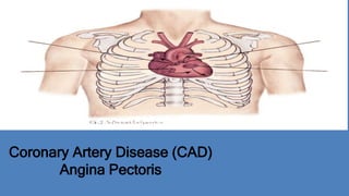
5. Coronary Artery Disease (CAD)
• DESCRIRTION:
• Is an insidious, progressive disease that result in coronary arterial
narrowing or complete occlusion.
• ETIOLOGY
- Atherosclerosis - Thrombosis - Spasm - Coronary dissection
- Aneurysm formation
8. Pathophysiology
• Atherosclerotic plaque narrows lumen of artery
• Angina: discrepancy between oxygen supply and demand causes
hypoxia
• Plaque rupture and coronary thrombosis
• Plaque regression is possible with change in risk factors
10. • Acute Coronary Syndrome
is used to describe the array of clinical presentation of CAD that
range from unstable angina to acute myocardial infarction
• ANGINA:
Is a blockage or spasm of a coronary artery leading to diminished
blood supply to the myocardium. ( coronary ischemia)
Acute Coronary Syndrome (ACS)
11. Type Of Angina Pectoris:
Stable Angina
Unstable Angina
Variant Angina
Angina Pectoris
12. Stable Angina
• Is predictable and caused by similar precipitating factors each time
such as exercise, emotional upset and tachycardia; is the result of
fixed lesions
• Pain control is achieved by rest and sublingual or intravenous
administration of the coronary artery vasodilator nitroglycerin
13. • More intense, different from stable angina. May awaken the person
from sleep or may necessitate more than nitrate for pain relief, and
the change in the level or frequency of symptom.
• PREINFARCTION OR CRESCENDO ANGINA : Severe angina
that persists for more than 15 minutes and is not relieved by three
nitroglycerin tablets
Unstable Angina
14. • Caused by coronary artery spasm with or without atherosclerotic
lesions.
• It commonly occurs when the individual is at rest and also can be
cyclic occurring at the same time every day. Smoking, alcohol and
cocaine use may also precipitate spasm.
• Drug of choice for variant angina are agents that vasodilate the
coronary arteries such as nitroglycerin or calcium channel
blockers.
Variant Angina (Prinzmetal Angina)
15. • Objective ECG evidence of myocardial ischemia (ST-segment
changes) without the patient experiencing any symptoms of
angina.
• Individual with co-existing CAD and diabetes mellitus are
particularly susceptible to silent ischemia and silent myocardial
infarction (MI).
Silent Ischemia
16. • Location : *beneath sternum , radiating to neck and jaw upper chest.*beneath
sternum , radiating down left arm.*upper chest*neck and jaw* epigastric
*epigastric radiating to neck, jaw and arm*left shoulder and intrascapular
• Duration: * 0.5 to 15 minutes (stable).* Duration longer than 15 min without relief
from rest or medication indicates unstable angina or preinfarction symptoms.
• Quality: *sensation of pressure or heavy weight on the chest. *Burning sensation *
shortness of breath , with feeling of suffocation*feeling of tightness.*most severe
pain ever experienced
Characteristics Of Angina Pectoris
17. • Radiation: * jaw * left shoulder * right arm * medial aspect of left arm.
• Precipitating factors: * Exertion/exercise* cold weather* exercising after a large
heavy meal *emotional upset* fright, anger* walking against the wind* coitus.
• Medication relief: Usually within 45 seconds to 5 minutes of sublingual nitroglycerin
administration
Characteristics Of Angina Pectoris
18. – The major goals of medical therapy for angina as an acute
coronary syndrome are:
1. Increase coronary artery perfusion to the myocardium
2. Prevent myocardial infarction disability or death
3. Actively intervene in acute coronary syndromes
Medical Management
19. – The pharmacologic treatment of choice are vasodilation by
nitroglycerin intravenous antiplatelet agents such as the
glycoprotein IIb/IIIa inhibitors, aspirin ,and IV heparin
– Another option is to take the patient directly to the cardiac
catheterization laboratory for direct visualization of the coronary
arteries and recanalization by the cardiologist.
Medical Management
20. • Nursing intervention focus on :
– Assessment of chest pain: location, duration, quality, radiation,
medication relief and precipitating
– Relief of pain: in the critical care unit control of angina pain is
achieved by a combination of supplemental oxygen, nitrates,
analgesia (morphine) and surveillance of the angina and of the
effects of pharmacologic therapy.
Nursing Management
21. – Maintain a calm environment: ensuring that the elements of a
calm environment that will alleviate the patient’s fear and anxiety
are maintained
– Coronary precautions: PATIENT BED REST
Nursing Management
22. – Doing ECG when ever the patient have chest pain
– Doing cardiac enzymes (CPK, CK-MB,T.I)
– Patient education : points to cover include risk factor
modification, signs and symptoms of angina, when to call the
physician, medications, and dealing with emotions and stress.
Nursing Management
24. • DESCRIRTION:
Irreversible myocardial necrosis due to an abrupt decrease or
total cessation of coronary blood flow to a specific area of the
myocardium
• The three mechanism that are primarily responsible for the acute
reduction in oxygen delivery to the myocardium are:
– Plaque rupture
– New coronary artery thrombosis
– Coronary artery spasm
Myocardial Infarction (MI)
25. • Zone of Ischemia:
The outer region of the
myocardium and is composed of
viable cells. (T-wave inversion)
• Zone of Injury:
The area surround the infarcted
zone but still potentially viable
tissue. (elevated ST segments)
Pathophysiology
26. • Zone of Infarction:
The area of cellular death and
muscle necrosis in the
myocardium.
( development of pathologic
Q waves)
Pathophysiology
27. MI are classified according to
their location on the myocardial
surface and the muscle layers
affected:
• Transmural MI (Pathologic
Q-wave MI):
involves all three muscle
layers the endocardium,
myocardium, and
epicardium
Classification of MI
28. • Nontransmural MI (Non-Q-wave MI):
are classified as either
Subendocardial : involving the
endocardium
Subepicardial : involving the
epicardium
• Some Myocardium may be
involved in nontransmural MI but
it is not a full thickness MI.
Generally abnormal Q wave are
not seen.
Classification of MI
29. • It begins with the end of QRS
complex, and extends to the
beginning of T wave
S-T segment
30. • If depressed or elevated,
indicates physiological or
organic changes
S-T segment
33. ST SEGMENT
NORMAL ST SEGMENT
ST segment < 2-3 small square (80 to 120 ms)
ST segment is isoelectric
and at the same level as
subsequent PR-interval
34. Variable Shapes Of ST Segment Elevations in AMI
Goldberger AL. Goldberger: Clinical Electrocardiography: A Simplified Approach. 7th
ed: Mosby Elsevier; 2006.
35. • The ECG manifestation that are used to diagnose an MI and pinpoint the
area of damaged ventricle include inverted T waves, ST-segment elevation,
and pathologic Q wave.
Surface of left ventricle ECG leads coronary artery usually involved
• Inferior II,III,aVf Right coronary artery (RCA)
• Lateral V5,V6,I,aVL Left Circumflex (LCX)
• Anterior V2,V3,V4 Left Anterior Descending (LAD)
• Septal V1,V2 Left Anterior Descending (LAD)
• Posterior V1,V2 (indirect) Left circumflex or RCA
V7,V8,V9(direct)
Myocardial Infarction Location
39. The definitive diagnosis of MI is based on a combination of
• Clinical symptoms: The most common is prolonged severe chest
pain (last 30 min or more), which often associated with nausea,
vomiting, and diaphoresis. Pain is located in the substernal or left
precordial area like an elephant sitting on my chest. The pain may
be radiate to the back, neck, jaw or left arm. Neither rest nor
nitrates relieve the pain
Assessment and Diagnosis
40. • 12-lead ECG changes: The ECG manifestation that are used to
diagnose an MI and pinpoint the area of damaged ventricle include
inverted T waves, ST-segment elevation, and pathologic Q waves
• Cardiac enzyme levels: To confirm the diagnosis of acute MI,
serum CK-MB isoenzymes, and Troponin I or Troponin T
Assessment and Diagnosis
41. • Bradycardias
• Bundle branch block
• Varying degrees of heart block
• Atrial and ventricular dysrhythmias
Dysrhythmias and Acute MI
42. • Tissue ischemia
• Hypoxemia
• Autonomic nervous system
influences
• Metabolic derangement
• Acid-base imbalances
• Hemodynamic abnormalities
• Drugs, especially digoxin toxicity
• Electrolyte imbalances
(K+ and Mg++)
• Fiber stretch (dilation and
cardiomyopathy)
Etiology of Dysrhythmias in MI
43. • Dysrhythmias
• Ventricular
aneurysm (see
picture)
• Ventricular septal
defect (see picture)
• Papillary muscle
rupture
• Pericarditis
• Cardiac rupture
• Sudden death
• Heart failure
• Pulmonary edema
• Cardiogenic shock
Complications of MI
46. Clinical Guideline Address the issue of
• Recanalization of the coronary artery: the essential immediate
interventions are fibrinolytic therapy or PCI to open occluded artery
for the patient with an acute STEMI
• Anticoagulation: in acute phase after STEMI, HEPARIN is
administered in combination with fibrinolytic therapy to open the
coronary artery.
• Dysrhythmia Interventions: the antidysrhytmic with the best safety
Medical Management
47. record after STEMI is amiodarone. Beta-blockers also are
recommended for all patients after STEMI.
• Tight Glucose Control: control Glucose during acute phase and
after MI improves survival
• Prevention of Ventricular Remodeling: Many patient are risk for
development Heart Failure (HF) after STEMI. Vasodilators drugs as
ACEIs or ARBs can stop or limit the ventricular remodeling that leads
to HF
Medical Management
48. Nursing intervention focus on :
• Patient assessment : monitoring the patient for dysrhythmia ;
evaluating vital signs for hemodynamic deterioration ; auscultating
breath sounds for signs of pulmonary congestion ; listening to heart
sounds for abnormalities ; evaluating side effects from the
medication
• Control of angina pain: continued ischemic pain represents
myocardium at risk; pain can be controlled by nitroglycerin and
morphine. If the patient is within 12 hour window in which the
Nursing Management
49. myocardium can be salvaged. PTCA or stent is the intervention
of choice. If cardiac catheterization is not available thrombolytic
therapy is used.
• Balance myocardial oxygen supply and demand and optimize
cardiac output:
Myocardial oxygen supply increased by the use of positive
inotropic drugs such as dopamine and dobutamine
Avoiding negative inotropic drugs such as beta blockers ;
Nursing Management
50. Give supplemental oxygen
To decrease cardiac work and myocardial oxygen consumption
bed rest with commode privileges is usually supplied during the
first 24-48 hrs.
• Prevent Complications:
Put the patient in upright position to foster better lung expansion
and also decreases venous return which lower preload and
decrease cardiac work ;
Nursing Management
51. Deep breathing decreases the risk of atelectasis ;
Avoid increasing intraabdominal pressure ( Valsalva maneuver)
and give stool softeners to lessen the risk of constipation from
analgesics and bed rest ;
Controls the critical care environment by decreasing noise ,
diminishing sensory overload , and allowing adequate rest periods ;
Because appetite is poor in such patients in first 24 hrs, Give a light
diet.
Nursing Management
52. • Patient education:
if the person arrives at the hospital after the window of time has
passed when the myocardium can be saved, the patient is
educated to clarify the reason for admission to the critical care
unit and the important of avoiding straining when coughing,
moving, or using the commode or bathroom.
Nursing Management