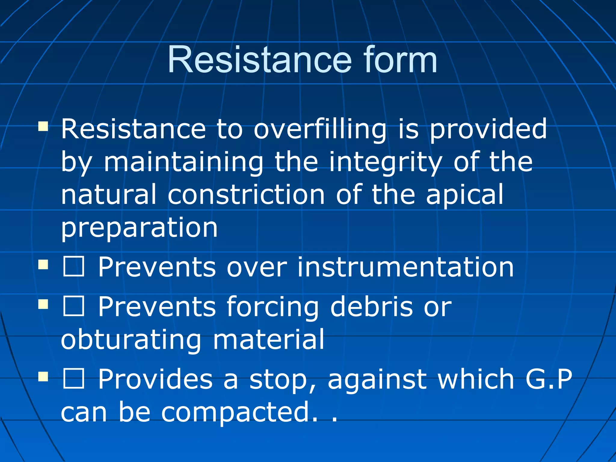
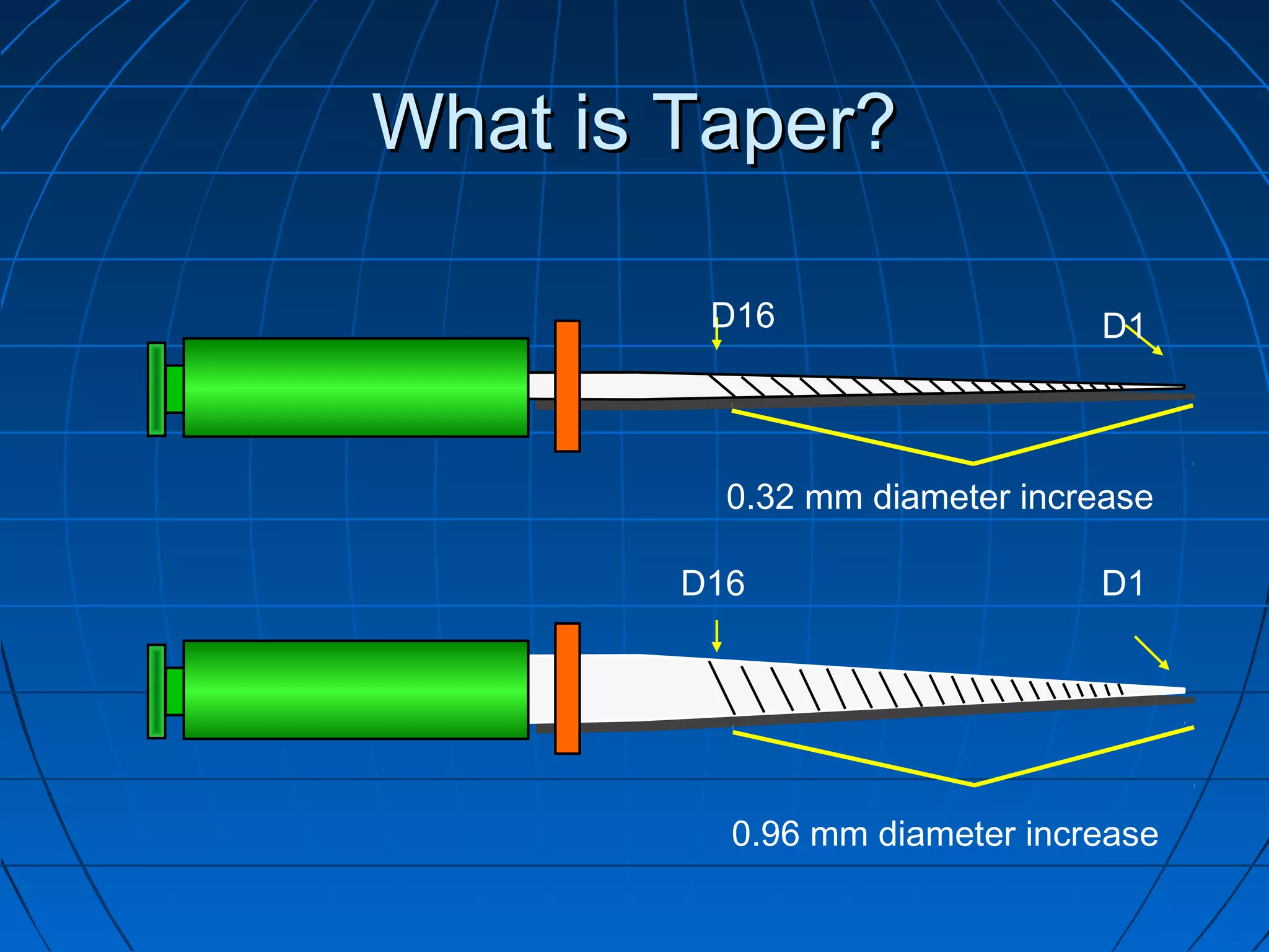
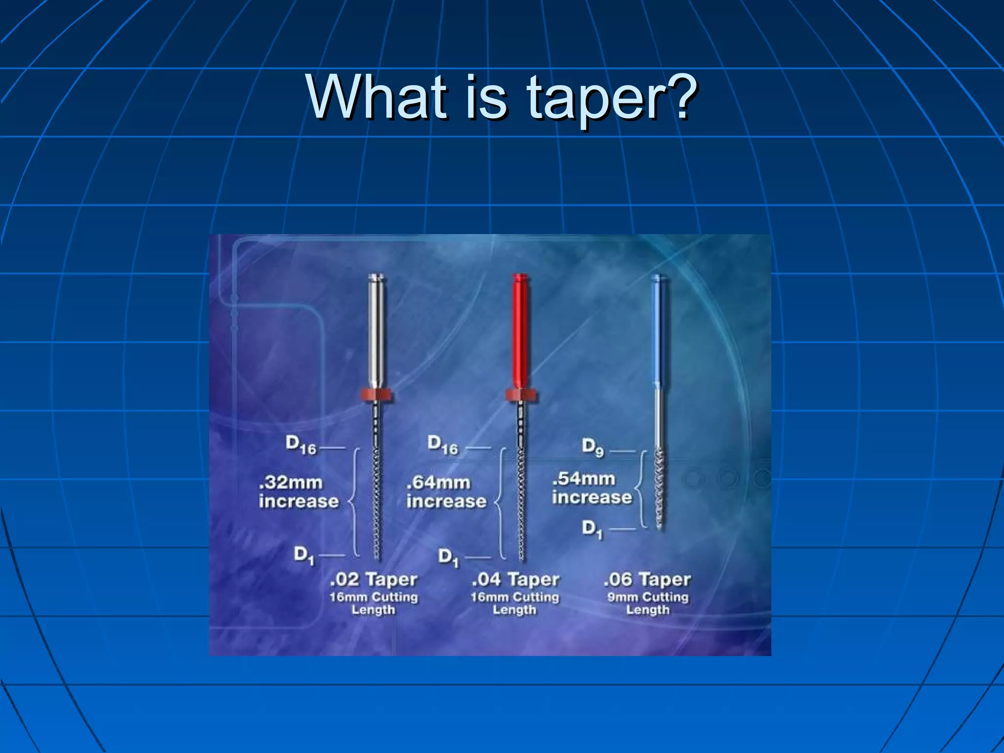
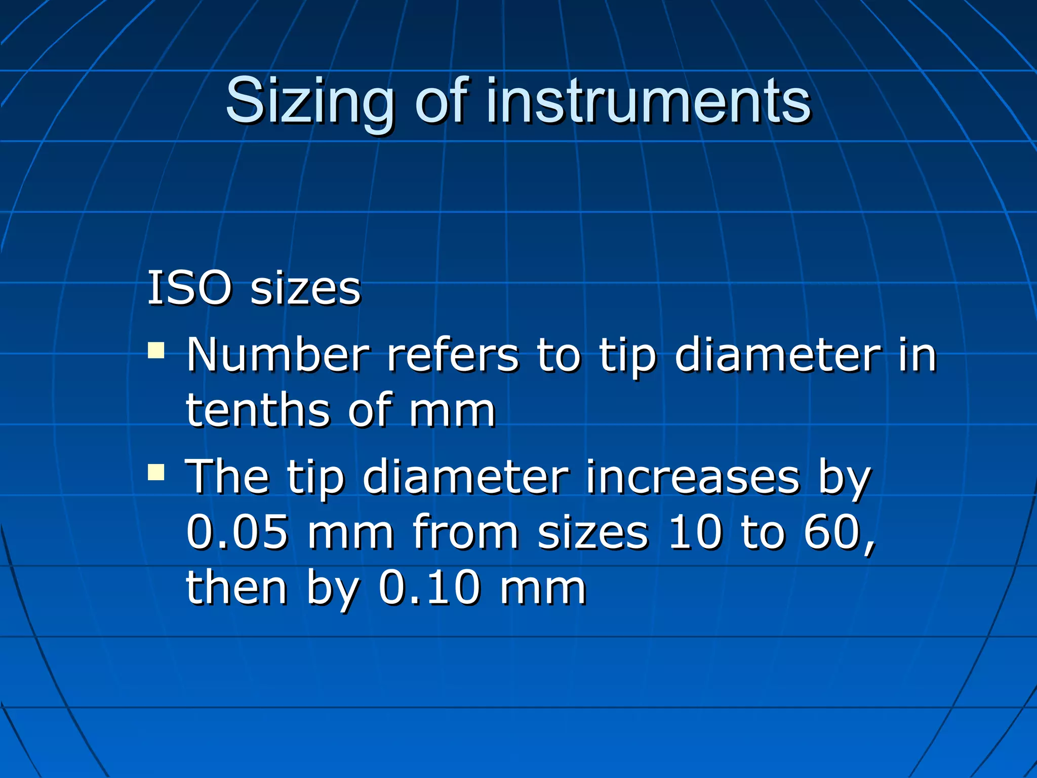
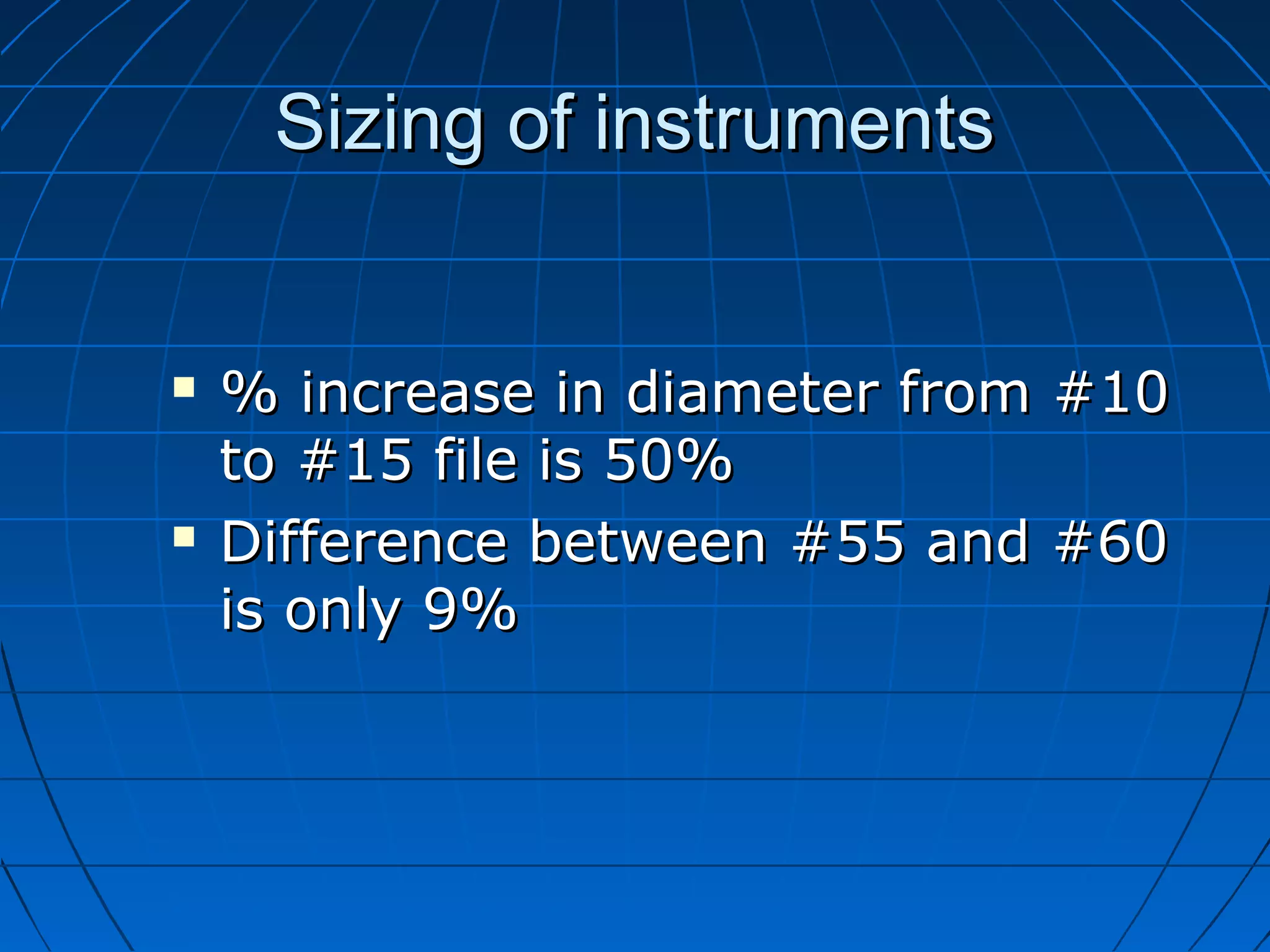
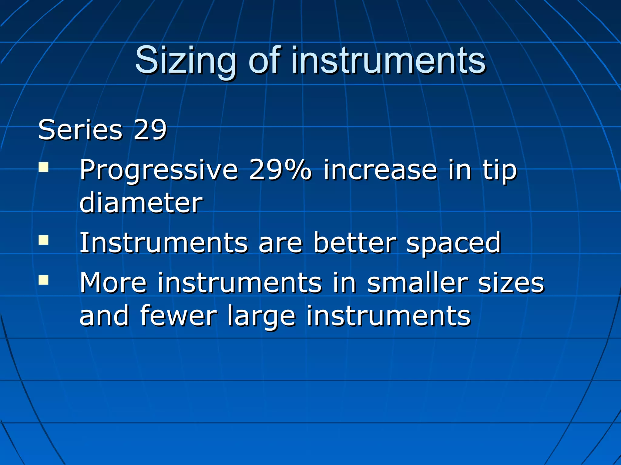
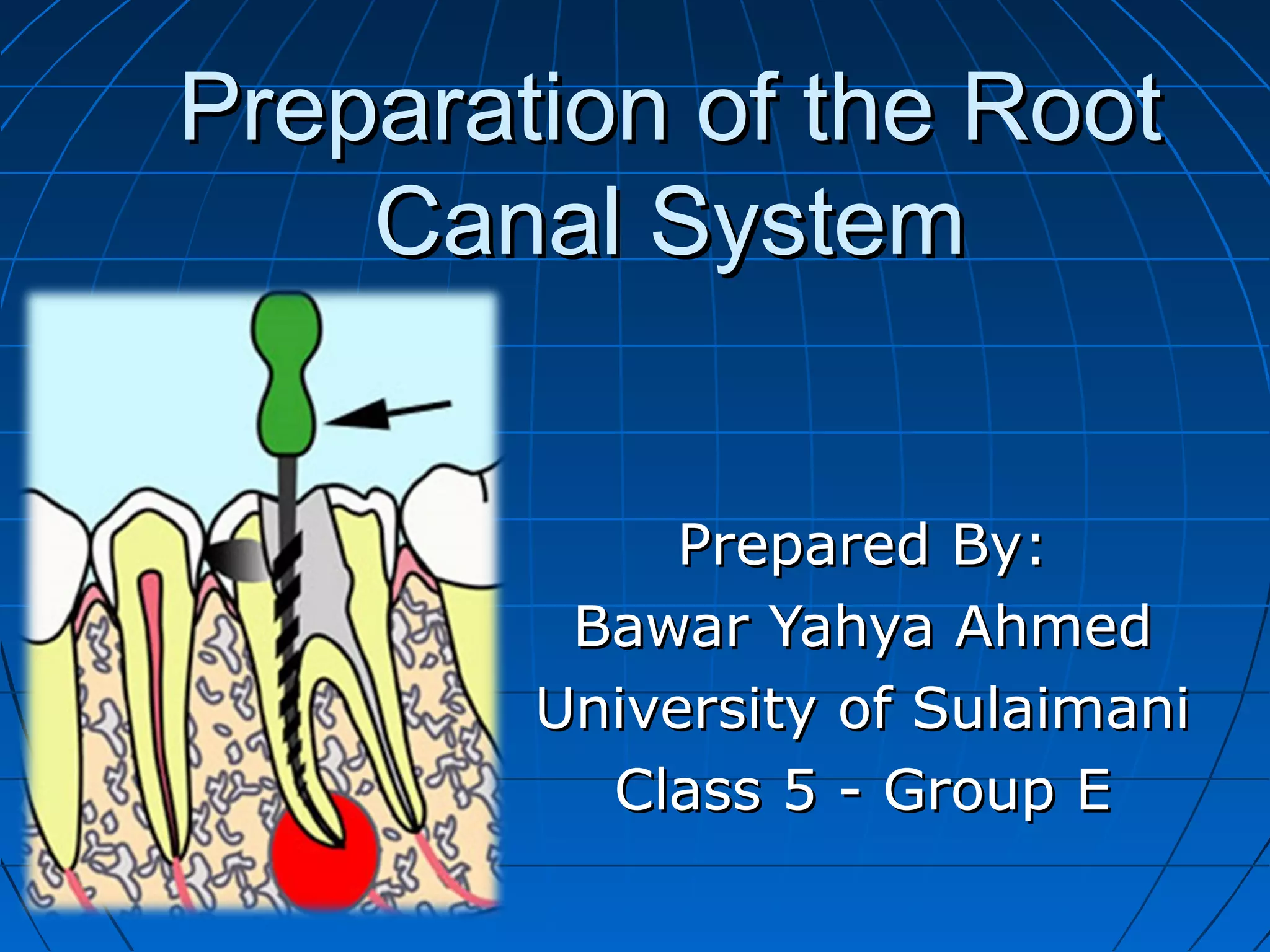
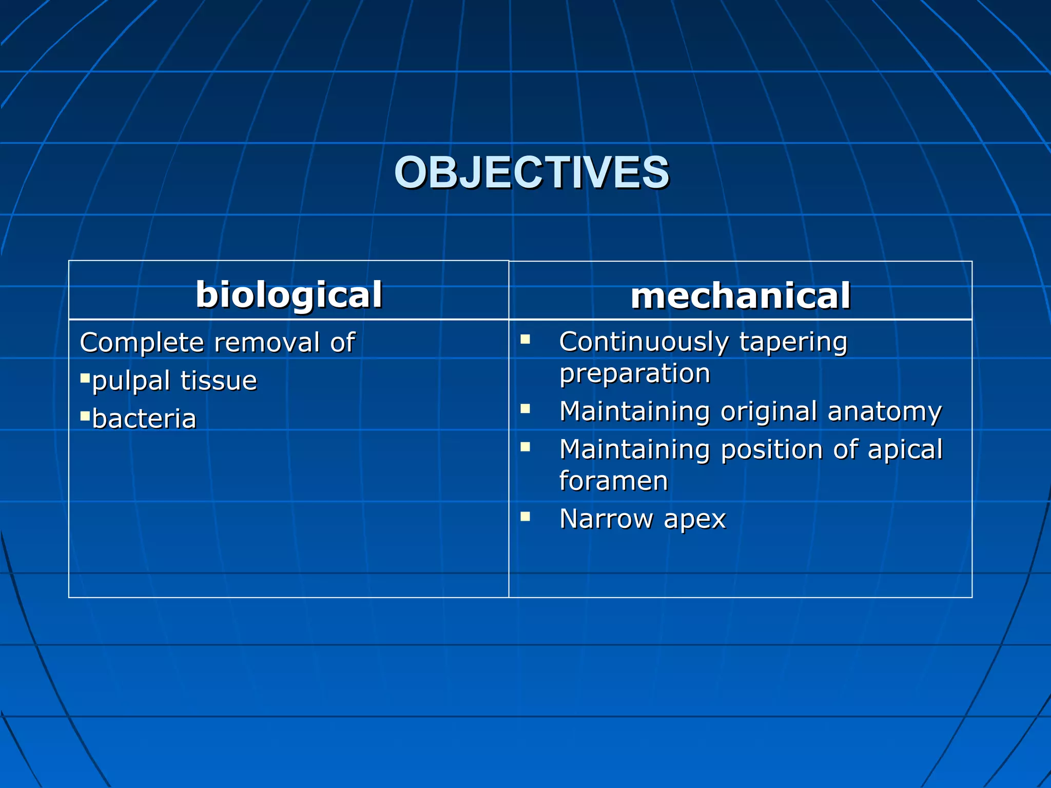
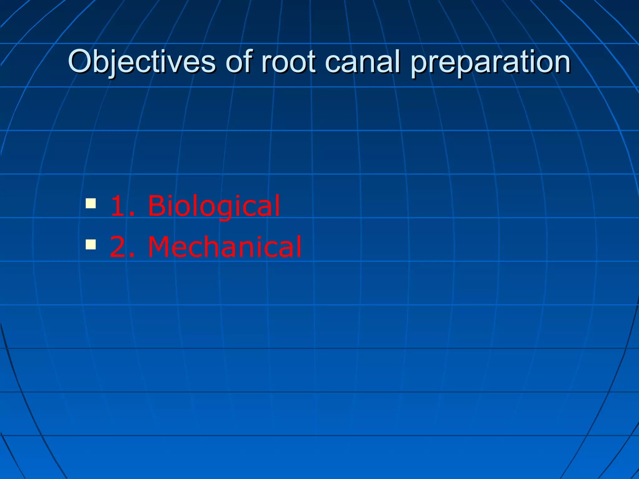
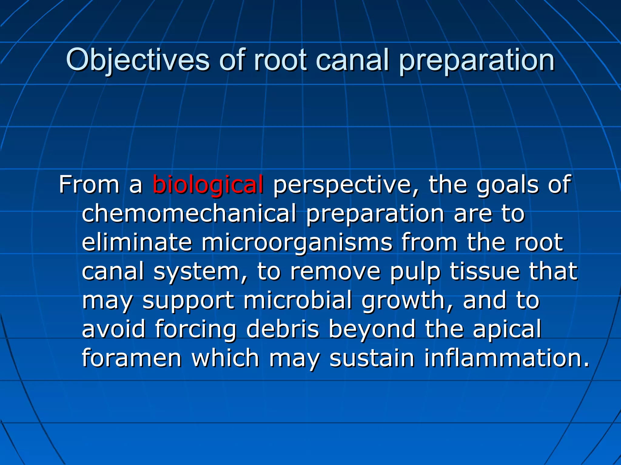
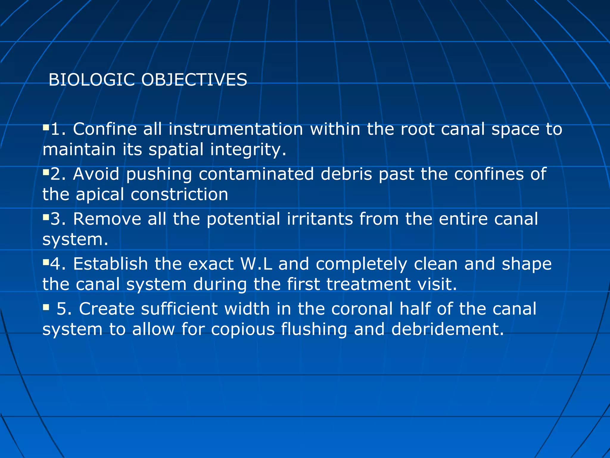
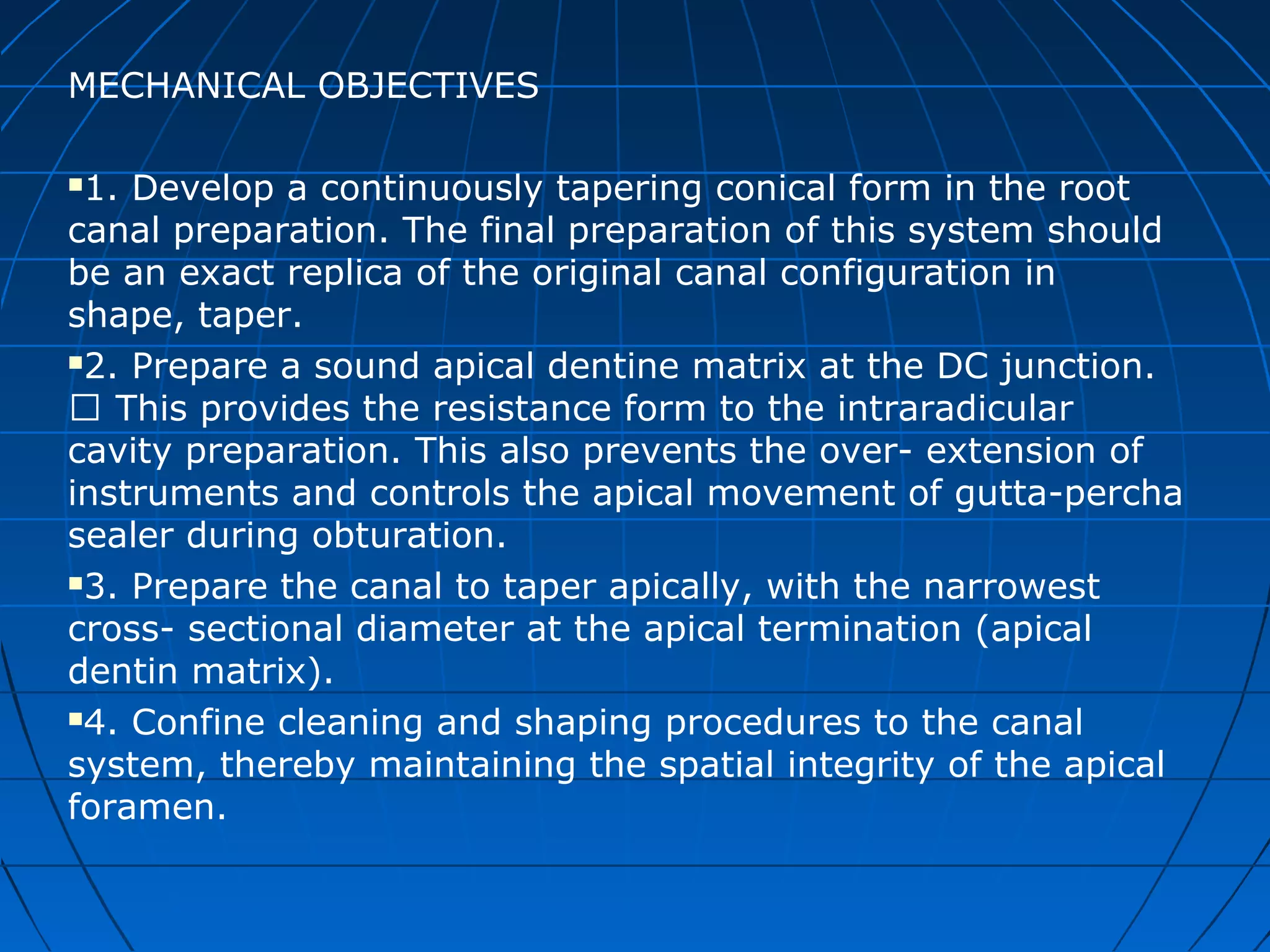
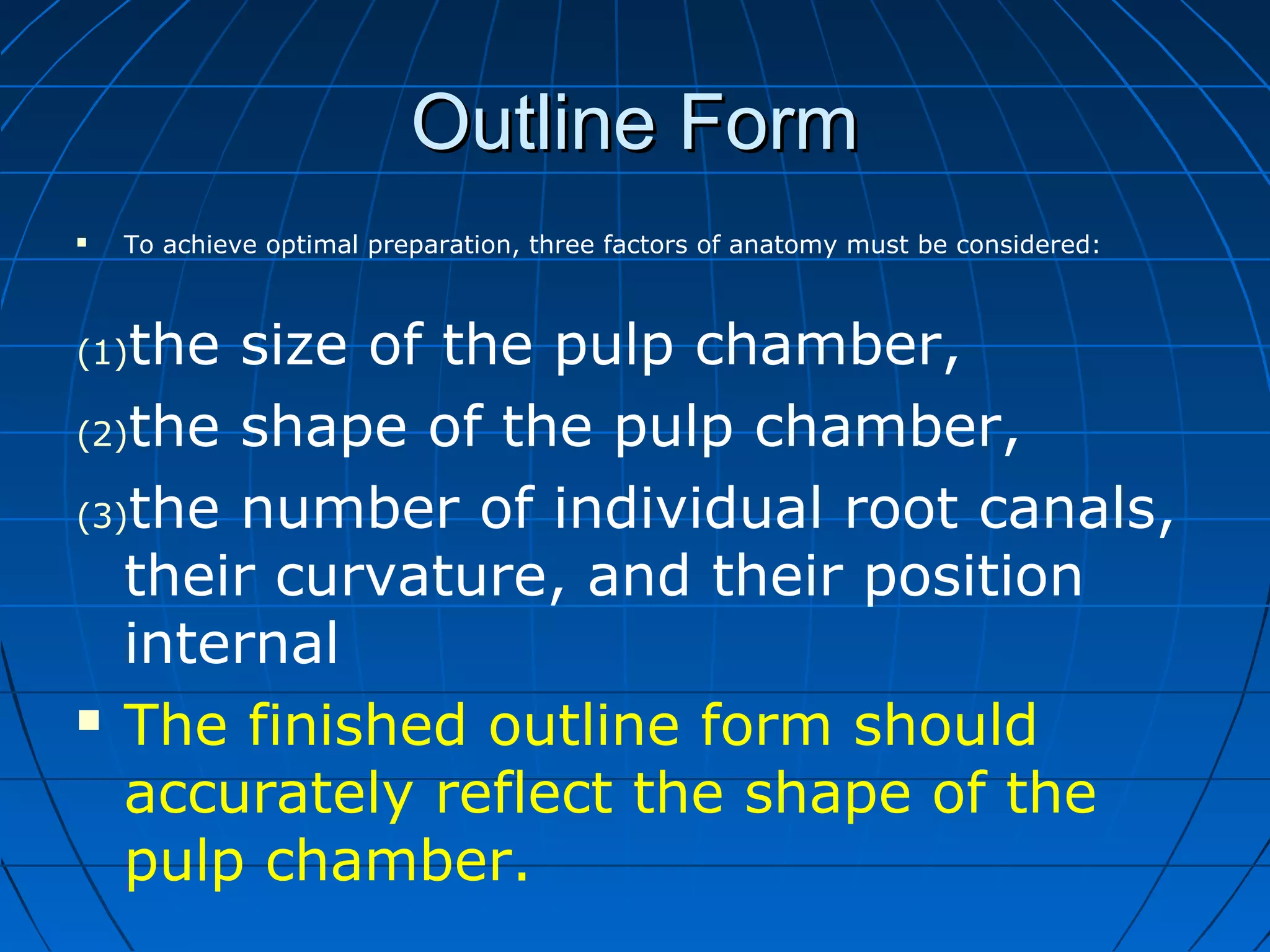
The document discusses the preparation of the root canal system through cleaning and shaping. It defines cleaning as the removal of contents from the root canal to eliminate bacteria, and shaping as the mechanical process of establishing a continuous taper to the canal to allow for better instrumentation, irrigation, and obturation. The objectives of preparation are to remove all irritants from the canal biologically and to develop a tapered conical form that maintains the original canal anatomy mechanically. Principles of preparation include outlining the canal shape, removing debris, and developing retention and resistance forms through appropriate tapers and diameters.

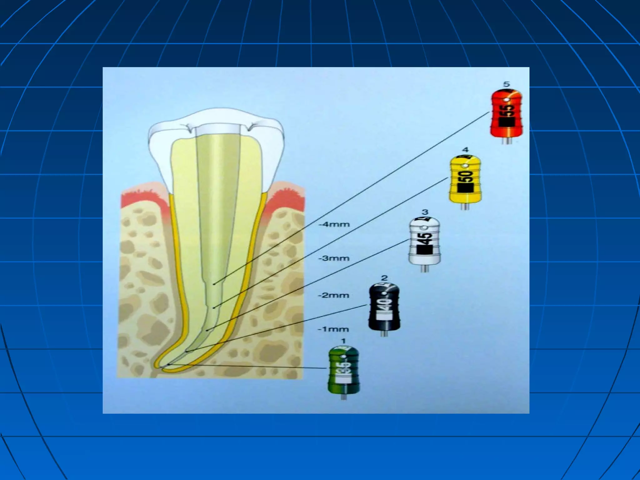
![Retention form
Near parallel walls in the apical 2-3
mm ensure a snugly fitting G.P
[ Apical TUG BACK ] .
Most crucial for preventing apical
leakage.](https://image.slidesharecdn.com/preparationoftherootcanalsystem-150422135640-conversion-gate02-181210174958/75/Preparation-of-the-root-canal-system-20-2048.jpg)