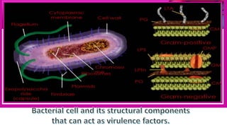
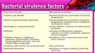
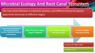
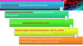
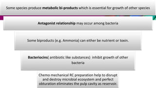
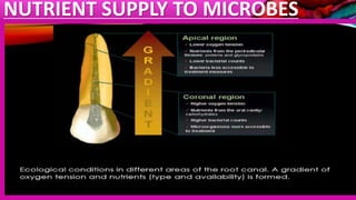
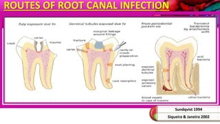
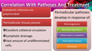
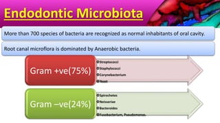
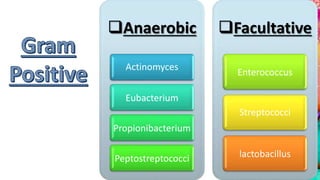
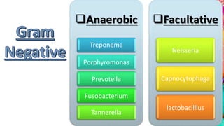
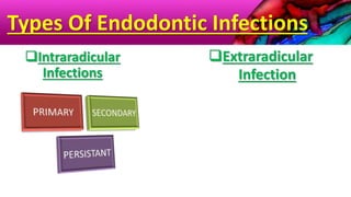
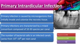
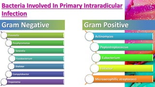
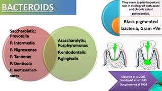
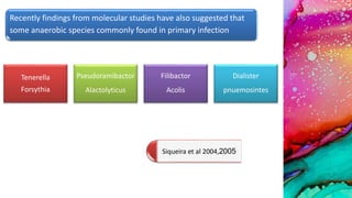
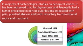
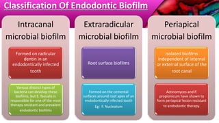
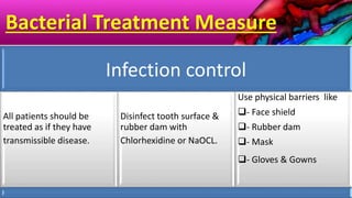
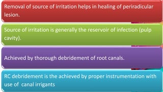
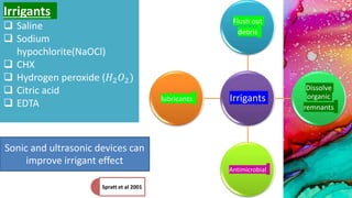
This document provides an overview of endodontic microbiology. It begins with an introduction to how microorganisms cause pulp and periradicular infections. It then discusses the mechanisms of microbial pathogenicity and virulence factors. It describes the microbial ecology of the root canal ecosystem and how the environment changes over time. It outlines the various routes of root canal infection and the typical microbes involved in primary, secondary, and persistent intraradicular infections. It also discusses extraradicular infections. The document covers biofilm formation and bacterial interactions. It concludes with discussing treatment measures and references.
This document discusses endodontic microbiology. It begins by explaining the importance of effective diagnosis and treatment of endodontic infections. It then discusses the various portals of entry for microorganisms into the root canal system. The document classifies bacteria found in endodontic infections and discusses their pathogenicity and virulence factors. It describes the different types of intra-radicular and extra-radicular infections and highlights Enterococcus faecalis as the bacteria most commonly associated with persistent infections. The final sections discuss biofilms in endodontic infections and methods to identify bacteria through culture, Gram staining, and molecular diagnostic techniques like DNA-DNA hybridization and polymerase chain reaction.
After reading this chapter, the student should be able to:
1. Understand the microbial etiology of apical
periodontitis.
2. Describe the routes of entry of microorganisms to the
pulp and periradicular tissues.
3. Recognize the different types of endodontic infections
and the main microbial species involved in each one.
4. Understand the bacterial diversity within infected root
canals.
5. Describe the factors involved with symptomatic
endodontic infections.
6. Understand the ecology of the endodontic microbiota
and the features of the endodontic ecosystem.
7. Discuss the role of microorganisms in the outcome of
endodontic treatment.
8. Understand the development and implications of
extraradicular infections.
Microbiology plays an important role in endodontic infections. Bacteria enter the root canal system through caries, periodontal disease, trauma, or cracks in the tooth. The root canal system becomes infected as bacteria colonize necrotic pulp tissue. Primary endodontic infections involve polymicrobial communities containing 10-30 bacterial species per canal, most of which are strict anaerobes. Key pathogens involved in endodontic disease include black-pigmented Prevotella and Porphyromonas bacteria, as well as Enterococcus faecalis, Fusobacterium, and Candida albicans. Bacterial virulence factors like lipopolysaccharide and capsules allow pathogens to evade the
1) Apical periodontitis is caused by microbial infection of the root canal system, usually after pulp necrosis. Bacteria are the primary cause, though fungi and archaea have also been implicated.
2) Bacteria enter the root canal system through caries, cracks, restorative procedures, or periodontal disease. They can also travel through dentinal tubules or directly expose the pulp.
3) Successful endodontic treatment aims to prevent or resolve apical periodontitis by thoroughly debriding and disinfecting the root canal system.
1. The document discusses the use of antibiotics in endodontic infections and summarizes various topics related to antibiotics including classification, endodontic microbiota, routes of infection, types of infection, commonly used antibiotics, and indications for systemic antibiotic use.
2. It classifies antibiotics based on chemical structure, mechanism of action, spectrum of activity, type of action, and source. It also discusses the endodontic microbiota and ecological factors that influence root canal infections.
3. The document outlines when systemic antibiotics may be indicated as an adjunct to endodontic treatment, such as for acute apical abscesses with systemic involvement, progressive infections, or persistent infections in medically compromised patients. It provides
This document discusses irrigation in endodontics. It provides an introduction to irrigation solutions and devices used, challenges of irrigation, and recent advances. It describes the ideal characteristics of endodontic irrigants and commonly used solutions such as sodium hypochlorite. Sodium hypochlorite is the current irrigant of choice and its properties, concentrations, effects on dentin, and safety considerations are discussed in detail. The document concludes by emphasizing the importance of irrigation in endodontic treatment.
Microbiology of Endodontic Infection.Mechanisms of MicrobialPathogenicity and Virulence Factors
Biofilm and Community-Based Microbial Pathogenesis
Biofilm and Bacterial Interactions
Biofilm Community Lifestyle
Quorum Sensing—Bacterial Intercommunication
Methods for Microbial Identification
Diversity of the Endodontic Microbiota
Primary Intraradicular Infection
Spatial Distribution of the Microbiota
Microbial Ecology and the Root Canal Ecosystem
Secondary/Persistent Infectionsand Treatment Failure
Biofilm is a microbial community characterized by cells attached to a surface and embedded in an extracellular matrix. Biofilms form in root canals and on materials placed in root canals. They are resistant to disinfection and prevent healing. Sodium hypochlorite, chlorhexidine, and MTAD are used to eradicate biofilms, but they often persist. Advanced techniques like lasers, photodynamic therapy, and ultrasound improve disinfection but sometimes biofilms still remain.
This document discusses the management of deep carious lesions. It begins by defining deep carious lesions as those that penetrate deeply into the dentin, potentially involving over half of the dentin thickness. It then covers the response of the pulpo-dentinal complex at different stages of carious lesion progression. This includes the cellular changes that occur as caries advances through enamel and into dentin. The document also discusses arrested caries, histopathology of carious dentin, effective depth of remaining dentin and its impact on pulpal response, prognosis of deep lesions, reparability of the pulpo-dentinal complex, and various treatment modalities including indirect pulp capping and stepwise excavation.
Biodentine is a new tricalcium silicate-based restorative cement that can be used as a dentin substitute with superior physical and biological properties compared to MTA. It sets faster than MTA due to the addition of calcium chloride as an accelerator. Upon setting, Biodentine releases calcium ions that stimulate reparative dentin formation and pulp healing. Studies show Biodentine forms a stronger bond to dentin and achieves higher mechanical strengths than MTA, making it suitable for various restorative, endodontic and pulp capping procedures.
Ultrasonics have various applications in endodontics. They can be used to refine root canal access and remove calcified deposits, detached pulp stones, and intracanal obstructions like broken instruments. Ultrasonics increase the flushing action of irrigants by generating acoustic streaming that more effectively cleans root canal walls and difficult anatomical features. They also aid in removing posts and silver points with minimal damage to tooth structure. Overall, ultrasonics provide a safe and effective method for various challenges in endodontic treatment.
Veneers are a conservative treatment to improve the appearance of teeth. They involve bonding thin facings of tooth-colored material to the front of teeth. Veneers can be made of composite resin, porcelain, or other ceramic materials. They are either bonded directly during a single appointment or indirectly with impressions taken and veneers fabricated by a dental laboratory over two appointments. Veneers can repair damage, close gaps, change the shape or length of teeth, or improve discoloration while minimizing the removal of tooth structure. The choice of material and technique depends on the specific needs and preferences of the patient.
Biomimetic materials used in conservative dentistry & endodonticsTirthankar Bhaumik
This document discusses biomimetic materials used in conservative dentistry and endodontics. It begins by defining biomimetics as materials and processes that mimic nature. Glass ionomer cement is highlighted as a key biomimetic material that acts as a dentin substitute. It has properties similar to dentin, such as elastic modulus and thermal expansion coefficient, and adheres chemically to tooth structure. The document outlines various uses of glass ionomer cement in restorations, luting, liners, and as a root canal sealer. While modifications have improved some properties, its strength and wear resistance remain lower than natural dentin. Overall, the document examines how glass ionomer cement biomimically replaces lost dentin structure for
Presentation about Internal, or non-vital bleaching, will updated more in the future hopefully.
This presentation aims to introduce basic principles, agents, and other details of internal bleaching, as it is one of the ways to achieve the conservative approach in modern dental care.
This document provides an overview of endodontics. It defines endodontics as the branch of dentistry concerned with the morphology, physiology, and pathology of the dental pulp and periradicular tissues. The scope of endodontics includes treating diseases of the pulp, such as pulpal inflammation and infection, through both nonsurgical and surgical root canal treatment and periradicular surgery. Pathogenesis is discussed, noting that while physical and chemical factors can induce inflammation, microbial infection is essential for progression of pulpal and periradicular disease. Routes of canal infection and the focal infection theory are also summarized.
This document discusses the sequelae of pulpitis, including necrotic pulp, acute and chronic apical periodontitis, periapical abscess, periapical granuloma, periapical cyst, osteomyelitis, cellulitis, and periostitis. It describes the clinical features, radiographic appearance, and treatment for each condition. Non-surgical management of apical lesions includes conservative root canal treatment, decompression techniques, aspiration and irrigation, use of calcium hydroxide, lesion sterilization and repair therapy, and the apexum procedure.
The document discusses various aspects of root canal obturation including definitions, purposes, techniques, and materials. Obturation involves filling and sealing the cleaned and shaped root canal using gutta-percha and a sealer. The goals are to achieve a fluid-tight seal, prevent microleakage and reinfection. Common techniques include cold lateral compaction, warm vertical compaction using heat carriers, continuous wave compaction, and thermoplasticized gutta-percha injection. Carrier-based techniques like Thermafil and SimpliFill are also described. Key factors for treatment success include absence of preoperative lesions, void-free fillings, obturation within 2mm of the apex, and adequate coronal restoration
The document discusses drugs used in endodontics, including classifications of drugs based on timing of administration and route of administration. It focuses on analgesics like NSAIDs and acetaminophen used to manage endodontic pain, as well as corticosteroids, which are potent anti-inflammatory drugs that can be administered intracanally or systemically to reduce post-treatment pain and inflammation when used as an adjunct to endodontic therapy. Clinical studies show corticosteroids significantly reduce the incidence and severity of post-operative endodontic pain within 24 hours when administered either intracanally or systemically.
This document discusses the process and effects of intracoronal bleaching. It involves placing bleaching agents such as superoxol and sodium perborate into the tooth chamber. The bleaching solution is then heated using a bleaching stick or light curing unit and repeated until the desired color is achieved. Some potential side effects include tooth sensitivity, effects on enamel and the pulp, decreased bond strength of composites, and toxicity from ingesting high concentrations of hydrogen peroxide.
This document discusses various obturation techniques. It introduces lateral compaction and warm vertical compaction as two primary methods. Warm vertical compaction involves vertically compacting gutta-percha into the prepared canal with heated pluggers. Variations include single/continuous wave compaction. Warm lateral compaction uses an electrically heated spreader to laterally condense softened gutta-percha. The document provides details on the procedure, advantages, and limitations of different obturation methods.
This document compares and contrasts microabrasion and macroabrasion techniques for treating tooth discoloration. Microabrasion involves using a paste of hydrochloric acid and pumice or silicon carbide to dissolve and abrade the enamel surface. It is a conservative treatment that removes only superficial stains. Macroabrasion uses diamond or carbide burs to remove defects at a faster rate but with less control than microabrasion. Both techniques can improve the appearance of teeth affected by conditions like fluorosis but microabrasion allows for better control and more conservative removal of tooth structure.
This document discusses the history and uses of calcium hydroxide in pediatric dentistry. It describes how calcium hydroxide has been used since the early 20th century for its antibacterial properties and ability to induce hard tissue formation. The document outlines key developments in the understanding and application of calcium hydroxide, including its introduction as a root canal filling material in 1920 and subsequent uses in pulp capping, pulpotomy, apexification, and as an intracanal medicament. The history section covers improved formulations and vehicles for calcium hydroxide from the 1930s-1960s.
Smear layer is a controversial topic in the field of operative dentistry and endodontics. This presentation includes composition, concepts, structure, advantages, disadvantages, and removal methods of smear layer.
The document discusses root canal irrigants and their importance in endodontic treatment. It provides a detailed history of irrigants used in endodontics from the early 20th century to present day. It describes the ideal properties and classifications of irrigants including chlorine-releasing agents, oxidizing agents, chelating agents, organic acids, and others. Sodium hypochlorite is discussed in depth, outlining its antimicrobial mechanisms of action, tissue dissolving properties, and recommended concentrations. The document emphasizes the critical role of irrigants to fully disinfect the complex root canal system.
Restoration of endodontically treated teethNivedha Tina
This document discusses various methods for restoring endodontically treated teeth. It covers the history of restoring pulpless teeth dating back to 1747. Key factors in selecting a restorative approach include the amount of residual root and crown structure remaining as well as the tooth's function and position. Methods discussed include direct composite restorations, indirect restorations, fiber posts, cast posts, and full crowns. The document also addresses the vulnerability of root filled teeth to fracture and emphasizes the importance of preserving tooth structure.
This document provides an overview of minimally invasive dentistry (MID), including:
1. MID aims to conserve healthy tooth structure using prevention, remineralization, and minimal intervention. It focuses on performing only necessary dentistry using long-lasting materials.
2. Key principles of MID include disease control, remineralization of early lesions, avoiding removal of excess tooth structure, and using strong, long-lasting materials.
3. Techniques discussed include caries diagnosis/risk assessment, various caries removal methods like air abrasion and lasers, fissure sealants, preventive resin restorations, ART, and chemo-mechanical caries removal.
Indian Dental Academy: will be one of the most relevant and exciting training center with best faculty and flexible training programs for dental professionals who wish to advance in their dental practice,Offers certified courses in Dental implants,Orthodontics,Endodontics,Cosmetic Dentistry, Prosthetic Dentistry, Periodontics and General Dentistry.
causes and mangment of post endodontic diseasepraveen_512
The document discusses causes and management of post-treatment endodontic disease. It identifies several potential causes of post-treatment apical periodontitis including persistent or reintroduced intraradicular microorganisms, extraradicular infection, foreign body reaction, and true cysts. Persistent intraradicular infections are the major cause, with bacteria able to survive through biofilm formation, entering dormant states, and activating stress response genes. Extraradicular infections may also develop through biofilm formation on root surfaces. While some case reports have suggested non-microbial causes, microbes are often difficult to rule out as a contributing factor.
Fortunately we are not alone and we provide residence to numerous microbial
communities comprising of bacterial species. The human body is made up of 10 14 cells of which
only 10% are mammalian and the remainder is contributed by the microorganisms that make up
the resident micro flora of the host. Normal microbial flora is a term that denotes the population
of microorganism that inhabit the skin and mucous membranes of healthy normal persons.
Numerous bacterial species colonize the mouth, upper airways, skin, vagina and intestinal tract
of humans where each one have a unique site specific fingerprint made of distinct microbe with
high level of diversity which is still unexplained.These microorganism are inhabit in the oral
cavity and their unavoidable interrelationships are essential component in maintaining
homeostasis between health and disease moreover the skin and mucous membrane always harbor
variety of organisms and they can be further categorized into two groups. One is the resident
flora which consists of relatively fixed type of microorganisms regularly found in given area at a
given age, and promptly gathers and re-establishes itself even if it is disturbed. The other one is
the transient flora, consist of non-pathogenic or potentially pathogenic microorganisms that
inhabit skin or mucous membrane for hours, days or week and it is derived from the environment
that does not produce disease and does not have the capacity to reestablish permanently on the
surface. However if the resident flora is disturbed, transient microorganisms may get colonized,
proliferate and produce disease. This indigenous microbiota plays an important role in health and
diseases of humans and contributing to the development of the immune system and provides
resistance to colonization by pathogenic microorganisms. Thereby the presentation is intended to
review on the importance and thrust areas of oral microbiome in health and disease.
ORAL MICROBIOME.pptx by UMNA FATIMA- BIOMEDumnajmi123
This PowerPoint presentation provides a thorough exploration of the oral microbiome and its significance in both maintaining health and contributing to disease. Beginning with an introduction to the oral microbiome, the presentation outlines its diverse composition and its crucial role in oral health. It further examines the concept of dysbiosis within the oral microbiome, highlighting the factors contributing to imbalance and its implications for oral and systemic health. The presentation also delves into emerging research linking oral microbiome dysbiosis to systemic diseases, shedding light on potential mechanisms and clinical implications. Methods for studying the oral microbiome are discussed, along with recent advancements in research methodologies and therapeutic strategies targeting microbial dysbiosis. Additionally, the presentation explores the evolving field of precision dentistry and its integration with oral microbiome analysis for personalized treatment approaches. Through case studies and examples, the audience gains insight into the practical applications of oral microbiome research. The presentation concludes with a summary of key points and an invitation for questions and discussion, emphasizing the importance of ongoing research in understanding and harnessing the potential of the oral microbiome for improving health outcomes.
This document provides an overview of endodontic microbiology. It discusses apical periodontitis as an infectious disease primarily caused by bacterial infection of the root canal system. The document outlines the various routes through which root canal infection can develop, such as through exposed dentin tubules or lateral/apical foramina. It also discusses the historical evidence demonstrating the role of bacteria in endodontic disease and pulpal pathology. Spatial distribution of microbiota within the root canal system and the development of endodontic biofilms are addressed.
Symposium oral micro flora /certified fixed orthodontic courses by Indian den...Indian dental academy
The Indian Dental Academy is the Leader in continuing dental education , training dentists in all aspects of dentistry and offering a wide range of dental certified courses in different formats.
Indian dental academy provides dental crown & Bridge,rotary endodontics,fixed orthodontics,
Dental implants courses.for details pls visit www.indiandentalacademy.com ,or call
0091-9248678078
1) The document discusses root canal microflora and provides details on the classification, sources, and types of microorganisms commonly found in endodontic infections.
2) It describes the stages of root canal infection and notes that primary endodontic infections typically involve a mixed consortium of 10-30 anaerobic bacterial species.
3) Virulence factors of pathogenic bacteria that can contribute to endodontic disease are discussed and include lipopolysaccharides, enzymes, fatty acids, and capsules.
Endodontic microbiology /certified fixed orthodontic courses by Indian denta...Indian dental academy
Welcome to Indian Dental Academy
The Indian Dental Academy is the Leader in continuing dental education , training dentists in all aspects of dentistry and offering a wide range of dental certified courses in different formats.
Indian dental academy has a unique training program & curriculum that provides students with exceptional clinical skills and enabling them to return to their office with high level confidence and start treating patients
State of the art comprehensive training-Faculty of world wide repute &Very affordable.
This document provides an overview of the microbiological and immunological aspects of the microbial-host interaction in periodontal disease. It discusses the various bacterial species involved, including the "red complex" bacteria Porphyromonas gingivalis, Treponema denticola, and Tannerella forsythia. It describes the virulence factors of these bacteria and how they evade host defenses. It also summarizes the innate and adaptive immune response phases, focusing on the acute inflammatory response and roles of neutrophils in controlling bacterial challenge through opsonization and phagocytosis.
This document discusses the microbial flora of the oral cavity. It describes the various microbial habitats in the mouth and the acquisition of normal flora beginning at birth. The major types of flora are discussed as well as factors that modulate microbial growth. The document also examines the oral microbiome's role in oral diseases and conditions like dental caries, periodontal disease, and infections. Additionally, it summarizes how orthodontic appliances can impact the oral microbiome by facilitating plaque accumulation and altering the microbial environment. Maintaining proper oral hygiene is emphasized when wearing orthodontic appliances.
This chapter discusses fungi that cause endodontic infections, particularly the yeast Candida albicans. It covers the characteristics of fungi, including their cellular structure and ability to grow in different morphological forms. It also discusses the prevalence and virulence factors of oral yeasts, especially C. albicans. Key virulence factors include the yeast's ability to transition between different growth forms, adhere to surfaces, and produce enzymes. The chapter examines the role of these factors in pathogenicity and reviews literature on the presence of yeasts in different dental tissues and their ability to infect root canals and surrounding areas. It concludes by assessing the antifungal activity of various endodontic irrigants and medications.
Anaerobic bacteria: Infection and Managementiosrjce
IOSR Journal of Dental and Medical Sciences is one of the speciality Journal in Dental Science and Medical Science published by International Organization of Scientific Research (IOSR). The Journal publishes papers of the highest scientific merit and widest possible scope work in all areas related to medical and dental science. The Journal welcome review articles, leading medical and clinical research articles, technical notes, case reports and others.
This document provides an overview of the microbiology of periodontal diseases. It begins with an introduction to the complex microbial flora found in the oral cavity and periodontal diseases as polymicrobial infections. The document then covers the historical perspectives on periodontal disease pathogenesis including the non-specific plaque hypothesis, specific plaque hypothesis, and updated hypotheses. Key microbial complexes and pathogens associated with periodontal diseases are discussed. The rest of the document focuses on dental plaque as a microbial biofilm, including its formation, structure, properties that contribute to pathogenicity, and gene regulation within biofilms.
This document discusses oral microbiology and the microorganisms associated with dental diseases. It begins by introducing the normal oral microflora and then discusses the microflora found in dental caries, root canals, and the periodontal pocket. It explores the importance of studying bacterial pathogenesis and how microbiology relates to dentistry. Key findings are that dental caries and periodontal disease are caused by shifts in the microbial environment that allow pathogenic bacteria like Streptococcus mutans and Porphyromonas gingivalis to dominate. Maintaining a balanced oral microbiome is important for oral health.
The primary goal of the present book is to produce a comprehensive text that fully integrates the latest concepts and techniques in management of odontogenic infections. The main aim is to provide the readers with an update information regarding pathophysiology, clinical and radiographic presentation, microbiology, diagnosis, management, and complications of odontogenic infections. Accordingly, the text has been divided into six chapters. Chapter one is concerned with oral microbiology and immunology. Chapter two is dealing with the pathophysiology of odontogenic infections. In chapter three, management of odontogenic infections is presented. In chapter four, antibiotic therapy of odontogenic infections is given. Chapter five deals with life-threatening complications. In chapter six osteomyelitis of the jaws is discussed.
1. Oral bacterial biofilms can enter the bloodstream through disruption caused by oral procedures like dental cleanings, tooth extractions, or chewing. This leads to transient bacteremia.
2. Viridans group streptococci and Actinomyces odontolyticus are among the oral bacteria most commonly found in the bloodstream, and they have attributes like adhesion that allow them to cause infections in other parts of the body.
3. Procedures that cause more tissue trauma like tooth extractions, periodontal probing, and endodontic treatments have a higher risk of resulting in bacteremia compared to less invasive activities like brushing or flossing. The presence of gum
The Indian Dental Academy is the Leader in continuing dental education , training dentists in all aspects of dentistry and
offering a wide range of dental certified courses in different formats.
The document discusses the historical evidence for bacteria playing a primary role in the etiology of periodontal disease. Early investigations from 1880-1930 suggested a variety of microorganisms could be involved, including amebae, spirochetes, fusiforms, and streptococci. While therapies targeting these organisms showed some success, their precise roles remained unclear due to limitations of detection techniques. Subsequently, stronger evidence accumulated from studies of acute infections, correlations of plaque/disease, antibiotic treatment trials, host immune responses, animal models, and more advanced microbiology approaches. Overall a bacterial etiology of periodontal disease became firmly established, though the field continues refining understanding of specific pathogen identities and disease associations.
The Indian Dental Academy is the Leader in continuing dental education , training dentists in all aspects of dentistry and
offering a wide range of dental certified courses in different formats.
This document provides an overview of histamines and antihistamines. It discusses how histamine is synthesized and its effects mediated through four types of histamine receptors. It then focuses on H1 receptor antagonists (antihistamines), describing their classification, pharmacological actions, and uses in treating allergic disorders and motion sickness. Newer non-sedating antihistamines are also covered. The document concludes by discussing applications of antihistamines in dentistry, such as in treating allergic reactions to local anesthetics.
Pain is an unpleasant sensory and emotional experience caused by actual or potential tissue damage. It serves a protective function by warning us of damage. Pain is transmitted through specialized pain pathways and is classified based on duration, location, and source. There are different types of pain receptors that detect various painful stimuli and transmit signals through different nerve fiber types to the central nervous system where pain is perceived.
This document discusses local anaesthesia complications including definitions, local complications like needle breakage, paresthesia, trismus, hematoma, pain on injection, infection, and ocular complications. It provides causes, prevention, and management strategies for each complication. Several case reports and studies are referenced that examine specific complications in more depth.
The document provides information on impression materials, including their history, classification, and properties. It discusses both reversible (agar) and irreversible (alginate) hydrocolloid impression materials. For agar, it describes the composition, mode of supply, uses, advantages, and disadvantages. For alginate, it outlines the composition, setting reaction, classification according to setting time, manipulation, and properties. The document thus summarizes the key types of impression materials and their characteristics.
This document provides information on various local anesthesia techniques used in dentistry. It begins with an introduction to regional anesthesia, including field blocks, nerve blocks, and local infiltration. It then describes different local anesthesia injection techniques such as supraperiosteal, intraligamentary, intraosseous, and intraseptal injections. The document proceeds to explain specific maxillary and mandibular injection techniques including posterior superior alveolar nerve block, anterior superior alveolar nerve block, greater palatine nerve block, and others. It concludes with a brief section on recent advancements in local anesthesia.
This document discusses root canal curvatures and methods for determining their degree. It begins with an introduction on the importance of understanding root canal anatomy for successful endodontic treatment. It then covers causes of canal curvatures, various classification systems, and techniques for assessing curvature using periapical radiographs, cone-beam computed tomography, and angular measurement methods. The relationship between curvature degree and ledge formation risk is also addressed, as are canal characteristics often invisible on routine radiographs. The document concludes by emphasizing the challenges of treating severely curved canals and the need for accurate preoperative assessment and proper instrumentation.
More from People's Dental Academy and Research Center (7)
Recomendações da OMS sobre cuidados maternos e neonatais para uma experiência pós-natal positiva.
Em consonância com os ODS – Objetivos do Desenvolvimento Sustentável e a Estratégia Global para a Saúde das Mulheres, Crianças e Adolescentes, e aplicando uma abordagem baseada nos direitos humanos, os esforços de cuidados pós-natais devem expandir-se para além da cobertura e da simples sobrevivência, de modo a incluir cuidados de qualidade.
Estas diretrizes visam melhorar a qualidade dos cuidados pós-natais essenciais e de rotina prestados às mulheres e aos recém-nascidos, com o objetivo final de melhorar a saúde e o bem-estar materno e neonatal.
Uma “experiência pós-natal positiva” é um resultado importante para todas as mulheres que dão à luz e para os seus recém-nascidos, estabelecendo as bases para a melhoria da saúde e do bem-estar a curto e longo prazo. Uma experiência pós-natal positiva é definida como aquela em que as mulheres, pessoas que gestam, os recém-nascidos, os casais, os pais, os cuidadores e as famílias recebem informação consistente, garantia e apoio de profissionais de saúde motivados; e onde um sistema de saúde flexível e com recursos reconheça as necessidades das mulheres e dos bebês e respeite o seu contexto cultural.
Estas diretrizes consolidadas apresentam algumas recomendações novas e já bem fundamentadas sobre cuidados pós-natais de rotina para mulheres e neonatos que recebem cuidados no pós-parto em unidades de saúde ou na comunidade, independentemente dos recursos disponíveis.
É fornecido um conjunto abrangente de recomendações para cuidados durante o período puerperal, com ênfase nos cuidados essenciais que todas as mulheres e recém-nascidos devem receber, e com a devida atenção à qualidade dos cuidados; isto é, a entrega e a experiência do cuidado recebido. Estas diretrizes atualizam e ampliam as recomendações da OMS de 2014 sobre cuidados pós-natais da mãe e do recém-nascido e complementam as atuais diretrizes da OMS sobre a gestão de complicações pós-natais.
O estabelecimento da amamentação e o manejo das principais intercorrências é contemplada.
Recomendamos muito.
Vamos discutir essas recomendações no nosso curso de pós-graduação em Aleitamento no Instituto Ciclos.
Esta publicação só está disponível em inglês até o momento.
Prof. Marcus Renato de Carvalho
www.agostodourado.com
ABDOMINAL TRAUMA in pediatrics part one.drhasanrajab
Abdominal trauma in pediatrics refers to injuries or damage to the abdominal organs in children. It can occur due to various causes such as falls, motor vehicle accidents, sports-related injuries, and physical abuse. Children are more vulnerable to abdominal trauma due to their unique anatomical and physiological characteristics. Signs and symptoms include abdominal pain, tenderness, distension, vomiting, and signs of shock. Diagnosis involves physical examination, imaging studies, and laboratory tests. Management depends on the severity and may involve conservative treatment or surgical intervention. Prevention is crucial in reducing the incidence of abdominal trauma in children.
Does Over-Masturbation Contribute to Chronic Prostatitis.pptxwalterHu5
In some case, your chronic prostatitis may be related to over-masturbation. Generally, natural medicine Diuretic and Anti-inflammatory Pill can help mee get a cure.
Promoting Wellbeing - Applied Social Psychology - Psychology SuperNotesPsychoTech Services
A proprietary approach developed by bringing together the best of learning theories from Psychology, design principles from the world of visualization, and pedagogical methods from over a decade of training experience, that enables you to: Learn better, faster!
These lecture slides, by Dr Sidra Arshad, offer a quick overview of the physiological basis of a normal electrocardiogram.
Learning objectives:
1. Define an electrocardiogram (ECG) and electrocardiography
2. Describe how dipoles generated by the heart produce the waveforms of the ECG
3. Describe the components of a normal electrocardiogram of a typical bipolar lead (limb II)
4. Differentiate between intervals and segments
5. Enlist some common indications for obtaining an ECG
6. Describe the flow of current around the heart during the cardiac cycle
7. Discuss the placement and polarity of the leads of electrocardiograph
8. Describe the normal electrocardiograms recorded from the limb leads and explain the physiological basis of the different records that are obtained
9. Define mean electrical vector (axis) of the heart and give the normal range
10. Define the mean QRS vector
11. Describe the axes of leads (hexagonal reference system)
12. Comprehend the vectorial analysis of the normal ECG
13. Determine the mean electrical axis of the ventricular QRS and appreciate the mean axis deviation
14. Explain the concepts of current of injury, J point, and their significance
Study Resources:
1. Chapter 11, Guyton and Hall Textbook of Medical Physiology, 14th edition
2. Chapter 9, Human Physiology - From Cells to Systems, Lauralee Sherwood, 9th edition
3. Chapter 29, Ganong’s Review of Medical Physiology, 26th edition
4. Electrocardiogram, StatPearls - https://www.ncbi.nlm.nih.gov/books/NBK549803/
5. ECG in Medical Practice by ABM Abdullah, 4th edition
6. Chapter 3, Cardiology Explained, https://www.ncbi.nlm.nih.gov/books/NBK2214/
7. ECG Basics, http://www.nataliescasebook.com/tag/e-c-g-basics
Histololgy of Female Reproductive System.pptxAyeshaZaid1
Dive into an in-depth exploration of the histological structure of female reproductive system with this comprehensive lecture. Presented by Dr. Ayesha Irfan, Assistant Professor of Anatomy, this presentation covers the Gross anatomy and functional histology of the female reproductive organs. Ideal for students, educators, and anyone interested in medical science, this lecture provides clear explanations, detailed diagrams, and valuable insights into female reproductive system. Enhance your knowledge and understanding of this essential aspect of human biology.
2. Contents
Introduction
Mechanisms of Microbial
Pathogenicity
Microbial Ecology and the Root
Canal Ecosystem
Routes of canal infection
Endodontic microbiota
Types of endodontic infections
Biofilm and bacterial interaction
Bacterial treatment measures
Conclusion
References
3. INTRODUCTION
Microorganisms cause virtually all pathoses of the
pulp and the periradicular tissues.
To effectively treat endodontic infections, clinicians
must recognize the cause and effect of microbial
invasion on the dental pulp space and the surrounding
periradicular tissues.
4. Mechanisms of Microbial Pathogenicity and
Virulence Factors
Pathogenicity
Virulence
Virulence factors
7. Microbial Ecology And Root Canal Ecosystem
In the very initial phases of
the pulpal infectious
process: facultative bacteria
predominate.
After a few days or weeks,
oxygen is depleted
loss of blood circulation in
the necrotic pulp.
Growth of obligate
anaerobic bacteria.
The root canal infection is a dynamic process, and different bacterial species
apparently dominate at different stages.
Sundqvist & Figdor 2003,
Sundqvist 1994
8. Necrotic pulp cavity becomes reservoir for microbes
Disintegrated tissue and fluids becomes nutrients for
microorganism
Nutrients are polypeptide and amino acids
Nutrient supply + Bacterial interactions + low 𝑶 𝟐 tension
determine the type of predominant bacteria.
Growth of anaerobes provides environment for
metabolization of peptides and amino acids
9. Some species produce metabolic bi-products which is essential for growth of other species
Antagonist relationship may occur among bacteria
Some biproducts (e.g. Ammonia) can either be nutrient or toxin.
Bacteriocins( antibiotic like substances) inhibit growth of other
bacteria
Chemo mechanical RC preparation help to disrupt
and destroy microbial ecosystem and perfect
obturation eliminates the pulp cavity as reservoir.
11. ROUTES OF ROOT CANAL INFECTION
Sundqvist 1994
Siqueira & Janeiro 2002
12. Correlation With Pathoses And Treatment
Endodontic infections are
polymicrobial
Periradicular tissues posses
Excellent collateral circulation
Lymphatic drainage.
Vast amount of undifferentiated
cells.
Periradicular pathoses
develop in response of
Microorganisms
Microbial biproducts
Inflammatory mediators
Instrument trauma
Chemicals
13. Endodontic Microbiota
More than 700 species of bacteria are recognized as normal inhabitants of oral cavity.
Root canal microflora is dominated by Anaerobic bacteria.
Streptococci
Staphylococci
Corynebacterium
Yeast
Gram +ve(75%)
Spirochetes
Neisseriae
Bacteroides
Fusobacterium, Pseudomonas.
Gram –ve(24%)
16. Types Of Endodontic Infections
Intraradicular
Infections
Extraradicular
Infection
17. Primary Intrardicular Infection
Primary infection is caused by microorganisms that
initially invade and colonize the necrotic tissue.
Primary infections are characterized by a mixed
consortium composed of 10-30 species per canal.
The number of bacterial cells in an infected canals
varies from 103-108 per root canal
Siqueira et al 2005
Sakamoto et al 2007
Vianna et al 2006
19. Saccharolytic;
Prevotella
P. Intermedia
P. Nigrescense
P. Tannerae
P. Denticola
P. multissachari-
vorax
Asaccharolytic;
Porphyromonas
P.endodontalis
P.gingivalis
BACTEROIDS
They seem to play important
role in etiology of both acute
and chronic apical
periodontitis.
Black pigmented
bacteria, Gram +Ve
Siqueira et al 2001
Sundqvist et al 1989
Dougherty et al 1998
20. Recently findings from molecular studies have also suggested that
some anaerobic species commonly found in primary infection
Tenerella
Forsythia
Pseudoramibactor
Alactolyticus
Filibactor
Acolis
Dialister
pnuemosintes
Siqueira et al 2004,2005
21. In majority of bacteriological studies on periapical lesions, it
has been observed that Porphyromonas and Prevotella had a
higher prevalence in periradicular lesions associated with
pain, purulent abscess and teeth refractory to conventional
root canal treatment.
Kiryu et al. 1994
Trowbridge & Stevens 1992
Bogen &Slots 1999
Yamasaki et al. 1998
22. Gram negative obligate nonmotile anaerobic bacteria.
Most common in symptomatic infection and abscess or
exacerbation lesion than asymptomatic infection.
5 subspecies of F. Nucleatum have been found which are different in genetic
data but no study found that which subspecies have more virulence factor.
Different type of subspecies are found in the same root canals.
Fusobacterium Nucleatum
Moraes et al 2002
Siqueira et al 2005
23. Spirochete
Although spirochetes have been frequently observed in samples by microscopy, they had
never been identified to the species level.
The application of molecular diagnosis to identification of spiral bacteria has been
overlooked the culture techniques.
They can be classified in 2 group; Saccharolytic and Asaccharolytic.
T. Denticola, T. Sokranskii have been detected in both symptomatic and asymptomatic
lesion.
Dewhirst et al 2000
Baumgartner et al 2007
Rocas et al 2003
24. Secondary Intraradicular Infections
If microorganisms are allowed to remain at the time of filling , there
is increased risk of adverse outcome of the endodontic treatment
Sjogren et al 1997
Waltimo et al 2005
26. Persistant Intraradicular Infection
Infections caused by microorganisms that resisted the
intracanal antimicrobial procedures.
Fungi and Gram positive facultative bacteria,
particularly Enterococcus faecalis, are predominant in
such cases.
27. E. Faecalis
Non spore forming, fermentative Facultative
anaerobic, Gram positive coccus.
This species has been found in low prevalence value in
case of primary infection and more relate in
asymptomatic cases than symptomatic cases.
Rocas et al 2005
28. E. feacalis has been found in root filled teeth evincing
persistent apical periodontitis in prevalence values ranging
from 30-90% of the cases.
This species can be inhibited by other members of mixed
bacterial consortium commonly present in primary
infection.
Sedgley et al 2006
29. Virulence factors of E. Faecalis
But its still not confirmed
which factor play an important
role in pathogenesis.
Kayaoglu et al 2004
Lytic enzymes Cytolysin Gelatinase Hyaluronidase
Pheromones
Lipoteichoic
acid
Adhesion
molecules
30. Why E. feacalis can survive in root filled
teeth?
Studies have revealed that E. feacalis has
ability to penetrate far into dentinal tubules
that can escape from intracanal
instrumentation and irrigants.
Haapasalo et al 1989
Siqueira et al 1996
31. E.feacalis has been shown to be able to form biofilms in
root canals and this ability can be important for bacterial
resistance and persistence in the instrumented canals.
Distel et al 2002
The ability to resist high pH value seems to be related to
functional proton pump to acidfy. So, is resistant to
calcium hydroxide.
Evans et al 2002
32. E.feacalis can enter a VBNC state which can survive
in adverse environmental condition, including
starvation.
They has the ability to survive in environments in scarcity of
nutrients and to flourish when the nutrient source is
reestablished and has a capacity to recover in root canal
treated teeth for 12 months without nutrients.
Figdor et al 2003
Sedgley et al 2005
33. Prevalence of E. Faecalis in samples from root canal treated teeth with apical periodontitis.
Data from Culture (Yellow bars) and data from Molecular (Blue bars) studies
34. Fungi
It is also possible that yeasts from oral cavity gain access to
root canal as contaminant during endodontic procedures.
Siren et al 1997
Siqueria & Sen et al 1997
They can overgrow after inefficient intracanal antimicrobial
procedures, which can cause imbalance in microbiota.
Siqueira et al 2004
E. faecalis and yeast, mainly C. albicans, has been repeatedly identified as
the species most commonly recovered from root canals undergoing
retreatment, in cases of failed endodontic therapy and canals with
persistent infections.
35.
36. Dentino phillic
microorganism due
to its ability to
colonize and invade
to dentin by their
hyphae.
Resist to some
intracanal
medication such as
calcium hydroxide.
Release hydrolytic
enzyme and can
form monoinfection
biofilm in root
canals.
Waltimo et al 1999
Matusow et al 1981
37. Ashraf et al 1997
Najzar & Fleger et al
CA in the root canal system of teeth with periapical lesions was more
noticeable and statistically significant compared to the teeth without
periapical lesions
55% of root canals contain Candida cell.
Presence of fungi in root canals more than expected and the knowledge of persistent periapical
infections may advocate the use of antifungal agents in modern endodontic therapy.
38. Prevalence of microorganism detected in root canal treated teeth with post treatment disease.
Data is from taxon-specific PCR assay
39. Extraradicular infections
Extra radicular infections are characterized by, microbial
invasion & proliferation in the inflamed peri radicular tissue as
a result of intra radicular infections.
The development of periradicular lesions creates a barrier with in the body
to prevent further spread of microorganisms. Bone tissue is resorbed and
substituted by a granulation tissue containing defense elements, such as
cells (phagocytes) and molecules (antibodies and complement molecules).
40. A dense wall composed of polymorphonuclear leucocytes, or less frequently an epithelial
plug, is usually present at the apical foramen, blocking the egress of microorganisms into
the periradicular tissues.
Very few endodontopathogens can advance through such barriers. However, microbial
products can diffuse through these defence barriers and are able to induce or perpetuate
periradicular pathosis
Vigil et al.1997
In recent years there is resurgence of the idea of extraradicular microbes in
apical periodontitis lesions with implied, controversial suggestion that
extraradicular infection is the cause of many failed endodontic treatment.
41. Extraradicular infection can be independent of Intraradicular infection.
Case of apical actinomyces in which non surgical endodontics, calcium
hydroxide, systemic erythromycin, and finally, a regimen of systemic
metronidazole failed to resolve the draining fistula associated with a
maxillary lateral incisor. but following periapical surgery, the lesion resolved.
Haapasalo et al.
1987
42. 16 periapical granuloma that were collected “during normal
periapical curettage, apecectomy, or retrograde filling”. It was
seen that most of the organism cultured were Veillonella species,
Streptococcus milleri, Streptococcus sanguis, Actinomycetes
naeslundii, Propionibacterium acnes and Bacteroides species
Iwu & Wallace 1990
43. Those extra radicular infections which are caused by intra
radicular bacteria are treated easily by RCT. Eg: Sinus Tract
If the extra radicular infection which are caused by outer side
are treated only by Endodontic Surgery. Eg: Actinomycosis
Cohen & Burns 2002
44. Remnants of
Dental pulp
Calculus
Caries on tooth crown
Leakage of rubber dam
Contamination of instrument
Irrigation solution
Intracanal medicaments
SOURCE OF
MICROBES
DURING
TREATMENT
45. Loss or leakage of
restorative material
Tooth fracture
SOUCE OF
MICROBES
DURING
APPOINTMENTS
46. Loss or leakage of restorative
material
Tooth fracture
Secondary caries
Delay in placement of
permanent filling or crown
prosthesis.
SOUCE OF
MICROBES
AFTER ROOT
CANAL
TREATMENT
47. Biofilm And Bacterial Interaction
Sessile multicellular
microbial community
characterized by cells that
are firmly attached to a
surface and embedded in a
self produced matrix of
extracellular polymeric
substances.
AAE, 2004
Defined as community of
microcolonies of
microorganisms in an
aqueous solution that is
surrounded by a matrix
made of glycocalyx, which
also attaches the bacterial
cell to a solid substratum.
Grossman,
13 edition
Definition
48. According to kishen
There are four distinct
stages in development of
biofilm
Formation of a
conditioning layer
Planktonic bacterial
cell attachment
Detachment (seeding
dispersal)
Bacterial growth and
biofilm expansion.
49.
50.
51. Autopoiesis
Ability to self
organize.
Homeostasis
Ability to resist
environmental
disturbances.
Synergy
More effective in
association with
fellow
microorganisms.
Communality
Response to
environmental
challenges as
combined unit.
Caldwell et al
The biofilm has following attributes that makes it resistance to
clinical
52. Communications between bacterial cells residing in a biofilm is
attained through signaling molecules by a process called as
Quorum sensing
Quorum sensing is mediated by low
molecular weight molecules- autoinducers
Qs leads to
Exchange of genetic
materials between species
Antibiotic resistance
Nutrient breakdown
Xenobiotic metabolism
Coordinated behaviour of
biofilm
53. Endodontic biofilms are therapeutically significant as they are one of the
basic survival methods for bacteria during starvation.
Endodontic biofilms help bacteria to survive because
Ability to protect
bacteria from
environment
Ability to entrap
nutrients for the
growth of microbial
constituent.
Offer a safe
environment to
exchange of genetic
material
Provide inherent
resistance to
antimicrobial agents
(irrigants & ICM)
Caldwell et al
54. Classification Of Endodontic Biofilm
Intracanal
microbial biofilm
Formed on radicular
dentin in an
endodontically infected
tooth
Various distinct types of
bacteria can develop these
biofilms, but E. faecalis is
responsible for one of the most
therapy resistant and prevalent
endodontic biofilms
Extraradicular
microbial biofilm
Root surface biofilms
Formed on the cemental
surfaces around root apex of an
endodontically infected tooth
Eg: F. Nucleatum
Periapical
microbial biofilm
Isolated biofilms
independent of internal
or external surface of the
root canal
Actinomyces and P.
propionicum have shown to
form periapical lesion resistant
to endodontic therapy.
55. Bacterial Treatment Measure
Infection control
All patients should be
treated as if they have
transmissible disease.
Disinfect tooth surface &
rubber dam with
Chlorhexidine or NaOCL.
Use physical barriers like
- Face shield
- Rubber dam
- Mask
- Gloves & Gowns
56. Removal of source of irritation helps in healing of periradicular
lesion.
Source of irritation is generally the reservoir of infection (pulp
cavity).
Achieved by thorough debridement of root canals.
RC debridement is the achieved by proper instrumentation with
use of canal irrigants
59. DRAINAGE
Key to manage an abscess and cellulitis
Achieved through the canal and incision
Reduces discomfort, toxins, pressure
Incision of indurated
swelling releases
Blood
Serous fluid
Bacteria &
biproducts
Inflammatory
mediators
Drainage removes these irritants
& improves local circulation.
60. Adjunctive Antibiotic therapy
Antibiotics are not a substitute for local
treatment.
The majority of endodontic cases can be
treated without antibiotics.
Pain & swelling of endodontic origin are
managed by debridement & drainage
Do not require antibiotics
Without systemic sign & symptoms
Symptomatic
pulpitis
Apical
periodontitis
Draining sinus tract
Localized swelling
Fouad et al.1996
Henry et al. 2001
Keenan et al. 2006
Cope et al. 2014
61. Prophylactic Antibiotics for Medically
Compromised Patients
Distant infection are high in case of transient bacteremia.
Bacteremia puts medically compromised patients at a
great risk.
Procedures that may produce bleeding induce bacteremia.
Procedures induces
bleeding
Rubber dam
Local injections
Surgical procedures
Over-instrumentation
ExtirpationPrior to surgical procedures gum &
mucosa should be disinfected with:
Chlorhexidine or iodine-glycerin
62. Medically compromised patients at great
risk of bacteremia include:
Rheumatic &
congenital
disease
Prosthetic cardiac
valves.
Valvular
prolapse&
regurgitation
Previous infective
endocarditis
Systemic
pulmonary shunts
Arterio-venous
shunts
Uncontrolled
diabetes.
Immunosuppressed
& immunologically
deficient cases
Medically
compromised patients
at risk of bacteremia
must receive a regimen
of antibiotics that
follows the
recommendations of
American Heart
Association (AHA)
63. Antibiotics
Antibiotics are prescribed in conjunction with endodontic
procedures.
Therefore antibiotics without endodontic procedures not
effective.
Antibiotic are prescribed when
Systemic involvement
Persistent infection
Spreading infection
Fever 38°c
Malaise
Trismus
Diffuse swelling
Cellulitis
Signs &
symptoms of
systemic
Involvement
& spread
infection
Alone or in
combination
Antibiotics should
be continued for 2
to 3 days after
disappearance of
signs & symptoms.
64. Selection of an Antibiotic Regimen
Penicillin remains the antibiotic of choice.
However, penicillin is allergic to approx. 10% of humans.
Adequate blood level of penicillin must be maintained.
Inexpensive
Antibiotics + proper endo procedure results in significant
improvement within 48 hours.
Effective against
many facultative
& strict
anaerobes.
Has low toxicity
Initial oral dose
of 1000mg
followed by 500
mg/6hours.
Penicillin
65. Alternative choice for patients allergic to penicillin
Ineffective against most anaerobes & serious infections.
GI upset on ingestion of milk while yogurt gives relief.
Transient deafness
Available as Erywell, Althrocin.
Effective against
facultative
bacteria.
Has low toxicity
Dose 1000mg
followed by
500mg/6 hours.
Erythromycin
66. Alternative choice for patients allergic to
penicillin
Greater antibacterial spectrum.
Less GI upset
Available as Clariwell, Klacid, Amicalri,
Claribid
Macrolide
Semisynthetic
derivative of
erythromycin
Has low
toxicity
Dose 500
mg/8-12
hours
Clarithromycin
67. Not recommended for penicillin allergic
patients.
broad spectrum but does not include
anaerobes.
long use pseudomembranous colitis.
Available as Climycin, Dalacin C, Myocin
Cefaclor(2nd
generation)
effective against
anaerobes.
Clindamycin effective
gm +ve & -ve
bacteria. Facultative
& strict anaerobes.
Dose 150 to 300
mg/6 hours.
Cephalosporin
68. Ineffective against aerobes
Metronidazole with Penicillin or other
antibiotic is effective in endo infection
Available as Flawell, Flagyl
Effective
against
anaerobes
Dose 250 to
500 mg/ 6
hours.
Metronidazole
69.
70. There is international concern about the overuse of
antibiotics and the emergence of antibiotic -resistant
bacterial strains
Dentists prescribe approximately 10% of antibiotics dispensed in primary
care, it is important not to underestimate the potential contribution of the
dental profession to the development of antibiotic resistant bacteria
In the UK, it has been reported that 40% of dentists prescribed
antibiotics at least three times each week, and 15% prescribed
antibiotics on a daily basis
Pallasch et
al. 2000
Cope et al.
2014
Lewis
2008
71. Furthermore, one Cochrane systematic review has found no evidence
to support the use of antibiotics for pain relief in irreversible pulpitis
Agnihotry et al. 2016
Thus, two systematic reviews concluded that infection must be systemic or the
patient must be febrile or immunocompromised to justify the need for antibiotics.
For these reasons, prescription of antibiotics by dentists should be limited
Matthews et al. 2003
Mohammadi 2009
Rodr íguez -Núñez et al. 2009
72. Antibiotic sensitivity of the bacteria found within the oral cavity is gradually decreasing, and a
growing number of resistant strains are being detected , in particular Porphyromonas spp. and
Prevotella spp .However, the phenomenon has also been reported for alpha haemolytic
streptococci (“Streptococcus viridans”) and for drugs such as macrolides, penicillin and
clindamycin.
Bresco -Salinas et al. 2006
Aracil et al. 2001
Groppo et al. 2004
Inappropriate use of antibiotics not only drives antibiotic resistance and misuses
resources; it also increases the risk of potentially fatal anaphylactic reactions and
exposes people to unnecessary side effects
Gonzales et al. 2001
Costelloe et al. 2010,
Cope et al. 2014
73. Culturing
Required when empirical use of antibiotics is not effective
Rubber dam isolation.
Disinfection with NaOCl or other disinfectant.
Access opening with sterile instruments
Microbial sampling with sterile paper points or aspiration.
74. Aspiration is done with 16 to 20 gauge needle.
In dry canals place a drop of a sterile solution before
sampling
Submucosal swellings should be sampled by
aspiration before incision
Samples are immediately placed in the media.
Antimicrobial irrigating solutions should not be used
before sampling.
75. Conclusion
Microbes seeking to establish in the root canal must leave the
nutritionally rich and diverse environment of the oral cavity,
breach enamel, invade dentine, overwhelm the immune
response of the pulp and settle in the remaining necrotic
tissue within the root canal.
During that time they have to compete in a limited space
with other microbes for the available nutrition.
The bacterial community profiles in treated cases vary from
individual to individual, indicating that distinct bacterial
combinations can play a role in treatment failure.
76. References
Kenneth M. Hargreaves, Stephen Cohen. Cohen’s Pathways of the
Pulp. 10th edition. Elsevier, Mosby.2011
Ingle, Bakland, Baumgartner. Ingle’s Endodontics 6. BC Decker. 2008
Sabeti M, Slots J: Herpesviral-bacterial coinfection in periapical
pathosis. J Endod 30:69, 2004
Siqueira JF, Jr, Rôças IN: Polymerase chain reaction-based analysis of
microorganisms associated with failed endodontic treatment. Oral
Surg Oral Med Oral Pathol Oral Radiol Endod 97:85, 2004.
Sakamoto M, Siqueira JF, Jr, Rôças IN, Benno Y: Molecular analysis of
the root canal microbiota associated with endodontic treatment
failures. Oral Microbiol Immunol 23:275–281, 2008.
77. Zoletti GO, Siqueira JF, Jr, Santos KR: Identification of Enterococcus
faecalis in root-filled teeth with or without periradicular lesions by
culture-dependent and –independent approaches. J Endod 32:722,
2006.
Rôças IN, Hulsmann M, Siqueira JF, Jr: Microorganisms in root canal-
treated teeth from a German population. J Endod 34:926, 2008.
Rôças IN, Siqueira JF, Jr, Aboim MC, Rosado AS: Denaturing gradient gel
electrophoresis analysis of bacterial communities associated with
failed endodontic treatment. Oral Surg Oral Med Oral Pathol Oral
Radiol Endod 98:741, 2004.
Kaufman B, Spangberg L, Barry J, Fouad AF: Enterococcus spp. in
endodontically treated teeth with and without periradicular lesions. J
Endod 31:851, 2005.
Aruna Kanaparthy, Rosaiah Kanaparthy:Biofilms-The Unforgiving Film in
Dentistry (Clinical Endodontic Biofilms) . Dentistry 2012, 2:7
Adalberto R. Vieira, Jose F. Siqueira, Domenico Ricucci. Dentinal Tubule
Infection as the Cause of Recurrent Disease and Late Endodontic
Treatment Failure: A Case Report. JOE — Volume 38, Number 2,
February 2012
78. Abou-Rass M, Bogen G(1998) Microorganisms in
closed periapical lesions. International Endodontic
Journal, 1 :39-47
Baumgartner JC, Falkler WA(1991) Bacteria in the
apical 5 mm of infected root canals. Journal of
Endodontics, 17:380–383.
Bogen G, Slots J(1999) Black-pigmented anaerobic
rods in closed periapical lesions. International
Endodontic Journal, 3:204-10.
Cohen S, Burns RC (2002) Pathways of the pulp, ed
8. Harcourt Asia Co., India,: 457-500.
Cruse WP, Bellizzi R (1980) A historic review of
endodontics, 1689–1963, part 1. Jornal of
Endodontics, 6: 495–499.
79. Dymock, D; Weightman, A J; Scully, C; Wade, W G (1996)
Molecular analysis of microflora associated with dentoalveolar
abscesses. Journal of Clinical Microbiology, 34 : 537-542
Goumas PD, Naxakis SS, Papavasiliou DA, Moschovakis ED,
Tsintsos SJ, Skoutelis A (1997) Periapical abscesses: causal
bacteria and antibiotic sensitivity. : Journal of Chemotherapy,
6:415-9.
G. Sundqvist & David Figdor (2003) Life as an endodontic
pathogen, Ecological differences between the untreated and
root-filled root canals. Endodontic Topics, 6: 3–28.
Haapasalo M, Ranta K, Ranta H (1987) Mixed anaerobic
periapical infection with sinus tract. Endodontics & dental
traumatology, 3; 83-5.
HedmanWJ (1951) An investigation into residual periapical
infection after pulp therapy. Oral Surgery Oral Medicine Oral
Pathology, 4 : 1173-9.
80. Henderson B, Wilson M (1998) Commensal communism and the
oral cavity. Journal of Dental Research, 77 : 1674–1683.
Hirshberg A, Tsesis I, Metzger Z, Kaplan I, Aviv T. Israel (2003)
Periapical actinomycosis: A clinicopathologic study. Oral Surgery
Oral Medicine Oral Pathology Oral Radiology Endodontics,
95:614-20.
Iwu C, Wallace T (1990) The microbiology of periapical granuloma.
Oral Surgery Oral Medicine Oral Pathology, 69: 502-5.
Jansen H-J, van der Hoeven JS (1997) Protein degradation by
Prevotella intermedia and Actinomyces meyeri supports the
growth of non-proteincleaving oral bacteria in serum. Journal of
Clinical Periodontology, 24: 346– 353.
Jansen H-J, van der Hoeven JS, Walji S, Goertz JH, Bakkeren JA
(1996) The importance of immunoglobulin breakdown supporting
the growth of bacteria in oral abscesses. Journal of Clinical
Periodontology, 23: 717–723.
Kiryu T, Hoshino E, Iwaku M (1994) Bacteria invading periapical
cementum. Bacteria invading periapical cementum. Journal of
Endodontics,4:169-72