Downloaded 523 times


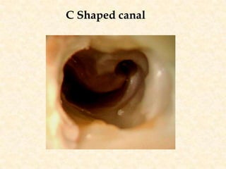
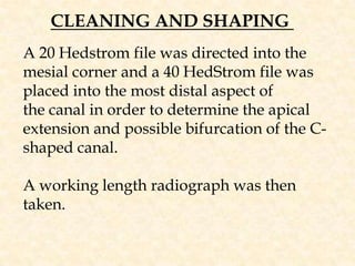
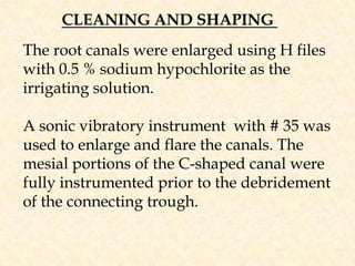
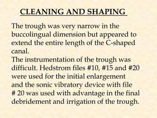
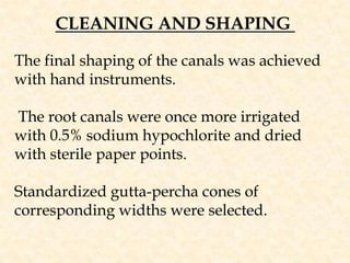


The document discusses the C-shaped canal, which occurs in approximately 1% of lower second molars. The C-shaped canal takes its name from its C-shaped appearance when viewed from above. Melton divided C-shaped canals into three types based on their shape and number of canals. The document then describes the cleaning and shaping process for a C-shaped canal, including using small files to determine the canal shape and irrigation with sodium hypochlorite. Master cones and additional gutta-percha cones are placed using guides to ensure proper placement within the C-shaped canal. Warm lateral condensation is then used to further adapt the filling to the canal anatomy.


