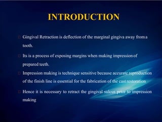
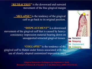
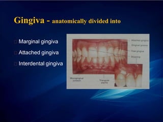
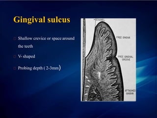
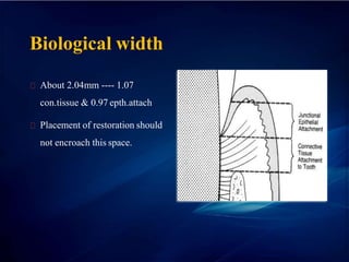
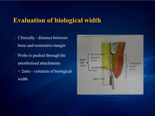
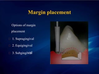
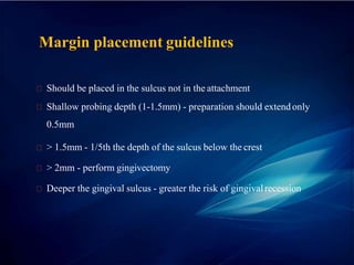
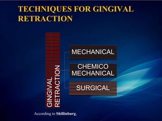
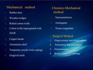
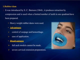
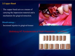
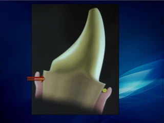
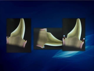
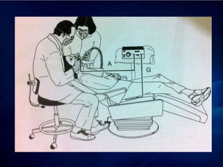
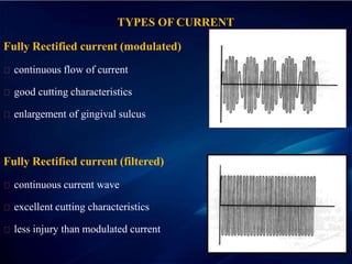
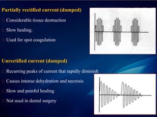
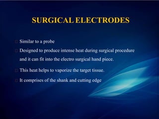
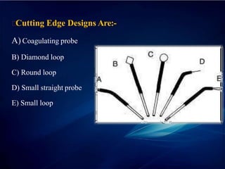
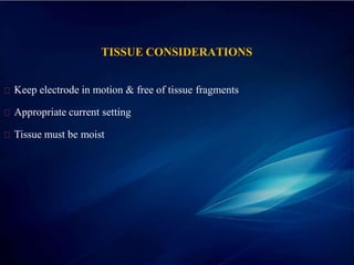
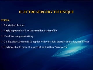
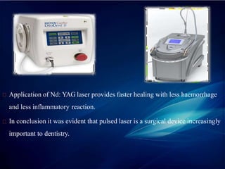
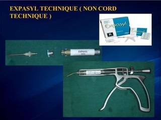
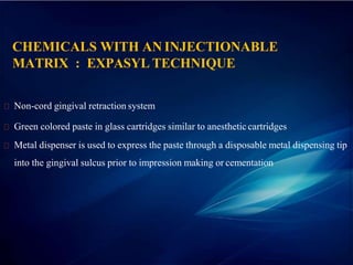
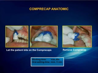
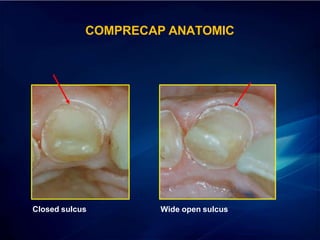
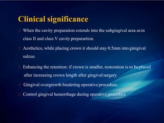
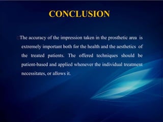
CONTENTS
Introduction
Gingival anatomy
Classification
Recent advances
Clinical significance
conclusion
INTRODUCTION
Gingival Retraction is deflection of the marginal gingiva away froma
tooth.
Its is a process of exposing margins when making impressionof
prepared teeth.
Impression making is technique sensitive because accurate reproduction
of the finish line is essential for the fabrication of the cast restoration
Hence it is necessary to retract the gingival sulcus prior to impression
making
NEED FOR GINGIVALRETRACTION
Contour of the future restoration
Patient’s comfort
Efficiency of impression material
Operators access and visibility
“RETRACTION” is the downward and outward
movement of the free gingival margin
“RELAPSE” is the tendency of the gingival
cuff to go back to its original position.
“DISPLACEMENT” is a downward
movement of the gingival cuff that is caused by heavy-
consistency impression material bearing down on
unsupported retracted gingival tissues.
“COLLAPSE” is the tendency of the
gingival cuff to flatten under forces associated with the
use of closely adapted customized impression trays
Gingival Retraction Techniques for Implants vs Teeth.
Bennani V
,Schwass D, Chandler N. J Am DentAssoc.2008;139:1354-63.
VARIOUS PHASES IN GINGIVAL DISPLACEMENT
During tooth preparation (Preparatory phase ) :-
plan the position of the cervical finish line in relation to the gingiva prior to tooth
preparation.
The gingiva must be displaced to give a clear view of the cervicalarea
During impression making ( working phase ) :-
An adequate access to the finish line should be obtained after tooth preparation is
done.
This displaces the gingiva apically and laterally to provide space for the impression
material to flow and record details.
During Cementation of Restoration (Maintenance phase ):-
The gingiva adjacent to the finish line must be displaced prior to cementation to
evaluate marginal fit and also to remove excess cement after cementation
CRITERIA FOR SELECTION OF AGINGIVAL
RETRACTION MATERIAL
According to Milford B.Reiman (1976), the gingival retraction material must
be effective enough to create a trough, free of blood and fluids and there must
be no damage to the gingiva in terms of inflammation or bleeding.
The resulting contours of the tissues must be predictable and tissue must recover
in a considerable period of time with minimal systemic or localized effects.
There are three criteria that must be satisfied by a gingival retraction
material:
- It should be effective in gingival retraction and achieve hemostasis
if necessary.
- There should be absence of systemic effects.
- No irreversible damage to gingival tissues with the material selected.
Shillinburg HT. Fundamentals of Tooth Preparation. 3rd Edition
Gingiva - anatomically divided into
Marginal gingiva
Attached gingiva
Interdental gingiva
Gingival sulcus
Shallow crevice or space around
the teeth
V- shaped
Probing depth ( 2-3mm)
Biological width
About 2.04mm ---- 1.07
con.tissue & 0.97 epth.attach
Placement of restoration should
not encroach this space.
Evaluation of biological width
Clinically - distance between
bone and restorative margin
Probe is pushed through the
anesthetised attachments
< 2mm - violation of biological
width
Margin placement
Options of margin
placement
1. Supragingival
2. Equigingival
3. Subgingival
Margin placement guidelines
Should be placed in the sulcus not in the attachment
Shallow probing depth (1-1.5mm) - preparation should extend only
0.5mm
> 1.5mm - 1/5th the depth of the sulcus below the crest
> 2mm - perform gingivectomy
Deeper the gingival sulcus - greater the risk of gingivalrecession
TECHNIQUES FOR GINGIVAL
RETRACTION
GINGIVAL
RETRACTION
MECHANICAL
CHEMICO
MECHANICAL
SURGICAL
According to Shillinburg,
Mechanical method
1. Rubber dam
2. Wooden wedges
3. Rolled cotton twills
4. Cotton twills impregnated with
ZnOE
5. Copper bands
6. Aluminium shell
7. Temporary acrylic resin copings
8. Gingival cords
Chemico-Mechanical
method
1. Vasoconstrictors
2. Astringents
3. Tissue coagulants
Surgical Method
1. Gingivectomy and Gingivoplasty
2. Periodontal flap procedures
3. Electrosurgery
4. Rotary Gingival Curettage
Mechanical Method
1.Rubber dam
It was introduced by S. C. Barnum (1864) , it produces retraction by
compression and is used when a limited number of teeth in one quadrant have
been prepared.
• Heavy weight rubber dams were used..
• Advantages
 control of seepage and hemorrhage.
 ease of application.
• Disadvantages
 full arch models cannot be made.
 severe cervical extension preparations.
Limitations :
Should not be used with polyvinyl siloxane impression material,
because the rubber dam will inhibit its polymerization.
Cannot be used to record subgingival preparation.
Shillinburg HT. Fundamentals of Tooth Preparation. 3rd Edition
2.Copper Band
The copper band acts as a means of
carrying the impression material and a
mechanism for gingival retraction.
Disadvantage :
Incisional injuries to gingival tissues
Shillinburg HT. Fundamentals of Tooth Preparation. 3rd Edition
Technique:-
 Selection of copper band.
 One surface of band may be perforated.
 Cervical end of the band may be trimmed in accordance with the
finish line.
 The band is filed with soft wax and seated on the tooth.
 The wax is chilled and impression is removed.
 The impression indicates over extension of the band.
 Adjustments if required may be made and second trial impression is made.
 The wax is melted and modelling compound is introduced.
Incisal or occlusal end gingival end
 Seat the band securely into its position.
 Pressure is applied on the compound directly.
 Chill the impression.
 A towel clamp may be used to remove the impression.
3. Cotton Twills With ZnoE Cement
• Employs gentle pressure over a period of time.
• ZnoE mixed into creamy consistency, Cotton twills are rolled into
this mass and then on a towel to gain compactness.
• This Prevents sticking of pack to the instruments and gives ease
in handling.
• Should reflect the tissue laterally.
• Pack held in place with fast setting ZnOE cement.
4.GINGIVAL CORD TECHNIQUE
It physically pushes the gingiva away from the finish line.
Its effectiveness is limited because pressure alone will not control sulcular
haemorrhage.
CLASSIFICATION OFCHORDS
Depending on the configuration
Plain
Twisted
Braided or Knitted
Depending on the surface finish
Waxed
Unwaxed
Depending on the chemical treatment
Plain
Impregnated
Twisted cord
Knitted
cord
Braided cord
Depending on the number of strands
• Single
• Double
Depending on the thickness (colour coded)
o Black 000
o Yellow 00
o Purple 0
o Blue 1
o Green 2
o Red 3
OTHER MATERIALS USED FOR PLAIN CORD TECHNIQUE
Nylon and polyester can be used for plain cord gingival retraction technique.
Cotton can also be used in conjunction with nylon and polyester.
Plain cotton cord yields maximum absorption capacity amongst all. The
diameter of these cords can range from 0.58-1.17mm.
FISCHER’S CORD PACKER
Serrated cord packer
Non-serrated cord packer
FORCE REQUIRED WHILE PLACING THE CORD INTO THE
GINGIVAL SULCUS
Epithelial attachment resistance:1 N/mm²
Pressure exerted in periodontal probing:1.31- 2.41N/mm²
Pressure exerted to insert the cord: 2.5-5 N/mm²
Hence for a marginal gingival opening of 0.5 mm in adults, requiers a
pressure of 0.1 N/mm² .
Barendregt DS. Van Der Velden U. Reiker L. Loos BG.
Journal of Clinical Periodontology 2001
TECHNIQUE FOR PLACEMENT OF
CORD INTO THE GINGIVALSULCUS
SINGLE CORD TECHNIQUE
••Simplest & least traumatic technique
••Indications
- when gingival tissue are healthy & do not bleed.
- For making impressions for 1 to 3 prepared teeth.
Procedure :-
Isolate the quadrant
Suitable length / diameter of cord selected.
Dip the cord in astringent solution and squeeze out the excess with gauzesquare
Push cord between tooth & gingiva on mesial aspect
Continue packing on lingual, distal & buccal aspects.
Leave 2 mm of cord in excess
Kept in place for 10 min
Krammer et al;DCNA 2004
DOUBLE CORD TECHNIQUE
Indication - gingival inflammation, increased hemorrhage.
Disadvantage - healing & re-attachment - unpredictable.
Procedure :
• An extra thin esp. # 00 size (0.3 mm dm) - placed
0.5 mm below finish line for 5 min;
• 2nd larger diameter impregnated cord is placed above
it for 8-10 mins for hemostasis.
• The 2nd cord is removed just before the impression is injected.
• 1St cord removed after temporization & cementation- to remove any
residual impression material in sulcus.
Krammer et al;DCNA 2004
Advantages:
Accurate and precise impression can be achieved showing the finish line clearly.
No chemical substances added to the sulcus.
Drawbacks of Retraction Cord technique
Risk of epithelial attachment injury.
Painful procedure requiring preventive anaesthesia.
It is technique sensitive.
Bleeding and seepage may occur.
Risk of irreversible gingival retraction.
CHEMICO-MECHANICALTECHNIQUE
Combining chemical action with pressure packing of the retraction cord.
Enlargement of gingival sulcus as well as control of fluids seeping from the walls
of the gingival sulcus cab be achieved.
Caustic Chemicals tried earlier:
Sulfuric acid
Trichloroacetic acid
Negatol (45% condensation product of meta cresol sulfonic acid and formaldehyde)
Zinc Chloride
Shillinburg HT. Fundamentals of Tooth Preparation. 3rd Edition
EFFECT OF THESEMEDICAMENTS:
Effective in shrinking the gingival tissues.
Zinc chloride is caustic and prolonged application or highconcentrations
cauterizes the tissue.
Negatol is highly acid and decalcifies the teeth.
An evaluation of the drugs used for gingival retraction. Woycheshin FF.
J of Prosthet Dent. 1964;14: 769-76
CHEMICALS USED ALONG WITH CORD
Hemostatic agents
ferric sulphate
Astringents { cause tissue contraction }
aluminium chloride
Aluminium sulphate
Vasoconstrictor
Epinephrine
EPINEPHRINE
Epinephrine (8%) has been documented as gingival retraction agent in 1980s
Shillinburg HT. Fundamentals of Tooth Preparation. 3rd
Edition.
Advantages of epinephrine :
Effectiveness in gingival displacement
Haemostasis
Absence of irreversible damage to gingiva
Disadvantages of epinephrine :
‘Epinephrine Syndrome’
Tachycardia
Rapid respiration
Elevated blood pressure
Anxiety
Postoperative depression
Contraindications of Epinephrine :
CVS Disease
Hypertension
Diabetes
Hyperthyroidism
Known Hypersensitivity to epinephrine
Patients on, Ganglionic Blockers or Epinephrine potentiating drugs
GELCORD
25% aluminium sulphate gel
Aids in hemostasis & tissue retraction
STAT GEL
15% ferric sulphate
Aids in hemostasis & tissue retraction
INFUSION TECHNIQUE
specialized instrument called a dento infusor is used to apply 15% or 20% ferric
sulphate in the sulcular area.
done with firm pressure with burnishing action.
cord is dipped in the ferric sulphate solution and packed into thesulcus.
left in the sulcus for 1 to 3 minutes
DENTO INFUSOR INSTRUMENT
NASAL AND OPHTHALMIC DECONGESTANTSFOR
GINGIVAL RETRACTION
Phenylephrine hydrochloride – 0.25%
Oxymetazoline hydrochloride – 0.05%
Tetrahydrozolin hydrochloride – 0.05%
Shillinburg HT. Fundamentals of Tooth Preparation. 3rd Edition
AMOUNT OF ABSORPTION OFMEDICAMENT
DEPENDS ON:
Exposure of the vascular bed
Length and concentration of the impregnated cord
Length of time of application
Donovan TE, Gandara BK, Nemetz H.
Review and survey of medicaments used with gingival retraction cords. J Prosthet
Dent. 1985;53:525-31.
SURGICAL METHOD
1. Gingivectomy and Gingivoplasty
2. Periodontal flap procedures
3. Electrosurgery
4. Rotary Gingival Curettage
ROTARY GINGIVALCURETTAGE
“Gingitage” or “Denttage”
Concept put forward by Amsterdam (1954)
Developed by Hansing and Ingraham
“Troughing technique”, the purpose of which is to produce limited removal
of epithelial tissue in the sulcus while a chamfer finish line is being created
in tooth structure
Shillinburg HT. Fundamentals of Tooth Preparation. 3rd Edition.
Shillinburg HT. Fundamentals of Tooth Preparation. 3rd
Edition.
CRITERIA FOR GINGIVALCURETTAGE:
Must be done on healthy and inflammation free tissue to prevent tissue
shrinkage that occurs when diseased tissue heals.
Absence of bleeding on probing.
Sulcus depth less than 3.0 mm.
Presence of adequate keratinized gingiva.
Shillinburg HT. Fundamentals of Tooth Preparation. 3rd Edition.
Procedure
In conjunction with axial reduction, a shoulder finish line is prepared at
the level of the gingival crest with a flat end tapered diamond.
Then a tapered diamond of 150 – 180 grit is used to extend the finish
line apically, one half to two thirds the depth of the sulcus converting the
finish line to a chamfer. Cord impregnated with aluminium chloride or alum is
gently placed to control hemorrhage and is removed after 4 – 8 minutes.
Disadvantages:
Poor tactile sensation when using diamonds in sulcular walls, can cause
deepening of the sulcus.
The technique also has the potential for destruction of periodontium if used
incorrectly.
Shillinburg HT. Fundamentals of Tooth Preparation. 3rd Edition.
ELECTROSURGERY (OR) SURGICAL DIATHERMY
Electrosurgery unit is a high frequency oscillator or radio transmitter
that uses either vaccum tube or a transistor to deliver a high
frequency electrical current of at least 1.0MHz.
History:
1891- Arsonval and Telsa: found that electricity at high
frequency can be passed through a body without producing a shock
or muscular response .
1924- William Clark: used dessication current for removal of
carcinomatous growths. He was known as father of American
Electrosurgery.
Principle:
Experiments of d’Arsonvol (1891) demonstrated that
electricity at high frequency can pass through a body without
producing a shock (pain or muscle spasm), producing instead an
increase in the internal temperature of the tissue.
eventual
Surgical
This discovery was used as the basis for
development of electrosurgery. It is also known as
Diathermy.
Mechanism of Action:
Controlled tissue destruction
Current flows through a small cutting electrode
Producing high current density and rapid temperature rise
Cells directly adjacent to electrode are destroyed due to temperature increase
The circuit is completed by contact between the patient and a ground
electrode
TYPES OF CURRENT
Fully Rectified current (modulated)
continuous flow of current
good cutting characteristics
enlargement of gingival sulcus
Fully Rectified current (filtered)
continuous current wave
excellent cutting characteristics
less injury than modulated current
Partially rectified current (damped)
Considerable tissue destruction
Slow healing.
Used for spot coagulation
Unrectified current (damped)
Recurring peaks of current that rapidly diminish
Causes intense dehydration and necrosis
Slow and painful healing
Not used in dental surgery
SURGICAL ELECTRODES
Similar to a probe
Designed to produce intense heat during surgical procedure
and it can fit into the electro surgical hand piece.
This heat helps to vaporize the target tissue.
It comprises of the shank and cutting edge
Cutting Edge Designs Are:-
A) Coagulating probe
B) Diamond loop
C) Round loop
D) Small straight probe
E) Small loop
TISSUE CONSIDERATIONS
Keep electrode in motion & free of tissue fragments
Appropriate current setting
Tissue must be moist
ELECTRO SURGERYTECHNIQUE
STEPS:
Anesthetize the area
Apply peppermint oil, at the vermilion border of lip
Check the equipment setting
Cutting electrode should be applied with very light pressure and quick, deftstrokes
Electrode should move at a speed of no less than 7mm/second
If it is necessary to retrace the path of a previous cut, 8 – 10 seconds
should be allowed to elapse before repeating the stroke.
Proper technique with the cutting electrode can be summed up in three
points:
Proper power setting
Quick passes with the electrode
Adequate time intervals between strokes
Advantages:
Clear operating area without or no bleeding.
Healing by primary intension.
Lack of pressure to incise tissue.
less tissue loss after healing
Disadvantages:
Unpleasant odour.
Slight loss of crestal bone
Burn mark on the root surface.
Not suitable for thin gingiva.
Adverse healing response
Heat is generated in tissues adjacent to electrosurgical incision
Alveolar bone is extremely sensitive to heat
Greater injury occurred after heating to 530C for aminute
Heating to 600C or more resulted in obvious bone tissuenecrosis
Theoretical upper limit is 560C, since alkaline phosphatase is known to denature at
this temperature.
Heat generated depends on
Waveform of the electrical current
Duration of current application
Power of the active tip electrode
Electrode size
Depth of electrode penetration
Contraindications
Should not be employed on patients with cardiac pace maker
Should not be used in the presence of flammable agents
There is slight danger with the use of nitrous oxide with electrosurgery.
Shillinburg HT. Fundamentals of Tooth Preparation. 3rd Edition
GINGIVALSULCUS ENLARGEMENT
To enlarge gingival sulcus, a small, straight or J-shaped electrode is selected. It is
used with wire parallel to the long axis of the tooth.
If the electrode is maintained in this direction the loss of gingival height will be
about 0.1mm.
Probe is run at a speed of 7mm per second to avoid lateral heat dissipation
Shillinburg HT. Fundamentals of Tooth Preparation. 3rd Edition
REMOVAL OF AN EDENTULOUSCUFF
Frequently the remnants of the interdental papilla adjacent to an edentulous
space will form a roll or cuff that will make it difficult to fabricate a pontic
with cleanable embrasure and strong connectors.
A large loop electrode is used for planning away the large roll of tissues.
CROWN LENGTHENING
There are circumstances in which it may be desirable to have a longer
clinical crown on a tooth than is present.
If there is sufficiently wide band of attached gingiva surrounding the tooth,
this can be accomplished with a clinical crown lengthening (gingivectomy)
using a diamond electrode.
When surgery leaves an extensive post-operative wound as in this case, it is
necessary to place a periodontal dressing, which should be changed in about
7 days.
Shillinburg HT. Fundamentals of Tooth Preparation. 3rd Edition
RECENT ADVANCES
LASER RETRACTION
Compared with other retraction techniques, diode lasers with a wavelength of
980 nanometers and neodymium: yttrium-aluminum-garnet(Nd:YAG) lasers
with a wavelength of 1,064 nm are less aggressive, cause less bleeding and
result in less recession around natural teeth (2.2% vs 10.0%)
Application of Nd: YAG laser provides faster healing with less haemorrhage
and less inflammatory reaction.
In conclusion it was evident that pulsed laser is a surgical device increasingly
important to dentistry.
EXPASYL TECHNIQUE ( NON CORD
TECHNIQUE )
CHEMICALS WITH AN INJECTIONABLE
MATRIX : EXPASYL TECHNIQUE
Non-cord gingival retraction system
Green colored paste in glass cartridges similar to anestheticcartridges
Metal dispenser is used to express the paste through a disposable metal dispensing tip
into the gingival sulcus prior to impression making or cementation
Visco-plastic product calculated to exert a stabilized pressure of
0.1N/mm².
The pressure depends on the viscosity of the product and on the speed
of the injection.
It is left in the place for 1-2 minutes and removed by rinsing.
Hemostasis is achieved by aluminum chloride.
Body is provided by kaolin and clay.
Principle of Expasyl Technique:
A paste product injected into the sulcus exerts a pressure of 0.1N/mm².
This pressure is too low to damage the epithelial attachment, but
sufficient to obtain a sulcus opening of 0.5mm for 2 minutes.
SULCUS OPENING WITH EXPASYL
Advantages:
Effectively achieves hemostasis
Little pressure – atraumatic
Less time consuming
Color makes easy to see
Easy removal
Easy to dispense with the gun
Disadvantages:
Expensive
Thickness of the paste makes it difficult to express into the sulcus.
Metal tips too big for interproximal areas
Tissue should be dried before placement
MAGIC FOAM CORD
non-hemostatic gingival retraction system Coltène/Whaledent.
expanding vinyl polysiloxane material
less time-consuming
Magic Foam Cord
Magic FoamCord is reportedly the first expanding vinyl polysiloxane material
designed for retraction of the gingival sulcus without the potentially traumatic
and time-consuming packing of retraction cord.
It is a non-traumatic method of temporary gingival retraction with easy and fast
application directly to the sulcus .It is not aimed to achieve hemostasis.
Procedure
Magic FoamCord material is syringed around the crown preparation margins
and a cap (Comprecap) is placed to reportedly maintain pressure.
After five minutes, the cap and foam are removed and the tooth is ready for the
final impression.
Pre-fitting of Comprecaps
Apply FoamCord around the
preparation
COMPRECAP ANATOMIC
Place Comprecap Anatomic
Let the patient bite on the Comprecaps Remove Comprecap
Working-time: max.60s
Oral-setting-time: mini. 5 min
COMPRECAP ANATOMIC
Closed sulcus Wide open sulcus
COMPRECAP ANATOMIC
Clinical significance
When the cavity preparation extends into the subgingival area asin
class II and class V cavity preparartion.
Aesthetics, while placing crown it should stay 0.5mm into ginigival
sulcus.
Enhancing the retention: if crown is smaller, restoration is to beplaced
after increasing crown length after gingivalsurgery.
Gingival overgrowth hindering operative procedure.
Control gingival hemorrhage during operativeprocedure
CONCLUSION
The accuracy of the impression taken in the prosthetic area is
extremely important both for the health and the aesthetics of
the treated patients. The offered techniques should be
patient-based and applied whenever the individual treatment
necessitates, or allows it.
gingival tissue management

gingival tissue management

  • 2.
  • 3.
    INTRODUCTION Gingival Retraction isdeflection of the marginal gingiva away froma tooth. Its is a process of exposing margins when making impressionof prepared teeth. Impression making is technique sensitive because accurate reproduction of the finish line is essential for the fabrication of the cast restoration Hence it is necessary to retract the gingival sulcus prior to impression making
  • 4.
    NEED FOR GINGIVALRETRACTION Contourof the future restoration Patient’s comfort Efficiency of impression material Operators access and visibility
  • 5.
    “RETRACTION” is thedownward and outward movement of the free gingival margin “RELAPSE” is the tendency of the gingival cuff to go back to its original position. “DISPLACEMENT” is a downward movement of the gingival cuff that is caused by heavy- consistency impression material bearing down on unsupported retracted gingival tissues. “COLLAPSE” is the tendency of the gingival cuff to flatten under forces associated with the use of closely adapted customized impression trays Gingival Retraction Techniques for Implants vs Teeth. Bennani V ,Schwass D, Chandler N. J Am DentAssoc.2008;139:1354-63.
  • 6.
    VARIOUS PHASES INGINGIVAL DISPLACEMENT During tooth preparation (Preparatory phase ) :- plan the position of the cervical finish line in relation to the gingiva prior to tooth preparation. The gingiva must be displaced to give a clear view of the cervicalarea During impression making ( working phase ) :- An adequate access to the finish line should be obtained after tooth preparation is done. This displaces the gingiva apically and laterally to provide space for the impression material to flow and record details. During Cementation of Restoration (Maintenance phase ):- The gingiva adjacent to the finish line must be displaced prior to cementation to evaluate marginal fit and also to remove excess cement after cementation
  • 7.
    CRITERIA FOR SELECTIONOF AGINGIVAL RETRACTION MATERIAL According to Milford B.Reiman (1976), the gingival retraction material must be effective enough to create a trough, free of blood and fluids and there must be no damage to the gingiva in terms of inflammation or bleeding. The resulting contours of the tissues must be predictable and tissue must recover in a considerable period of time with minimal systemic or localized effects.
  • 8.
    There are threecriteria that must be satisfied by a gingival retraction material: - It should be effective in gingival retraction and achieve hemostasis if necessary. - There should be absence of systemic effects. - No irreversible damage to gingival tissues with the material selected. Shillinburg HT. Fundamentals of Tooth Preparation. 3rd Edition
  • 9.
    Gingiva - anatomicallydivided into Marginal gingiva Attached gingiva Interdental gingiva
  • 10.
    Gingival sulcus Shallow creviceor space around the teeth V- shaped Probing depth ( 2-3mm)
  • 11.
    Biological width About 2.04mm---- 1.07 con.tissue & 0.97 epth.attach Placement of restoration should not encroach this space.
  • 12.
    Evaluation of biologicalwidth Clinically - distance between bone and restorative margin Probe is pushed through the anesthetised attachments < 2mm - violation of biological width
  • 13.
    Margin placement Options ofmargin placement 1. Supragingival 2. Equigingival 3. Subgingival
  • 14.
    Margin placement guidelines Shouldbe placed in the sulcus not in the attachment Shallow probing depth (1-1.5mm) - preparation should extend only 0.5mm > 1.5mm - 1/5th the depth of the sulcus below the crest > 2mm - perform gingivectomy Deeper the gingival sulcus - greater the risk of gingivalrecession
  • 15.
  • 16.
    Mechanical method 1. Rubberdam 2. Wooden wedges 3. Rolled cotton twills 4. Cotton twills impregnated with ZnOE 5. Copper bands 6. Aluminium shell 7. Temporary acrylic resin copings 8. Gingival cords Chemico-Mechanical method 1. Vasoconstrictors 2. Astringents 3. Tissue coagulants Surgical Method 1. Gingivectomy and Gingivoplasty 2. Periodontal flap procedures 3. Electrosurgery 4. Rotary Gingival Curettage
  • 17.
  • 18.
    1.Rubber dam It wasintroduced by S. C. Barnum (1864) , it produces retraction by compression and is used when a limited number of teeth in one quadrant have been prepared. • Heavy weight rubber dams were used.. • Advantages  control of seepage and hemorrhage.  ease of application. • Disadvantages  full arch models cannot be made.  severe cervical extension preparations.
  • 19.
    Limitations : Should notbe used with polyvinyl siloxane impression material, because the rubber dam will inhibit its polymerization. Cannot be used to record subgingival preparation. Shillinburg HT. Fundamentals of Tooth Preparation. 3rd Edition
  • 20.
    2.Copper Band The copperband acts as a means of carrying the impression material and a mechanism for gingival retraction. Disadvantage : Incisional injuries to gingival tissues Shillinburg HT. Fundamentals of Tooth Preparation. 3rd Edition
  • 21.
    Technique:-  Selection ofcopper band.  One surface of band may be perforated.  Cervical end of the band may be trimmed in accordance with the finish line.  The band is filed with soft wax and seated on the tooth.  The wax is chilled and impression is removed.  The impression indicates over extension of the band.  Adjustments if required may be made and second trial impression is made.
  • 22.
     The waxis melted and modelling compound is introduced. Incisal or occlusal end gingival end  Seat the band securely into its position.  Pressure is applied on the compound directly.  Chill the impression.  A towel clamp may be used to remove the impression.
  • 24.
    3. Cotton TwillsWith ZnoE Cement • Employs gentle pressure over a period of time. • ZnoE mixed into creamy consistency, Cotton twills are rolled into this mass and then on a towel to gain compactness. • This Prevents sticking of pack to the instruments and gives ease in handling. • Should reflect the tissue laterally. • Pack held in place with fast setting ZnOE cement.
  • 25.
    4.GINGIVAL CORD TECHNIQUE Itphysically pushes the gingiva away from the finish line. Its effectiveness is limited because pressure alone will not control sulcular haemorrhage.
  • 26.
    CLASSIFICATION OFCHORDS Depending onthe configuration Plain Twisted Braided or Knitted Depending on the surface finish Waxed Unwaxed Depending on the chemical treatment Plain Impregnated
  • 27.
  • 28.
    Depending on thenumber of strands • Single • Double Depending on the thickness (colour coded) o Black 000 o Yellow 00 o Purple 0 o Blue 1 o Green 2 o Red 3
  • 29.
    OTHER MATERIALS USEDFOR PLAIN CORD TECHNIQUE Nylon and polyester can be used for plain cord gingival retraction technique. Cotton can also be used in conjunction with nylon and polyester. Plain cotton cord yields maximum absorption capacity amongst all. The diameter of these cords can range from 0.58-1.17mm.
  • 30.
    FISCHER’S CORD PACKER Serratedcord packer Non-serrated cord packer
  • 31.
    FORCE REQUIRED WHILEPLACING THE CORD INTO THE GINGIVAL SULCUS Epithelial attachment resistance:1 N/mm² Pressure exerted in periodontal probing:1.31- 2.41N/mm² Pressure exerted to insert the cord: 2.5-5 N/mm² Hence for a marginal gingival opening of 0.5 mm in adults, requiers a pressure of 0.1 N/mm² . Barendregt DS. Van Der Velden U. Reiker L. Loos BG. Journal of Clinical Periodontology 2001
  • 32.
    TECHNIQUE FOR PLACEMENTOF CORD INTO THE GINGIVALSULCUS
  • 33.
    SINGLE CORD TECHNIQUE ••Simplest& least traumatic technique ••Indications - when gingival tissue are healthy & do not bleed. - For making impressions for 1 to 3 prepared teeth. Procedure :- Isolate the quadrant Suitable length / diameter of cord selected. Dip the cord in astringent solution and squeeze out the excess with gauzesquare Push cord between tooth & gingiva on mesial aspect Continue packing on lingual, distal & buccal aspects. Leave 2 mm of cord in excess Kept in place for 10 min Krammer et al;DCNA 2004
  • 36.
    DOUBLE CORD TECHNIQUE Indication- gingival inflammation, increased hemorrhage. Disadvantage - healing & re-attachment - unpredictable. Procedure : • An extra thin esp. # 00 size (0.3 mm dm) - placed 0.5 mm below finish line for 5 min; • 2nd larger diameter impregnated cord is placed above it for 8-10 mins for hemostasis. • The 2nd cord is removed just before the impression is injected. • 1St cord removed after temporization & cementation- to remove any residual impression material in sulcus. Krammer et al;DCNA 2004
  • 39.
    Advantages: Accurate and preciseimpression can be achieved showing the finish line clearly. No chemical substances added to the sulcus. Drawbacks of Retraction Cord technique Risk of epithelial attachment injury. Painful procedure requiring preventive anaesthesia. It is technique sensitive. Bleeding and seepage may occur. Risk of irreversible gingival retraction.
  • 40.
    CHEMICO-MECHANICALTECHNIQUE Combining chemical actionwith pressure packing of the retraction cord. Enlargement of gingival sulcus as well as control of fluids seeping from the walls of the gingival sulcus cab be achieved. Caustic Chemicals tried earlier: Sulfuric acid Trichloroacetic acid Negatol (45% condensation product of meta cresol sulfonic acid and formaldehyde) Zinc Chloride Shillinburg HT. Fundamentals of Tooth Preparation. 3rd Edition
  • 41.
    EFFECT OF THESEMEDICAMENTS: Effectivein shrinking the gingival tissues. Zinc chloride is caustic and prolonged application or highconcentrations cauterizes the tissue. Negatol is highly acid and decalcifies the teeth. An evaluation of the drugs used for gingival retraction. Woycheshin FF. J of Prosthet Dent. 1964;14: 769-76
  • 42.
    CHEMICALS USED ALONGWITH CORD Hemostatic agents ferric sulphate Astringents { cause tissue contraction } aluminium chloride Aluminium sulphate Vasoconstrictor Epinephrine
  • 43.
    EPINEPHRINE Epinephrine (8%) hasbeen documented as gingival retraction agent in 1980s Shillinburg HT. Fundamentals of Tooth Preparation. 3rd Edition.
  • 44.
    Advantages of epinephrine: Effectiveness in gingival displacement Haemostasis Absence of irreversible damage to gingiva Disadvantages of epinephrine : ‘Epinephrine Syndrome’ Tachycardia Rapid respiration Elevated blood pressure Anxiety Postoperative depression
  • 45.
    Contraindications of Epinephrine: CVS Disease Hypertension Diabetes Hyperthyroidism Known Hypersensitivity to epinephrine Patients on, Ganglionic Blockers or Epinephrine potentiating drugs
  • 46.
    GELCORD 25% aluminium sulphategel Aids in hemostasis & tissue retraction
  • 47.
    STAT GEL 15% ferricsulphate Aids in hemostasis & tissue retraction
  • 48.
    INFUSION TECHNIQUE specialized instrumentcalled a dento infusor is used to apply 15% or 20% ferric sulphate in the sulcular area. done with firm pressure with burnishing action. cord is dipped in the ferric sulphate solution and packed into thesulcus. left in the sulcus for 1 to 3 minutes
  • 49.
  • 50.
    NASAL AND OPHTHALMICDECONGESTANTSFOR GINGIVAL RETRACTION Phenylephrine hydrochloride – 0.25% Oxymetazoline hydrochloride – 0.05% Tetrahydrozolin hydrochloride – 0.05% Shillinburg HT. Fundamentals of Tooth Preparation. 3rd Edition
  • 51.
    AMOUNT OF ABSORPTIONOFMEDICAMENT DEPENDS ON: Exposure of the vascular bed Length and concentration of the impregnated cord Length of time of application Donovan TE, Gandara BK, Nemetz H. Review and survey of medicaments used with gingival retraction cords. J Prosthet Dent. 1985;53:525-31.
  • 52.
    SURGICAL METHOD 1. Gingivectomyand Gingivoplasty 2. Periodontal flap procedures 3. Electrosurgery 4. Rotary Gingival Curettage
  • 53.
    ROTARY GINGIVALCURETTAGE “Gingitage” or“Denttage” Concept put forward by Amsterdam (1954) Developed by Hansing and Ingraham “Troughing technique”, the purpose of which is to produce limited removal of epithelial tissue in the sulcus while a chamfer finish line is being created in tooth structure Shillinburg HT. Fundamentals of Tooth Preparation. 3rd Edition.
  • 54.
    Shillinburg HT. Fundamentalsof Tooth Preparation. 3rd Edition.
  • 55.
    CRITERIA FOR GINGIVALCURETTAGE: Mustbe done on healthy and inflammation free tissue to prevent tissue shrinkage that occurs when diseased tissue heals. Absence of bleeding on probing. Sulcus depth less than 3.0 mm. Presence of adequate keratinized gingiva. Shillinburg HT. Fundamentals of Tooth Preparation. 3rd Edition.
  • 56.
    Procedure In conjunction withaxial reduction, a shoulder finish line is prepared at the level of the gingival crest with a flat end tapered diamond. Then a tapered diamond of 150 – 180 grit is used to extend the finish line apically, one half to two thirds the depth of the sulcus converting the finish line to a chamfer. Cord impregnated with aluminium chloride or alum is gently placed to control hemorrhage and is removed after 4 – 8 minutes. Disadvantages: Poor tactile sensation when using diamonds in sulcular walls, can cause deepening of the sulcus. The technique also has the potential for destruction of periodontium if used incorrectly. Shillinburg HT. Fundamentals of Tooth Preparation. 3rd Edition.
  • 57.
    ELECTROSURGERY (OR) SURGICALDIATHERMY Electrosurgery unit is a high frequency oscillator or radio transmitter that uses either vaccum tube or a transistor to deliver a high frequency electrical current of at least 1.0MHz. History: 1891- Arsonval and Telsa: found that electricity at high frequency can be passed through a body without producing a shock or muscular response . 1924- William Clark: used dessication current for removal of carcinomatous growths. He was known as father of American Electrosurgery.
  • 58.
    Principle: Experiments of d’Arsonvol(1891) demonstrated that electricity at high frequency can pass through a body without producing a shock (pain or muscle spasm), producing instead an increase in the internal temperature of the tissue. eventual Surgical This discovery was used as the basis for development of electrosurgery. It is also known as Diathermy.
  • 59.
    Mechanism of Action: Controlledtissue destruction Current flows through a small cutting electrode Producing high current density and rapid temperature rise Cells directly adjacent to electrode are destroyed due to temperature increase The circuit is completed by contact between the patient and a ground electrode
  • 61.
    TYPES OF CURRENT FullyRectified current (modulated) continuous flow of current good cutting characteristics enlargement of gingival sulcus Fully Rectified current (filtered) continuous current wave excellent cutting characteristics less injury than modulated current
  • 62.
    Partially rectified current(damped) Considerable tissue destruction Slow healing. Used for spot coagulation Unrectified current (damped) Recurring peaks of current that rapidly diminish Causes intense dehydration and necrosis Slow and painful healing Not used in dental surgery
  • 63.
    SURGICAL ELECTRODES Similar toa probe Designed to produce intense heat during surgical procedure and it can fit into the electro surgical hand piece. This heat helps to vaporize the target tissue. It comprises of the shank and cutting edge
  • 64.
    Cutting Edge DesignsAre:- A) Coagulating probe B) Diamond loop C) Round loop D) Small straight probe E) Small loop
  • 65.
    TISSUE CONSIDERATIONS Keep electrodein motion & free of tissue fragments Appropriate current setting Tissue must be moist
  • 66.
    ELECTRO SURGERYTECHNIQUE STEPS: Anesthetize thearea Apply peppermint oil, at the vermilion border of lip Check the equipment setting Cutting electrode should be applied with very light pressure and quick, deftstrokes Electrode should move at a speed of no less than 7mm/second
  • 67.
    If it isnecessary to retrace the path of a previous cut, 8 – 10 seconds should be allowed to elapse before repeating the stroke. Proper technique with the cutting electrode can be summed up in three points: Proper power setting Quick passes with the electrode Adequate time intervals between strokes
  • 68.
    Advantages: Clear operating areawithout or no bleeding. Healing by primary intension. Lack of pressure to incise tissue. less tissue loss after healing Disadvantages: Unpleasant odour. Slight loss of crestal bone Burn mark on the root surface. Not suitable for thin gingiva.
  • 69.
    Adverse healing response Heatis generated in tissues adjacent to electrosurgical incision Alveolar bone is extremely sensitive to heat Greater injury occurred after heating to 530C for aminute Heating to 600C or more resulted in obvious bone tissuenecrosis Theoretical upper limit is 560C, since alkaline phosphatase is known to denature at this temperature. Heat generated depends on Waveform of the electrical current Duration of current application Power of the active tip electrode Electrode size Depth of electrode penetration
  • 70.
    Contraindications Should not beemployed on patients with cardiac pace maker Should not be used in the presence of flammable agents There is slight danger with the use of nitrous oxide with electrosurgery. Shillinburg HT. Fundamentals of Tooth Preparation. 3rd Edition
  • 71.
    GINGIVALSULCUS ENLARGEMENT To enlargegingival sulcus, a small, straight or J-shaped electrode is selected. It is used with wire parallel to the long axis of the tooth. If the electrode is maintained in this direction the loss of gingival height will be about 0.1mm. Probe is run at a speed of 7mm per second to avoid lateral heat dissipation
  • 73.
    Shillinburg HT. Fundamentalsof Tooth Preparation. 3rd Edition
  • 74.
    REMOVAL OF ANEDENTULOUSCUFF Frequently the remnants of the interdental papilla adjacent to an edentulous space will form a roll or cuff that will make it difficult to fabricate a pontic with cleanable embrasure and strong connectors. A large loop electrode is used for planning away the large roll of tissues.
  • 75.
    CROWN LENGTHENING There arecircumstances in which it may be desirable to have a longer clinical crown on a tooth than is present. If there is sufficiently wide band of attached gingiva surrounding the tooth, this can be accomplished with a clinical crown lengthening (gingivectomy) using a diamond electrode. When surgery leaves an extensive post-operative wound as in this case, it is necessary to place a periodontal dressing, which should be changed in about 7 days.
  • 76.
    Shillinburg HT. Fundamentalsof Tooth Preparation. 3rd Edition
  • 77.
  • 78.
    LASER RETRACTION Compared withother retraction techniques, diode lasers with a wavelength of 980 nanometers and neodymium: yttrium-aluminum-garnet(Nd:YAG) lasers with a wavelength of 1,064 nm are less aggressive, cause less bleeding and result in less recession around natural teeth (2.2% vs 10.0%)
  • 80.
    Application of Nd:YAG laser provides faster healing with less haemorrhage and less inflammatory reaction. In conclusion it was evident that pulsed laser is a surgical device increasingly important to dentistry.
  • 81.
    EXPASYL TECHNIQUE (NON CORD TECHNIQUE )
  • 82.
    CHEMICALS WITH ANINJECTIONABLE MATRIX : EXPASYL TECHNIQUE Non-cord gingival retraction system Green colored paste in glass cartridges similar to anestheticcartridges Metal dispenser is used to express the paste through a disposable metal dispensing tip into the gingival sulcus prior to impression making or cementation
  • 83.
    Visco-plastic product calculatedto exert a stabilized pressure of 0.1N/mm². The pressure depends on the viscosity of the product and on the speed of the injection. It is left in the place for 1-2 minutes and removed by rinsing. Hemostasis is achieved by aluminum chloride. Body is provided by kaolin and clay.
  • 84.
    Principle of ExpasylTechnique: A paste product injected into the sulcus exerts a pressure of 0.1N/mm². This pressure is too low to damage the epithelial attachment, but sufficient to obtain a sulcus opening of 0.5mm for 2 minutes.
  • 86.
  • 87.
    Advantages: Effectively achieves hemostasis Littlepressure – atraumatic Less time consuming Color makes easy to see Easy removal Easy to dispense with the gun Disadvantages: Expensive Thickness of the paste makes it difficult to express into the sulcus. Metal tips too big for interproximal areas Tissue should be dried before placement
  • 88.
    MAGIC FOAM CORD non-hemostaticgingival retraction system Coltène/Whaledent. expanding vinyl polysiloxane material less time-consuming
  • 89.
    Magic Foam Cord MagicFoamCord is reportedly the first expanding vinyl polysiloxane material designed for retraction of the gingival sulcus without the potentially traumatic and time-consuming packing of retraction cord. It is a non-traumatic method of temporary gingival retraction with easy and fast application directly to the sulcus .It is not aimed to achieve hemostasis.
  • 90.
    Procedure Magic FoamCord materialis syringed around the crown preparation margins and a cap (Comprecap) is placed to reportedly maintain pressure. After five minutes, the cap and foam are removed and the tooth is ready for the final impression.
  • 91.
    Pre-fitting of Comprecaps ApplyFoamCord around the preparation COMPRECAP ANATOMIC Place Comprecap Anatomic
  • 92.
    Let the patientbite on the Comprecaps Remove Comprecap Working-time: max.60s Oral-setting-time: mini. 5 min COMPRECAP ANATOMIC
  • 93.
    Closed sulcus Wideopen sulcus COMPRECAP ANATOMIC
  • 94.
    Clinical significance When thecavity preparation extends into the subgingival area asin class II and class V cavity preparartion. Aesthetics, while placing crown it should stay 0.5mm into ginigival sulcus. Enhancing the retention: if crown is smaller, restoration is to beplaced after increasing crown length after gingivalsurgery. Gingival overgrowth hindering operative procedure. Control gingival hemorrhage during operativeprocedure
  • 95.
    CONCLUSION The accuracy ofthe impression taken in the prosthetic area is extremely important both for the health and the aesthetics of the treated patients. The offered techniques should be patient-based and applied whenever the individual treatment necessitates, or allows it.