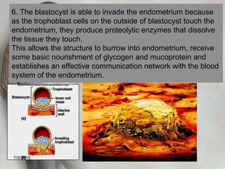
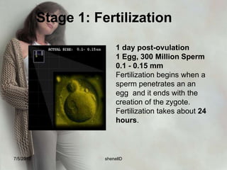
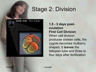
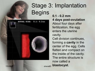
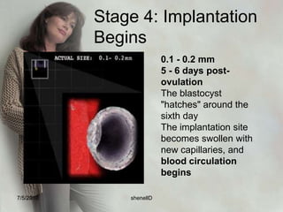
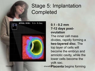
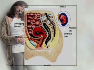
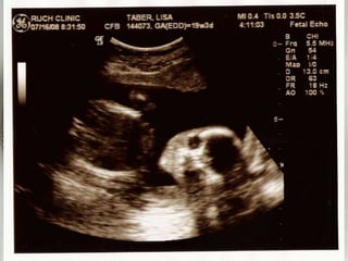
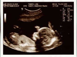
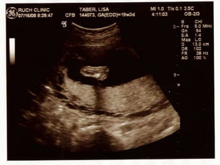
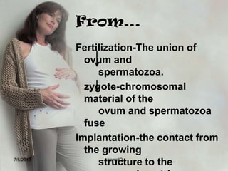
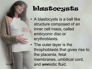
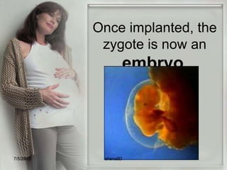
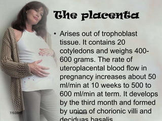
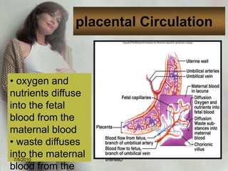
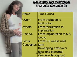
PREGNANCYBy: shenellD7/4/2010shenellD
PregnancyObjectives:What happens to an egg after fertiliZation?How does a baby develop in the uterus?7/4/2010shenellD
Fertilization The union of ovum and spermatozoa.
 Fertilization occurs in the outer third of the fallopian tube – the ampullar portion.
other terms are conception, impregnation, or fecundation.
The critical time span during which fertilization may occur is about 72 hours.7/4/2010shenellD
Steps in fertilization7/4/2010shenellD
1.Following ovulation, as the ovum is extruded from the graafian follicle, it is surrounded by a ring of mucopolysaccharide fluid (zonapellucida) and a circle of cells (corona radiata). These structures increase the bulk of the ovum, facilitating it’s migration to the uterus. 7/4/2010shenellD
2. The ovum and surroundings cells are propelled, into the fallopian tube by the fimbriae, the fine, hair-like structures that line the openings of the fallopian tubes.7/4/2010shenellD
3. Only one ovum reaches maturity a month, a normal ejaculation of semen averages 2.5 ml of fluid containing 50 to 200 million spermatozoa per ml. or averages of 300-400 million per ejaculation. To promote the possibility of a sperm reaching the ovum, there is a reduction in the viscosity of cervical mucus at the time of ovulation.7/4/2010shenellD
4. Spermatozoa deposited in the vagina reaches the cervix of uterus within 90 seconds after deposition ant the outer end of the fallopian tube in 5 minutes. The functional life of spermatozoa is 48 hours.7/4/2010shenellD
5. Spermatozoa move by means of their flagella (tails) and uterine contraction through the cervix, the body of uterus toward the waiting ovum. All the spermatozoa that reaches the ovum cluster around the ovum’s protective layer of corona cells7/4/2010shenellD
6. Hyaluronidase(a proteolytic enzyme) is released by the spermatozoa which acts to dissolve the layer of cells protecting the ovum.7/4/2010shenellD
7. Only one spermatozoa is able to penetrate the cell membrane of the ovum. After it has done, cell membrane becomes impervious to other spermatozoa. 7/4/2010shenellD
8. After penetration, the chromosomal material of the ovum and spermatozoa fuse and the structure is called zygote.Sperm (23)               +             Egg (23)                    =     Fertilized Cell (46)7/4/2010shenellD
implantationoccurs on the seventh day after fertilization
Is the contact between the growing structure and the uterine endometrium7/4/2010shenellD
1. Once of fertilization is complete, the zygote migrate for 3 to 4 days to reach the body of uterus. This time mitotic cell division or cleavage begins. The first cleavage occurs at about 24 hours7/4/2010shenellD
2. As the zygote reaches the uterus it consists of 16 to 50 cells. Its bumpy outward appearance is termed morula (from Latin word morus meaning “mulberry.”)7/4/2010shenellD
3. The morula continues to multiply as it floats free in the uterine cavity for 3 or 4 more days. Large cells tend to mass at the periphery of the ball, leaving a fluid space surrounding an inner cell mass. The structure is now termed blastocyst. 7/4/2010shenellD
4. The cells in the outer ring are known as trophoblast cells. They are the part of the structure that forms the placenta and membrane the inner cell called erythroblast cells is the portion that forms the embryo.7/4/2010shenellD
5. After the 4th day of free floating, the residues of corona and zonapellucida are shed by growing structure. The blastocyst brushes against the rich uterine endometrium a process termed apposition. It attaches to the surface of the endometrium (termed adhesion) and settles down into soft folds (invasion)7/4/2010shenellD
6. The blastocyst is able to invade the endometrium because as the trophoblast cells on the outside of blastocyst touch the endometrium, they produce proteolytic enzymes that dissolve the tissue they touch. This allows the structure to burrow into endometrium, receive some basic nourishment of glycogen and mucoprotein and establishes an effective communication network with the blood system of the endometrium.7/4/2010shenellD
stages7/4/2010shenellD
Stage 1: Fertilization1 day post-ovulation1 Egg, 300 Million Sperm0.1 - 0.15 mmFertilization begins when a sperm penetrates an an egg  and it ends with the creation of the zygote. Fertilization takes about 24 hours.7/4/2010shenellD
Stage 2: Division1.5 - 3 days post-ovulationFirst Cell DivisionWhen cell division produces sixteen cells, the zygote becomes mulberry shaped. It leaves the fallopian tube and three to four days after fertilization7/4/2010shenellD
Stage 3: Implantation Begins0.1 - 0.2 mm4 days post-ovulationAbout four days after fertilization, the egg enters the uterine cavity.Cell division continues, forming a cavity in the center of the egg. Cells flatten and compact on the inside of the cavity.The entire structure is now called a blastocyst.7/4/2010shenellD
Stage 4: Implantation Begins0.1 - 0.2 mm5 - 6 days post-ovulationThe blastocyst "hatches" around the sixth dayThe implantation site becomes swollen with new capillaries, and blood circulation begins7/4/2010shenellD
Stage 5: Implantation Completed0.1 - 0.2 mm7-12 days post-ovulationThe inner cell mass divides, rapidly forming a two-layered disc. The top layer of cells will become the embryo and amniotic cavity, while the lower cells become the yolk sac.Placenta begins forming7/4/2010shenellD
7/4/2010shenellD
7/4/2010shenellD
7/4/2010shenellD
7/4/2010shenellD
7/4/2010shenellD
Thank you!!!7/4/2010shenellD
From…Fertilization-The union of ovum and 			spermatozoa.	zygote-chromosomal material of the 			ovum and spermatozoa fuse Implantation-the contact from the growing 		structure to the 				endometrium.7/4/2010shenellD
blastocystsA blastocysts is a ball like structure composed of an inner cell mass, called embryonic disc or erythroblasts. The outer layer is the throphoblasts that gives rise to the placenta, fetal membranes, umbilical cord, and amniotic fluid.7/4/2010shenellD
the embryonic disc gives rise to the three primary layers which are:Ectoderm- gives rise to the skin, hair, nails, sense organs, nervous system, mucous membrane of the mouth and anus.Mesoderm- kidney musculoskeletal system, reproductive system and cardiovascular systemEntoderm-bladder,lining of the gastrointestinal tract, tonsils, thyroid gland, and respiratory system. 7/4/2010shenellD
7/4/2010shenellD
trophoblastsThe important functions of the trophoblasts is to absorb nutrients from the  endometrium and secrete hormone HCG or human chorionic gonadotropin, necessary in prolonging the life of the corpus luteum7/4/2010shenellD
7/4/2010shenellD
Once implanted, the zygote is now an embryo.7/4/2010shenellD
Embryonic and fetal structures7/4/2010shenellD
The deciduaDecidua- latin word means “falling off”After implantation, the endometrium is referred to as decidua, the specialized endometrium of pregnancy. It is composed of 3 layers:DeciduaveraDeciduabasalisDaciduacapsularis7/4/2010shenellD
DeciduaBasalispart of the endometrium lying directly under the embryo and where trophoblast cells are establishing communication with maternal blood vessels.7/4/2010shenellD
DeciduaCapsularisStretches or encapsulates the surfaces of the trophoblast7/4/2010shenellD
Decidua Verathe remaining portion of the uterine liningIt fuses with deciduacapsularis when the gestational rings grows enough to occupy the entire  uterine cavity.Like a blanket of the embryoAt birth the entire surface of the uterus is stripped away, leaving the organ susceptible to hemorrhage and infection.7/4/2010shenellD
7/4/2010shenellD
the chorionic villiChorionic villi- miniature villi, or probing “fingers” that reach out from the single layer of cells into the uterine endometrium.Two distinct layers:Syncytiotrophoblast or syncytial layerCytotrophoblast or langhans’ layer7/4/2010shenellD
Syncytiotrophoblast or Syncytial Layerouter layer responsible in the production of HCG, Somatomammotropin (human placental lactogen), estrogen and progesterone.7/4/2010shenellD
Cytotrophoblastor Langhan’s LayerInner layer that protects the growing embryo and fetus from infections organisms such as spirochete of syphilis.7/4/2010shenellD
The chorionic villi in contact with deciduabasalis proliferate rapidly because they will receive rich blood supply from the uterus.Responsible for absorbing nutrients and oxygen from maternal blood stream and disposing fetal waste products including carbon dioxide.7/4/2010shenellD
7/4/2010shenellD
The placentaPlacenta- latin for pancake, because of the appearance.It covers about half of  the surface area of the internal uterus.It serves as the fetal lungs, kidneys, and gastrointestinal tract and a separate endocrine gland throughout the pregnancy.7/4/2010shenellD
The placentaArises out of trophoblast tissue. It contains 20 cotyledons and weighs 400-600 grams. The rate of uteroplacental blood flow in pregnancy increases about 50 ml/min at 10 weeks to 500 to 600 ml/min at term. It develops by the third month and formed by union of chorionic villi and deciduas basalis. 7/4/2010shenellD
PlacentaConsists of an embryonic portion and a maternal portion7/4/2010shenellD
placental Circulation oxygen and nutrients diffuse into the fetal blood from the maternal blood
 waste diffuses into the maternal blood from the fetal blood7/4/2010shenellD
What is the function of yolk sac?Yolk sac appears to supply the nourishment only until implantation.After which, its main purpose is to provide a source of red blood cells until the embryo’s hematopoetic system is mature enough to perform this function.So, circulation starts as early as 16th day of life and heart beat as early as the 24th day.7/4/2010shenellD
7/4/2010shenellD
7/4/2010shenellD
7/4/2010shenellD
The umbilical cordFormed as chronic villi begins to function, initiating circulatory communication with the maternal blood pools joined together into larger veins and arteries; about 21 inches in length at term and 2cm in thicknessContains one vein and two arteries ( AVA)7/4/2010shenellD
FunctionsThe bulk of the cord is a gelatinous mucopolysaccharide called Wharton’s jelly which gives the cord body and protects therein and arteries from pressure To transport oxygen and nutrients to the fetus from the placenta.Smooth muscle is abundant in the arteries of the cord and the construction of these muscles after birth contributes to homeostasis and helps prevent hemorrhage of the newborn.7/4/2010shenellD
Fetal membranesFetal Membranes – membranes that surround the fetus and what give placenta the shiny appended 7/4/2010shenellD
Amniotic Fluid Amniotic Fluid – forms within the amniotic cavity and surrounds the embryo. Consist of 800 to 1200 ml of fluid at the end of pregnancy; contains fetal urine, lanugo from fetal skin, epithelial cells and subaqueous materials.pH – 7.2; specific gravity – 1.005 – 1.025 7/4/2010shenellD
Functions:Provides a cushion against injuryProtects the fetus from changes in temperatureProtects the umbilical cord from pressure, protecting fetal oxygenationAids muscular developmentExcretion collection systemThe fetus drinks the fluid 7/4/2010shenellD
7/4/2010shenellD
TERMS TO DENOTE FETAL GROWTH7/4/2010shenellD
Care of the pregnant woman Physiologic change of pregnancy7/4/2010shenellD
Local changeFace – Chloasma – darkening patches of the face due to melanocyte stimulating hormone.“Mask of Pregnancy”7/4/2010shenellD
7/4/2010shenellD
Breast – the areola darkens in color and diameter increase from 3.5 to 5 cm; formation of secondary areola.blue veins become prominent and the sebaceous glands of areola (Montgomery’s tubercles) enlarge and become protuberantby the 16 week- colostrums, a thin, watery, high protein fluid may be expelled from the nipples7/4/2010shenellD
AbdomenDiastasis; due to overstretching of tissue to accommodate growing fetus and separation of rectus muscles. Bluish groove at the site of separation.Linea Nigra: a brown line running from the umbilicus to the symphysis pubisStriaeGravidarum: pink or reddish streaks on the sides of eh abdomen wall and on thigh due to rupture and atrophy of small segment of connective layer of the skin.Spider hemangiomas.7/4/2010shenellD
7/4/2010shenellD
Vagina – Chadwick’s sign – purplish discoloration Leukorrhea– thick whitish vaginal discharge without signs of itching.7/4/2010shenellD
Cervix –Goodell’s sign – softening of the cervix - formation of mucus plug (operculum) to seal out bacteria7/4/2010shenellD
7/4/2010shenellD
Uterus -Hegar’s sign – softening of lower uterine segment Braxton Hick’s Contractions: occurs through out pregnancyAmenorrheaBallottement: during the 16th to 20th week of pregnancy, a sudden push of the fetus7/4/2010shenellD
Bi-manual pelvic exam to palpate uterus7/4/2010shenellD
7/4/2010shenellD
Signs and Symptoms of Pregnancy 7/4/2010shenellD
Presumptive Signs – largely subjective that are experienced by the woman but cannot be documented by the examiner least indicative of pregnancy7/4/2010shenellD
A -   amenorrhea B -   breast changesC -   color changes			S-striaegravidarum			M-melasma			L-lineanigraF-   fatigue, nausea, vomitingU-  urinary frequency, uterine enlargement, leukorrheaQ-  quickening7/4/2010shenellD
Probable signs –(objective) Can be documented by the examiner but not considered positive diagnostic \findings.7/4/2010shenellD
(+)   positive pregnancy testH  -  Hegar’s signC  -  Chadwick’s signG  -  Goodell’s signB  -  ballotementF  -   Fetal parts as felt by the examiner7/4/2010shenellD
Positive Signs – signs that confirm pregnancyFetal Heart sounds (16th week)Fetal Movements felt by the examinerFetal Movement on SonogramFetal Outline (UTZ)7/4/2010shenellD
Systemic Change during pregnancy7/4/2010shenellD
Cardiovascular System:Heart rate increase 10-15 beats/ minute.Blood pressure decrease slightly in the second trimester due to lowered peripheral resistance to circulation but rises in the third trimester. Cardiac output increase 20% -30% during first and second trimester to meet increase tissue demands7/4/2010shenellD
Supine hypotension Syndrome – the woman experience light-headedness, faintness and heart palpitation as the woman lies supine, the weight of uterus presses the vena cava, obstruction  to the blood flow.7/4/2010shenellD
Pseudoanemia - as the plasma volume first increase, the concentration of hemoglobin and erythrocytes may decline – Increase in RBC creating Normal levels of RBC again (Inc. Iron Ferrous sulfate) S04.7/4/2010shenellD
*Women need iron supplement300-400 ml blood loss from normal delivery800-1000 ml blood loss from cesarean delivery7/4/2010shenellD
Respiratory SystemDiaphragm rises as much as 1inch; slight dyspnea may occur until lightening .Increased vital capacity, tidal volume, respiratory minute volume to supply maternal and fetal needs.7/4/2010shenellD
Digestive SystemSlowed gastrointestinal motility and digestion.Tooth loss due to demineralizationDisplacement of intestine and compression  of stomach.HYPERPTYALISM – increase salivation formation – increase  level of estrogen.7/4/2010shenellD
Common problems:1. Morning Sickness – nausea and vomiting early in the morning. HCG and progesterone begin  to rise. 2. Heartburn - Pyrosis- reflux of stomach content into esophagus due to displacement of  the stomach.- decrease gastric motility; relieved by eating small meals frequently and not lying down immediately after eating, to help prevent reflux.2. Pica	- eating non-food substance.	- abnormal craving for substance 	- The most common is craving for ice cube	- Often accompanies iron deficiency anemia	*Encourage to take iron supplements7/4/2010shenellD
3. Constipation - because of reduced activity with GIT and pressure of growing fetus, and	placental hormone relaxing contribute to decreased gastric motility. 4. Flatulence 5. Bleeding gums7/4/2010shenellD
Urinary System Increased urinary frequency on the first and third trimester because of pressure on bladderGlomerular filtration rate increased 50%7/4/2010shenellD
Glycosuria - because of increased excretion of sugar by lowered renal threshold.- presence of sugar in the urine. Lower specific gravity – a result of increased urinary outputPolyuria –increase urine output – additional sodium and therefore additional water.7/4/2010shenellD
Endocrine System Thyroid activity in increasedHCG reaches a peak in the third monthSecretions of oxytocin which stimulates uterine contractions coupled with the drop in progesterone brings about laborUterine contractions increase in frequency and intensity culminating in fetal expulsion7/4/2010shenellD
Skeletal System Gradual softening of pelvic ligaments and joints to facilitate passage of the body Lordosis(forward curvature of the lumber spinal standing with the shoulders back and abdomen forward in order to change center of gravity and make ambulation easier. “The Pride of Pregnancy”7/4/2010shenellD
Discomforts of Pregnancy and its Management 7/4/2010shenellD
Nausea and VomitingEat five or six small, frequent meals; in between meals, have crackers without fluid. Avoid foods high in carbohydrates, fried and greasy or strong odor.7/4/2010shenellD
Fatigue Take frequent rest periods during the day. A good resting position is a modified Sim’s position with top of the fetus on bed, not on the woman, and allows good circulation in the lower extremities 7/4/2010shenellD
Frequency of UrinationKegel’s exercise (alternately contracting and relaxing perineal muscles) helps to strength urinary control and decrease the possibility of stress incontinence and strength of perineal muscles for delivery7/4/2010shenellD
Breast tendernessEncourage to wear a bra with a wide shoulder strap for support and to dress to avoid cold drafts. 7/4/2010shenellD
Flatulence Maintain daily bowel movement; avoid gas-forming foodsHeartburn Avoid fatty, fried and highly spiced foods; small frequent feedings; 7/4/2010shenellD
Constipation Drink sufficient fluids;  Eat fruits and foods high in fiber and roughage;  Exercise moderately;  Avoid using mineral oil.  (It interferes with the absorption of fat – soluble vitamins needed for good fetal growth and material health.7/4/2010shenellD
Hemorrhoids Apply ointments, suppositories, warm compresses; Avoid constipation.InsomniaPrevent prolonged nap time, offer milk, encourage warm bath.7/4/2010shenellD
Backaches Rest and improve posture; use a firm mattres; Use a good abdominal support; wear comfortable shoes; Do exercises such as squatting, sitting, and pelvic rock.7/4/2010shenellD
Varicosities, legs and vulvaAvoid long periods of standing or sitting with legs crossed.  Sit or lie with feet and hips elevated.  Move about while standing to improve circulation; Wear support hose; avoid tight garters.7/4/2010shenellD
Edema of legs and feet Elevate feet while standing or lying down; Avoid standing or sitting in one position for long periods.7/4/2010shenellD
Muscle cramps Extend cramped leg and flex ankles, pushing foot upward with toes pointed toward knee; Increased calcium intake elevating the lower extremities frequently during the day. 7/4/2010shenellD
DyspneaSit up.  Lie on back with arms extended above bed. Uses 2 or more pillows to sleep at night.7/4/2010shenellD
Supine Hypotensive SyndromeChange position to left side to relieve pressure of uterus on interior vena cava.7/4/2010shenellD
Leukorrhea (vaginal discharge)Practice proper cleansing an d hygiene; Avoid douche unless recommended by physician; A daily bath or shower to wash away secretions; Observe for signs of vaginal infection common in pregnancy.7/4/2010shenellD
NEED A BREAK?7/4/2010shenellD
PsychologicChanges of PregnancyMaternal Adaptations to pregnancy   7/4/2010shenellD
First Trimester: Initial ambivalence about pregnancy; pregnant woman places main focus upon self.Mother is self centered, baby is part of her.Grandparents are usually the first relatives to be told of pregnancy.  Accept the biological fact of pregnancy “I am pregnant”7/4/2010shenellD
Second Trimester: Acceptance of reality of pregnancy; increased awareness and interesting fetus; introversion and feeling of well – being.Fantasizes about unborn child.Accept the growing fetus as distinct from self and as a person to care of ….. “I am going to have a baby.”7/4/2010shenellD
Third Trimester: Anticipation of labor and delivery and assuming mothering role, viewing infant as reality vs. fantasy; fears , fantasies and dreams about labor are common, “nesting” behaviors like preparing layette.Prepare realistically for the birth and parenting of the child…..  “I am going to be a mother.”7/4/2010shenellD
Paternal Reactions to Pregnancy7/4/2010shenellD
   First Trimester: Ambivalence and anxiety about role change; concern or identification with wife’s discomfort (couvades) 7/4/2010shenellD
Second Trimester:  Increased confidence and interest in mother care; difficulty relating to fetus; jealousy. 7/4/2010shenellD
   Third Trimester	Changing self-concept; concern about body change; active involvement common fears about delivery, mutilation, or death of partner or fetus.7/4/2010shenellD
Prenatal Period 7/4/2010shenellD
Prenatal VisitSchedule of Visit if no complications:Every 4 weeks, up to 32 weeksEvery 2 weeks from 32-36 weeks (more frequently if problems exist.0Every week from 36-40 weeks7/4/2010shenellD
History Taking Assessment of Risk Factors:Age: Under 16 or 35 (greater risk over 40)Pregnant adolescence have a higher incidence of prematurity, pregnancy induced hypertension, cephalopelvic disproportion, poor nutrition and inadequate antepartal care.Women over 35 year old at risk for chromosomal disorder in infants, pregnancy – induced hypertension, and cesarean delivery; those over 35 years for first pregnancy may be at increased risk.7/4/2010shenellD
TerminologyGravidity#of current and completed pregnancies of any kindParity# of completed pregnancies ≥ 20 weeksnot delivered infants (e.g. twins)7/4/2010shenellD
Primigravida – a woman who is pregnant for the 1st childPrimipara – a woman who had delivered, live born child; a woman who is pregnant for the first time.7/4/2010shenellD
Multigravida– a woman who has been pregnant previously.Multipara – a woman who has delivered 1 or more children previouslyNulligravida– a woman who has never been pregnant.7/4/2010shenellD
Parity (TPAL)T= Number of Term BirthsP= Number of Premature birthsA= Number of AbortionsL= Number of living children7/4/2010shenellD
G3/1-0-1-1:Terminology7/4/2010shenellD
G3/1-0-1-1:3rd Pregnancy1 Term delivery0 Preterm deliveries1 Abortion1 Living childTerminology7/4/2010shenellD
G5/2-1-1-0:Terminology7/4/2010shenellD
G5/2-1-1-0:5th Pregnancy2 Term deliveries1 Preterm delivery1 Abortion0 Living childrenTerminology7/4/2010shenellD
G2/0-2-0-3:Terminology7/4/2010shenellD
G2/0-2-0-3:2nd Pregnancy0 Term deliveries2 Preterm deliveries 0 Abortions2 Living childrenTerminology7/4/2010shenellD
Physical AssessmentLEOPOLD’S MANUEVERa systematic method of observation and palpation to determine the presenting part, fetal position, presentation and engorgement. The woman should be in supine position with her knees flexed slightly.7/4/2010shenellD
1st ManeuverPalpate the superior surface of the fundusFacing the head part, palpate for fetal part found in the fundus Leopold_first.flv7/4/2010shenellD
2nd Maneuver 	Palpate the sides of uterus to determine where the fetal back is facing	The left hand is left stationary on the left side of the uterus while the right hand palpates opposite side of the uterus from the top to bottom.	Next, hold right hand stationary to immobilize the uterus, and palpate top to bottom on the left sideLeopold_second.flv7/4/2010shenellD
3rd Maneuver	Palpate to discover what is at the inlet of the pelvis.	Grasp the lower portion of the abdomen just above the symphysis pubis between the thumb and index finger and try to press the thumb and finger togetherThe presenting part is not engaged if the presenting part moves upward so an examiner’s hand can be pressed together.Leopold_third.flv7/4/2010shenellD
4th ManeuverPalpate to determine the fetal attitudePlace fingers on both sides of the uterus 2 inches above inguinal ligaments. Press downward and inwardThe fingers of one hand will slide along the uterine contour and meet no obstruction; this is the fetal neck.The other hand will meet an obstruction and inch or so above the ligament, this is the fetal brow.Leopold_final.flv7/4/2010shenellD
7/4/2010shenellD
Estimating Expected Date of Confinement (EDC) 7/4/2010shenellD
Bartholomew’s Rule Estimate AOG:3rd month(12 weeks)- fundus is slightly above symphisis pubis5th month(20 weeks)-  fundusia at level of umbilicus8th month(32 weeks)- below the xyphoid process9th month(36 weeks)- same level7/4/2010shenellD
Naegels’sRuleFormula: 3 months + 7 days + 1 yearExample Last Menstrual Period 	April 20		1995- 3 mos+7 days+1 yearJanuary    271996 EDC: January 27, 1996 7/4/2010shenellD
Example: solve the EDCLMP September 15, 2009LMP July 20, 2006LMP August 5, 2000LMP April 16, 2014LMP January 01, 2009  7/4/2010shenellD
Mc Donald’s FormulaAge of Gestation Formula: Fundicheight in cmx2/7 = AOG in months X 4 weeks = AOG in weeksExample: Fundic heights is 21 cm21 cm x 2 = 42 /7=6 months x 4 weeks = 24 weeksfundal_height.flv7/4/2010shenellD
Example: solve for AOGFundic height is 18 cmFundic height is 24 cmFundic height is 32 cmFundic height is 16 cmFundic height is 20 cm7/4/2010shenellD
Haase’sRuleFetal Length Formula: 1 to 5 months = months (squared)6 to 10 months = months x 5Examples5 months = 5 mos. = 25 cm length8 months = 8 mos. x 5 = 40 cm length7/4/2010shenellD
Example: solve for fetal length6 mos. And 2 weeks4 months3 mos. And 3 weeks8 mos. And 1 week2 mos. And 2 weeks7/4/2010shenellD
Johnson’s Rule (grams)Fetal Weight Formula:Fundic Height (cm) – n x kN = 	11 if part is not engaged12 if part is engaged K = 155 grams (standard value)Example: Fundicheight = 21 cm not engaged21 – 11 = 10 x 155 = 1, 550 grams7/4/2010shenellD
Example: solve for fetal weightFH is 24 cm engagedFH is 18 cm not engagedFH is 20 cm engagedFH is 16 cm not engagedFH is 22 cm engaged7/4/2010shenellD
NUTRITION DURING PREGNANCYWeight gain- variable,but 25 lbs usually appropriate for average woman with single pregnancy.Recommended weight gain during pregnancy:2-4 lbs in the first trimester11-14 lbs in the second trimester8-11 lbs in the third trimester (0.5 lb weekly)7/4/2010shenellD
Weight gain in pregnancy occurs from the both growth of fetus and accumulation of maternal stores:Breast 			1.5 –3 lbsFetus			7 lbsPlacenta			1.5 lbsUterus 			2.5 lbsAmniotic fluid	    	2 lbsBlood volume		3.5 lbs7/4/2010shenellD
NUTRITION DURING PREGNANCYB. Specific nutrition needsCalories: +300 kcal/day. Never < 1800 kcal/day
Protein: +30 g/day to ensure intake of 74-76 g/day
Iron: provide 100-200 mg/tab daily
Calcium: 1200 mg/day7/4/2010shenellD
Sexual activity during pregnancyBasically sex is permitted on 2nd trimester as long as your comfortable and you don’t have complications.Avoid breast massage since it may stimulate early uterine contractions.Side by side or woman on top position.7/4/2010shenellD
Different Types of ExerciseTAILOR SITTINGIt strengthens the thigh and stretches perineal muscles. The woman should not put one ankle on top of the other but should place one leg in front of the other gently push on her knees (pushing them toward the floor until she feels her perineum “stretch”7/4/2010shenellD
SQUATTING Helps to stretch the muscle of the pelvic floor. It should be done for 15 minutes day. The woman must keep her feet flat on the floor.7/4/2010shenellD

Pregnancy new ppt

  • 1.
  • 2.
    PregnancyObjectives:What happens toan egg after fertiliZation?How does a baby develop in the uterus?7/4/2010shenellD
  • 3.
    Fertilization The unionof ovum and spermatozoa.
  • 4.
    Fertilization occursin the outer third of the fallopian tube – the ampullar portion.
  • 5.
    other terms areconception, impregnation, or fecundation.
  • 6.
    The critical timespan during which fertilization may occur is about 72 hours.7/4/2010shenellD
  • 7.
  • 8.
    1.Following ovulation, asthe ovum is extruded from the graafian follicle, it is surrounded by a ring of mucopolysaccharide fluid (zonapellucida) and a circle of cells (corona radiata). These structures increase the bulk of the ovum, facilitating it’s migration to the uterus. 7/4/2010shenellD
  • 9.
    2. The ovumand surroundings cells are propelled, into the fallopian tube by the fimbriae, the fine, hair-like structures that line the openings of the fallopian tubes.7/4/2010shenellD
  • 10.
    3. Only oneovum reaches maturity a month, a normal ejaculation of semen averages 2.5 ml of fluid containing 50 to 200 million spermatozoa per ml. or averages of 300-400 million per ejaculation. To promote the possibility of a sperm reaching the ovum, there is a reduction in the viscosity of cervical mucus at the time of ovulation.7/4/2010shenellD
  • 11.
    4. Spermatozoa depositedin the vagina reaches the cervix of uterus within 90 seconds after deposition ant the outer end of the fallopian tube in 5 minutes. The functional life of spermatozoa is 48 hours.7/4/2010shenellD
  • 12.
    5. Spermatozoa moveby means of their flagella (tails) and uterine contraction through the cervix, the body of uterus toward the waiting ovum. All the spermatozoa that reaches the ovum cluster around the ovum’s protective layer of corona cells7/4/2010shenellD
  • 13.
    6. Hyaluronidase(a proteolyticenzyme) is released by the spermatozoa which acts to dissolve the layer of cells protecting the ovum.7/4/2010shenellD
  • 14.
    7. Only onespermatozoa is able to penetrate the cell membrane of the ovum. After it has done, cell membrane becomes impervious to other spermatozoa. 7/4/2010shenellD
  • 15.
    8. After penetration,the chromosomal material of the ovum and spermatozoa fuse and the structure is called zygote.Sperm (23) + Egg (23) = Fertilized Cell (46)7/4/2010shenellD
  • 16.
    implantationoccurs on theseventh day after fertilization
  • 17.
    Is the contactbetween the growing structure and the uterine endometrium7/4/2010shenellD
  • 18.
    1. Once offertilization is complete, the zygote migrate for 3 to 4 days to reach the body of uterus. This time mitotic cell division or cleavage begins. The first cleavage occurs at about 24 hours7/4/2010shenellD
  • 19.
    2. As thezygote reaches the uterus it consists of 16 to 50 cells. Its bumpy outward appearance is termed morula (from Latin word morus meaning “mulberry.”)7/4/2010shenellD
  • 20.
    3. The morulacontinues to multiply as it floats free in the uterine cavity for 3 or 4 more days. Large cells tend to mass at the periphery of the ball, leaving a fluid space surrounding an inner cell mass. The structure is now termed blastocyst. 7/4/2010shenellD
  • 21.
    4. The cellsin the outer ring are known as trophoblast cells. They are the part of the structure that forms the placenta and membrane the inner cell called erythroblast cells is the portion that forms the embryo.7/4/2010shenellD
  • 22.
    5. After the4th day of free floating, the residues of corona and zonapellucida are shed by growing structure. The blastocyst brushes against the rich uterine endometrium a process termed apposition. It attaches to the surface of the endometrium (termed adhesion) and settles down into soft folds (invasion)7/4/2010shenellD
  • 23.
    6. The blastocystis able to invade the endometrium because as the trophoblast cells on the outside of blastocyst touch the endometrium, they produce proteolytic enzymes that dissolve the tissue they touch. This allows the structure to burrow into endometrium, receive some basic nourishment of glycogen and mucoprotein and establishes an effective communication network with the blood system of the endometrium.7/4/2010shenellD
  • 24.
  • 25.
    Stage 1: Fertilization1day post-ovulation1 Egg, 300 Million Sperm0.1 - 0.15 mmFertilization begins when a sperm penetrates an an egg and it ends with the creation of the zygote. Fertilization takes about 24 hours.7/4/2010shenellD
  • 26.
    Stage 2: Division1.5- 3 days post-ovulationFirst Cell DivisionWhen cell division produces sixteen cells, the zygote becomes mulberry shaped. It leaves the fallopian tube and three to four days after fertilization7/4/2010shenellD
  • 27.
    Stage 3: ImplantationBegins0.1 - 0.2 mm4 days post-ovulationAbout four days after fertilization, the egg enters the uterine cavity.Cell division continues, forming a cavity in the center of the egg. Cells flatten and compact on the inside of the cavity.The entire structure is now called a blastocyst.7/4/2010shenellD
  • 28.
    Stage 4: ImplantationBegins0.1 - 0.2 mm5 - 6 days post-ovulationThe blastocyst "hatches" around the sixth dayThe implantation site becomes swollen with new capillaries, and blood circulation begins7/4/2010shenellD
  • 29.
    Stage 5: ImplantationCompleted0.1 - 0.2 mm7-12 days post-ovulationThe inner cell mass divides, rapidly forming a two-layered disc. The top layer of cells will become the embryo and amniotic cavity, while the lower cells become the yolk sac.Placenta begins forming7/4/2010shenellD
  • 30.
  • 31.
  • 32.
  • 33.
  • 34.
  • 35.
  • 36.
    From…Fertilization-The union ofovum and spermatozoa. zygote-chromosomal material of the ovum and spermatozoa fuse Implantation-the contact from the growing structure to the endometrium.7/4/2010shenellD
  • 37.
    blastocystsA blastocysts isa ball like structure composed of an inner cell mass, called embryonic disc or erythroblasts. The outer layer is the throphoblasts that gives rise to the placenta, fetal membranes, umbilical cord, and amniotic fluid.7/4/2010shenellD
  • 38.
    the embryonic discgives rise to the three primary layers which are:Ectoderm- gives rise to the skin, hair, nails, sense organs, nervous system, mucous membrane of the mouth and anus.Mesoderm- kidney musculoskeletal system, reproductive system and cardiovascular systemEntoderm-bladder,lining of the gastrointestinal tract, tonsils, thyroid gland, and respiratory system. 7/4/2010shenellD
  • 39.
  • 40.
    trophoblastsThe important functionsof the trophoblasts is to absorb nutrients from the endometrium and secrete hormone HCG or human chorionic gonadotropin, necessary in prolonging the life of the corpus luteum7/4/2010shenellD
  • 41.
  • 42.
    Once implanted, thezygote is now an embryo.7/4/2010shenellD
  • 43.
    Embryonic and fetalstructures7/4/2010shenellD
  • 44.
    The deciduaDecidua- latinword means “falling off”After implantation, the endometrium is referred to as decidua, the specialized endometrium of pregnancy. It is composed of 3 layers:DeciduaveraDeciduabasalisDaciduacapsularis7/4/2010shenellD
  • 45.
    DeciduaBasalispart of theendometrium lying directly under the embryo and where trophoblast cells are establishing communication with maternal blood vessels.7/4/2010shenellD
  • 46.
    DeciduaCapsularisStretches or encapsulatesthe surfaces of the trophoblast7/4/2010shenellD
  • 47.
    Decidua Verathe remainingportion of the uterine liningIt fuses with deciduacapsularis when the gestational rings grows enough to occupy the entire uterine cavity.Like a blanket of the embryoAt birth the entire surface of the uterus is stripped away, leaving the organ susceptible to hemorrhage and infection.7/4/2010shenellD
  • 48.
  • 49.
    the chorionic villiChorionicvilli- miniature villi, or probing “fingers” that reach out from the single layer of cells into the uterine endometrium.Two distinct layers:Syncytiotrophoblast or syncytial layerCytotrophoblast or langhans’ layer7/4/2010shenellD
  • 50.
    Syncytiotrophoblast or SyncytialLayerouter layer responsible in the production of HCG, Somatomammotropin (human placental lactogen), estrogen and progesterone.7/4/2010shenellD
  • 51.
    Cytotrophoblastor Langhan’s LayerInnerlayer that protects the growing embryo and fetus from infections organisms such as spirochete of syphilis.7/4/2010shenellD
  • 52.
    The chorionic villiin contact with deciduabasalis proliferate rapidly because they will receive rich blood supply from the uterus.Responsible for absorbing nutrients and oxygen from maternal blood stream and disposing fetal waste products including carbon dioxide.7/4/2010shenellD
  • 53.
  • 54.
    The placentaPlacenta- latinfor pancake, because of the appearance.It covers about half of the surface area of the internal uterus.It serves as the fetal lungs, kidneys, and gastrointestinal tract and a separate endocrine gland throughout the pregnancy.7/4/2010shenellD
  • 55.
    The placentaArises outof trophoblast tissue. It contains 20 cotyledons and weighs 400-600 grams. The rate of uteroplacental blood flow in pregnancy increases about 50 ml/min at 10 weeks to 500 to 600 ml/min at term. It develops by the third month and formed by union of chorionic villi and deciduas basalis. 7/4/2010shenellD
  • 56.
    PlacentaConsists of anembryonic portion and a maternal portion7/4/2010shenellD
  • 57.
    placental Circulation oxygenand nutrients diffuse into the fetal blood from the maternal blood
  • 58.
    waste diffusesinto the maternal blood from the fetal blood7/4/2010shenellD
  • 59.
    What is thefunction of yolk sac?Yolk sac appears to supply the nourishment only until implantation.After which, its main purpose is to provide a source of red blood cells until the embryo’s hematopoetic system is mature enough to perform this function.So, circulation starts as early as 16th day of life and heart beat as early as the 24th day.7/4/2010shenellD
  • 60.
  • 61.
  • 62.
  • 63.
    The umbilical cordFormedas chronic villi begins to function, initiating circulatory communication with the maternal blood pools joined together into larger veins and arteries; about 21 inches in length at term and 2cm in thicknessContains one vein and two arteries ( AVA)7/4/2010shenellD
  • 64.
    FunctionsThe bulk ofthe cord is a gelatinous mucopolysaccharide called Wharton’s jelly which gives the cord body and protects therein and arteries from pressure To transport oxygen and nutrients to the fetus from the placenta.Smooth muscle is abundant in the arteries of the cord and the construction of these muscles after birth contributes to homeostasis and helps prevent hemorrhage of the newborn.7/4/2010shenellD
  • 65.
    Fetal membranesFetal Membranes– membranes that surround the fetus and what give placenta the shiny appended 7/4/2010shenellD
  • 66.
    Amniotic Fluid AmnioticFluid – forms within the amniotic cavity and surrounds the embryo. Consist of 800 to 1200 ml of fluid at the end of pregnancy; contains fetal urine, lanugo from fetal skin, epithelial cells and subaqueous materials.pH – 7.2; specific gravity – 1.005 – 1.025 7/4/2010shenellD
  • 67.
    Functions:Provides a cushionagainst injuryProtects the fetus from changes in temperatureProtects the umbilical cord from pressure, protecting fetal oxygenationAids muscular developmentExcretion collection systemThe fetus drinks the fluid 7/4/2010shenellD
  • 68.
  • 69.
    TERMS TO DENOTEFETAL GROWTH7/4/2010shenellD
  • 70.
    Care of thepregnant woman Physiologic change of pregnancy7/4/2010shenellD
  • 71.
    Local changeFace –Chloasma – darkening patches of the face due to melanocyte stimulating hormone.“Mask of Pregnancy”7/4/2010shenellD
  • 72.
  • 73.
    Breast – theareola darkens in color and diameter increase from 3.5 to 5 cm; formation of secondary areola.blue veins become prominent and the sebaceous glands of areola (Montgomery’s tubercles) enlarge and become protuberantby the 16 week- colostrums, a thin, watery, high protein fluid may be expelled from the nipples7/4/2010shenellD
  • 74.
    AbdomenDiastasis; due tooverstretching of tissue to accommodate growing fetus and separation of rectus muscles. Bluish groove at the site of separation.Linea Nigra: a brown line running from the umbilicus to the symphysis pubisStriaeGravidarum: pink or reddish streaks on the sides of eh abdomen wall and on thigh due to rupture and atrophy of small segment of connective layer of the skin.Spider hemangiomas.7/4/2010shenellD
  • 75.
  • 76.
    Vagina – Chadwick’ssign – purplish discoloration Leukorrhea– thick whitish vaginal discharge without signs of itching.7/4/2010shenellD
  • 77.
    Cervix –Goodell’s sign– softening of the cervix - formation of mucus plug (operculum) to seal out bacteria7/4/2010shenellD
  • 78.
  • 79.
    Uterus -Hegar’s sign– softening of lower uterine segment Braxton Hick’s Contractions: occurs through out pregnancyAmenorrheaBallottement: during the 16th to 20th week of pregnancy, a sudden push of the fetus7/4/2010shenellD
  • 80.
    Bi-manual pelvic examto palpate uterus7/4/2010shenellD
  • 81.
  • 82.
    Signs and Symptomsof Pregnancy 7/4/2010shenellD
  • 83.
    Presumptive Signs –largely subjective that are experienced by the woman but cannot be documented by the examiner least indicative of pregnancy7/4/2010shenellD
  • 84.
    A - amenorrhea B - breast changesC - color changes S-striaegravidarum M-melasma L-lineanigraF- fatigue, nausea, vomitingU- urinary frequency, uterine enlargement, leukorrheaQ- quickening7/4/2010shenellD
  • 85.
    Probable signs –(objective)Can be documented by the examiner but not considered positive diagnostic \findings.7/4/2010shenellD
  • 86.
    (+) positive pregnancy testH - Hegar’s signC - Chadwick’s signG - Goodell’s signB - ballotementF - Fetal parts as felt by the examiner7/4/2010shenellD
  • 87.
    Positive Signs –signs that confirm pregnancyFetal Heart sounds (16th week)Fetal Movements felt by the examinerFetal Movement on SonogramFetal Outline (UTZ)7/4/2010shenellD
  • 88.
    Systemic Change duringpregnancy7/4/2010shenellD
  • 89.
    Cardiovascular System:Heart rateincrease 10-15 beats/ minute.Blood pressure decrease slightly in the second trimester due to lowered peripheral resistance to circulation but rises in the third trimester. Cardiac output increase 20% -30% during first and second trimester to meet increase tissue demands7/4/2010shenellD
  • 90.
    Supine hypotension Syndrome– the woman experience light-headedness, faintness and heart palpitation as the woman lies supine, the weight of uterus presses the vena cava, obstruction to the blood flow.7/4/2010shenellD
  • 91.
    Pseudoanemia - asthe plasma volume first increase, the concentration of hemoglobin and erythrocytes may decline – Increase in RBC creating Normal levels of RBC again (Inc. Iron Ferrous sulfate) S04.7/4/2010shenellD
  • 92.
    *Women need ironsupplement300-400 ml blood loss from normal delivery800-1000 ml blood loss from cesarean delivery7/4/2010shenellD
  • 93.
    Respiratory SystemDiaphragm risesas much as 1inch; slight dyspnea may occur until lightening .Increased vital capacity, tidal volume, respiratory minute volume to supply maternal and fetal needs.7/4/2010shenellD
  • 94.
    Digestive SystemSlowed gastrointestinalmotility and digestion.Tooth loss due to demineralizationDisplacement of intestine and compression of stomach.HYPERPTYALISM – increase salivation formation – increase level of estrogen.7/4/2010shenellD
  • 95.
    Common problems:1. MorningSickness – nausea and vomiting early in the morning. HCG and progesterone begin to rise. 2. Heartburn - Pyrosis- reflux of stomach content into esophagus due to displacement of the stomach.- decrease gastric motility; relieved by eating small meals frequently and not lying down immediately after eating, to help prevent reflux.2. Pica - eating non-food substance. - abnormal craving for substance - The most common is craving for ice cube - Often accompanies iron deficiency anemia *Encourage to take iron supplements7/4/2010shenellD
  • 96.
    3. Constipation -because of reduced activity with GIT and pressure of growing fetus, and placental hormone relaxing contribute to decreased gastric motility. 4. Flatulence 5. Bleeding gums7/4/2010shenellD
  • 97.
    Urinary System Increased urinaryfrequency on the first and third trimester because of pressure on bladderGlomerular filtration rate increased 50%7/4/2010shenellD
  • 98.
    Glycosuria - becauseof increased excretion of sugar by lowered renal threshold.- presence of sugar in the urine. Lower specific gravity – a result of increased urinary outputPolyuria –increase urine output – additional sodium and therefore additional water.7/4/2010shenellD
  • 99.
    Endocrine System Thyroid activityin increasedHCG reaches a peak in the third monthSecretions of oxytocin which stimulates uterine contractions coupled with the drop in progesterone brings about laborUterine contractions increase in frequency and intensity culminating in fetal expulsion7/4/2010shenellD
  • 100.
    Skeletal System Gradual softeningof pelvic ligaments and joints to facilitate passage of the body Lordosis(forward curvature of the lumber spinal standing with the shoulders back and abdomen forward in order to change center of gravity and make ambulation easier. “The Pride of Pregnancy”7/4/2010shenellD
  • 101.
    Discomforts of Pregnancyand its Management 7/4/2010shenellD
  • 102.
    Nausea and VomitingEatfive or six small, frequent meals; in between meals, have crackers without fluid. Avoid foods high in carbohydrates, fried and greasy or strong odor.7/4/2010shenellD
  • 103.
    Fatigue Take frequent restperiods during the day. A good resting position is a modified Sim’s position with top of the fetus on bed, not on the woman, and allows good circulation in the lower extremities 7/4/2010shenellD
  • 104.
    Frequency of UrinationKegel’sexercise (alternately contracting and relaxing perineal muscles) helps to strength urinary control and decrease the possibility of stress incontinence and strength of perineal muscles for delivery7/4/2010shenellD
  • 105.
    Breast tendernessEncourage towear a bra with a wide shoulder strap for support and to dress to avoid cold drafts. 7/4/2010shenellD
  • 106.
    Flatulence Maintain daily bowelmovement; avoid gas-forming foodsHeartburn Avoid fatty, fried and highly spiced foods; small frequent feedings; 7/4/2010shenellD
  • 107.
    Constipation Drink sufficientfluids; Eat fruits and foods high in fiber and roughage; Exercise moderately; Avoid using mineral oil. (It interferes with the absorption of fat – soluble vitamins needed for good fetal growth and material health.7/4/2010shenellD
  • 108.
    Hemorrhoids Apply ointments,suppositories, warm compresses; Avoid constipation.InsomniaPrevent prolonged nap time, offer milk, encourage warm bath.7/4/2010shenellD
  • 109.
    Backaches Rest andimprove posture; use a firm mattres; Use a good abdominal support; wear comfortable shoes; Do exercises such as squatting, sitting, and pelvic rock.7/4/2010shenellD
  • 110.
    Varicosities, legs andvulvaAvoid long periods of standing or sitting with legs crossed. Sit or lie with feet and hips elevated. Move about while standing to improve circulation; Wear support hose; avoid tight garters.7/4/2010shenellD
  • 111.
    Edema of legsand feet Elevate feet while standing or lying down; Avoid standing or sitting in one position for long periods.7/4/2010shenellD
  • 112.
    Muscle cramps Extend crampedleg and flex ankles, pushing foot upward with toes pointed toward knee; Increased calcium intake elevating the lower extremities frequently during the day. 7/4/2010shenellD
  • 113.
    DyspneaSit up. Lie on back with arms extended above bed. Uses 2 or more pillows to sleep at night.7/4/2010shenellD
  • 114.
    Supine Hypotensive SyndromeChangeposition to left side to relieve pressure of uterus on interior vena cava.7/4/2010shenellD
  • 115.
    Leukorrhea (vaginal discharge)Practiceproper cleansing an d hygiene; Avoid douche unless recommended by physician; A daily bath or shower to wash away secretions; Observe for signs of vaginal infection common in pregnancy.7/4/2010shenellD
  • 116.
  • 117.
    PsychologicChanges of PregnancyMaternalAdaptations to pregnancy  7/4/2010shenellD
  • 118.
    First Trimester: Initialambivalence about pregnancy; pregnant woman places main focus upon self.Mother is self centered, baby is part of her.Grandparents are usually the first relatives to be told of pregnancy. Accept the biological fact of pregnancy “I am pregnant”7/4/2010shenellD
  • 119.
    Second Trimester: Acceptanceof reality of pregnancy; increased awareness and interesting fetus; introversion and feeling of well – being.Fantasizes about unborn child.Accept the growing fetus as distinct from self and as a person to care of ….. “I am going to have a baby.”7/4/2010shenellD
  • 120.
    Third Trimester: Anticipationof labor and delivery and assuming mothering role, viewing infant as reality vs. fantasy; fears , fantasies and dreams about labor are common, “nesting” behaviors like preparing layette.Prepare realistically for the birth and parenting of the child….. “I am going to be a mother.”7/4/2010shenellD
  • 121.
    Paternal Reactions toPregnancy7/4/2010shenellD
  • 122.
    First Trimester: Ambivalence and anxiety about role change; concern or identification with wife’s discomfort (couvades) 7/4/2010shenellD
  • 123.
    Second Trimester: Increased confidence and interest in mother care; difficulty relating to fetus; jealousy. 7/4/2010shenellD
  • 124.
    Third Trimester Changing self-concept; concern about body change; active involvement common fears about delivery, mutilation, or death of partner or fetus.7/4/2010shenellD
  • 125.
  • 126.
    Prenatal VisitSchedule ofVisit if no complications:Every 4 weeks, up to 32 weeksEvery 2 weeks from 32-36 weeks (more frequently if problems exist.0Every week from 36-40 weeks7/4/2010shenellD
  • 127.
    History Taking Assessmentof Risk Factors:Age: Under 16 or 35 (greater risk over 40)Pregnant adolescence have a higher incidence of prematurity, pregnancy induced hypertension, cephalopelvic disproportion, poor nutrition and inadequate antepartal care.Women over 35 year old at risk for chromosomal disorder in infants, pregnancy – induced hypertension, and cesarean delivery; those over 35 years for first pregnancy may be at increased risk.7/4/2010shenellD
  • 128.
    TerminologyGravidity#of current andcompleted pregnancies of any kindParity# of completed pregnancies ≥ 20 weeksnot delivered infants (e.g. twins)7/4/2010shenellD
  • 129.
    Primigravida – awoman who is pregnant for the 1st childPrimipara – a woman who had delivered, live born child; a woman who is pregnant for the first time.7/4/2010shenellD
  • 130.
    Multigravida– a womanwho has been pregnant previously.Multipara – a woman who has delivered 1 or more children previouslyNulligravida– a woman who has never been pregnant.7/4/2010shenellD
  • 131.
    Parity (TPAL)T= Numberof Term BirthsP= Number of Premature birthsA= Number of AbortionsL= Number of living children7/4/2010shenellD
  • 132.
  • 133.
    G3/1-0-1-1:3rd Pregnancy1 Termdelivery0 Preterm deliveries1 Abortion1 Living childTerminology7/4/2010shenellD
  • 134.
  • 135.
    G5/2-1-1-0:5th Pregnancy2 Termdeliveries1 Preterm delivery1 Abortion0 Living childrenTerminology7/4/2010shenellD
  • 136.
  • 137.
    G2/0-2-0-3:2nd Pregnancy0 Termdeliveries2 Preterm deliveries 0 Abortions2 Living childrenTerminology7/4/2010shenellD
  • 138.
    Physical AssessmentLEOPOLD’S MANUEVERasystematic method of observation and palpation to determine the presenting part, fetal position, presentation and engorgement. The woman should be in supine position with her knees flexed slightly.7/4/2010shenellD
  • 139.
    1st ManeuverPalpate thesuperior surface of the fundusFacing the head part, palpate for fetal part found in the fundus Leopold_first.flv7/4/2010shenellD
  • 140.
    2nd Maneuver Palpatethe sides of uterus to determine where the fetal back is facing The left hand is left stationary on the left side of the uterus while the right hand palpates opposite side of the uterus from the top to bottom. Next, hold right hand stationary to immobilize the uterus, and palpate top to bottom on the left sideLeopold_second.flv7/4/2010shenellD
  • 141.
    3rd Maneuver Palpate todiscover what is at the inlet of the pelvis. Grasp the lower portion of the abdomen just above the symphysis pubis between the thumb and index finger and try to press the thumb and finger togetherThe presenting part is not engaged if the presenting part moves upward so an examiner’s hand can be pressed together.Leopold_third.flv7/4/2010shenellD
  • 142.
    4th ManeuverPalpate todetermine the fetal attitudePlace fingers on both sides of the uterus 2 inches above inguinal ligaments. Press downward and inwardThe fingers of one hand will slide along the uterine contour and meet no obstruction; this is the fetal neck.The other hand will meet an obstruction and inch or so above the ligament, this is the fetal brow.Leopold_final.flv7/4/2010shenellD
  • 143.
  • 144.
    Estimating Expected Dateof Confinement (EDC) 7/4/2010shenellD
  • 145.
    Bartholomew’s Rule EstimateAOG:3rd month(12 weeks)- fundus is slightly above symphisis pubis5th month(20 weeks)- fundusia at level of umbilicus8th month(32 weeks)- below the xyphoid process9th month(36 weeks)- same level7/4/2010shenellD
  • 146.
    Naegels’sRuleFormula: 3 months+ 7 days + 1 yearExample Last Menstrual Period April 20 1995- 3 mos+7 days+1 yearJanuary 271996 EDC: January 27, 1996 7/4/2010shenellD
  • 147.
    Example: solve theEDCLMP September 15, 2009LMP July 20, 2006LMP August 5, 2000LMP April 16, 2014LMP January 01, 2009 7/4/2010shenellD
  • 148.
    Mc Donald’s FormulaAgeof Gestation Formula: Fundicheight in cmx2/7 = AOG in months X 4 weeks = AOG in weeksExample: Fundic heights is 21 cm21 cm x 2 = 42 /7=6 months x 4 weeks = 24 weeksfundal_height.flv7/4/2010shenellD
  • 149.
    Example: solve forAOGFundic height is 18 cmFundic height is 24 cmFundic height is 32 cmFundic height is 16 cmFundic height is 20 cm7/4/2010shenellD
  • 150.
    Haase’sRuleFetal Length Formula:1 to 5 months = months (squared)6 to 10 months = months x 5Examples5 months = 5 mos. = 25 cm length8 months = 8 mos. x 5 = 40 cm length7/4/2010shenellD
  • 151.
    Example: solve forfetal length6 mos. And 2 weeks4 months3 mos. And 3 weeks8 mos. And 1 week2 mos. And 2 weeks7/4/2010shenellD
  • 152.
    Johnson’s Rule (grams)FetalWeight Formula:Fundic Height (cm) – n x kN = 11 if part is not engaged12 if part is engaged K = 155 grams (standard value)Example: Fundicheight = 21 cm not engaged21 – 11 = 10 x 155 = 1, 550 grams7/4/2010shenellD
  • 153.
    Example: solve forfetal weightFH is 24 cm engagedFH is 18 cm not engagedFH is 20 cm engagedFH is 16 cm not engagedFH is 22 cm engaged7/4/2010shenellD
  • 154.
    NUTRITION DURING PREGNANCYWeightgain- variable,but 25 lbs usually appropriate for average woman with single pregnancy.Recommended weight gain during pregnancy:2-4 lbs in the first trimester11-14 lbs in the second trimester8-11 lbs in the third trimester (0.5 lb weekly)7/4/2010shenellD
  • 155.
    Weight gain inpregnancy occurs from the both growth of fetus and accumulation of maternal stores:Breast 1.5 –3 lbsFetus 7 lbsPlacenta 1.5 lbsUterus 2.5 lbsAmniotic fluid 2 lbsBlood volume 3.5 lbs7/4/2010shenellD
  • 156.
    NUTRITION DURING PREGNANCYB.Specific nutrition needsCalories: +300 kcal/day. Never < 1800 kcal/day
  • 157.
    Protein: +30 g/dayto ensure intake of 74-76 g/day
  • 158.
  • 159.
  • 160.
    Sexual activity duringpregnancyBasically sex is permitted on 2nd trimester as long as your comfortable and you don’t have complications.Avoid breast massage since it may stimulate early uterine contractions.Side by side or woman on top position.7/4/2010shenellD
  • 161.
    Different Types ofExerciseTAILOR SITTINGIt strengthens the thigh and stretches perineal muscles. The woman should not put one ankle on top of the other but should place one leg in front of the other gently push on her knees (pushing them toward the floor until she feels her perineum “stretch”7/4/2010shenellD
  • 162.
    SQUATTING Helps to stretchthe muscle of the pelvic floor. It should be done for 15 minutes day. The woman must keep her feet flat on the floor.7/4/2010shenellD
  • 163.
    PELVIC FLOOR CONTRACTIONS(KEGEL’S EXCERISE)Promotes perineal healing, increases sexual responsiveness and prevents stress, incontinence. While sitting at her desk or working around the house, the woman can tighten the muscles surrounding her vagina, relax tighten the muscles surrounding her rectum, relax, tighten her perineum, relax. It can be done 50-100 times daily 7/4/2010shenellD
  • 164.
    ABDOMINAL MUSCLE CONTRACTIONS:Helpstrengthen abdominal muscles during pregnancy and prevents constipation in the postpartal period. It can be done in a standing or lying position. The woman tightens her abdominal muscles, then relaxes and she can repeat the exercise as often as she wishes.7/4/2010shenellD
  • 165.
    PELVIC ROCKING:Helps relievebackache during pregnancy. It can be done on hands and knees, lying down, sitting or standing. If the woman lies supine, she tightens her buttocks and flattens her lower back against the floor trying to lengthen her spine. She holds the position for 1 minute, then hollows her back or raises the lumbar spine of the floor.7/4/2010shenellD
  • 166.
  • 167.
  • 168.
    on the 1stmonth of pregnancy a“plus sign“ came to me…A missed pill brings a baby.On the 2nd month of pregnancy my body said to me…two sore boobs!On the 3rd month of pregnancy my husband said to me….3 months of no SEX!On the 4th month of pregnancy my belly said to me…..4 bowls of ice cream7/4/2010shenellD
  • 169.
    On the 5th month of pregnancy my husband brought to me…. 5 pickle pizzas!On the 6th month of pregnancy my husband bought for me… 6 bars of chocolatesOn the 7th month of pregnancy my shower brought to me… 7 identical strollersOn the 8th month of pregnancy my husband said to me… 8th months of hormones!On the 9th month of pregnancy a bill was sent to me… 9 thousand dollars!7/4/2010shenellD
  • 170.
    Thank you! Goodluck on your prelim exam7/4/2010shenellD