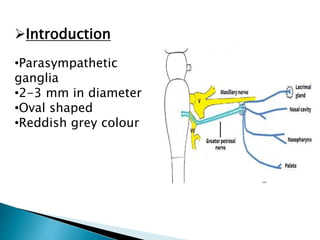
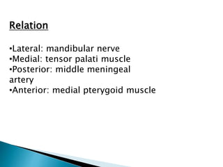
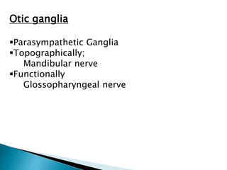
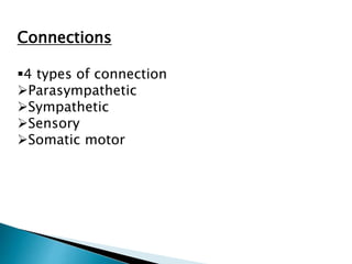
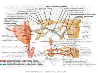
By:- Pradeep Tyagi
MBBS 1st Year
Roll no :- 63
CONtENTS
INTRODUCTION
LOCATION AND RELATIONS
TOPOGRAPHIC AND FUNCTIONAL RELATION
ROOTS OR CONNECTIONS
CLINICAL SIGNIFICANCE
Introduction
•Parasympathetic
ganglia
•2-3 mm in diameter
•Oval shaped
•Reddish grey colour
Location
Infratemporal fossa
Just below foramen
ovale, between
tensor palati and
mandibular nerve.
Relation
•Lateral: mandibular nerve
•Medial: tensor palati muscle
•Posterior: middle meningeal
artery
•Anterior: medial pterygoid muscle
Otic ganglia
Parasympathetic Ganglia
Topographically;
Mandibular nerve
Functionally
Glossopharyngeal nerve
Connections
4 types of connection
Parasympathetic
Sympathetic
Sensory
Somatic motor
Sympathetic
Post ganglionic fibres arising in
Superior cervical ganglia.
Sympathetic root derived from
plexus around Middle meningeal
artery.
Fibres cannot relay in otic ganglia
and reach parotid gland via
Auricotemporal nerve.
Fibres are vasomotor in function.
 The tympanic plexus innervates the mucous
membrane lining the walls and content of middle ear.
 Formed by tympanic nerve, a branch of
glossopharyngeal nerve.
Sensory and
somatic motor
 Sensory root comes
from Auricotemporal
nerve.
 Motor fibres pass
through otic ganglion
without relay and
supply following
muscles
- tensor palati
- tensor tympani
Other connections
 Alternate pathway of
taste
 Buccal nerve
Clinical significance
Frey’s Syndrome
Due to damage of
Auricotemporal nerve.
Salvation induce
perspiration(sweating).
Accompanied with
erythema(reddening of
skin).
Reference
Gray’s Anatomy
Vishram Singh
B.D. Chaurasia
otic ganglion.pptx

otic ganglion.pptx