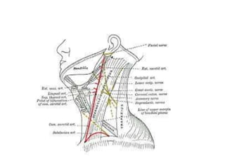
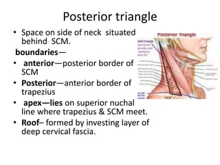
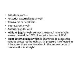
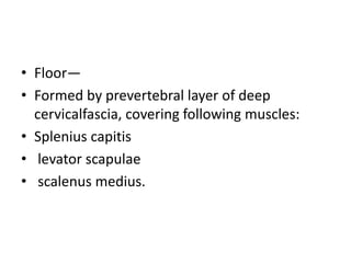
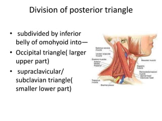
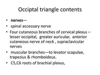
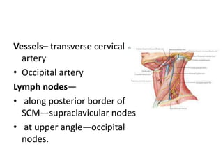
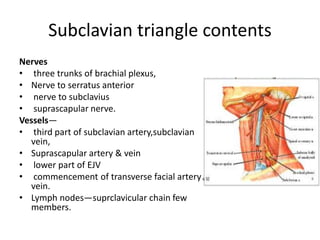
Triangles of neck
By:CHIRANJEEV SINGH
Posterior triangle
• Space on side of neck situated
behind SCM.
boundaries—
• anterior—posterior border of
SCM
• Posterior—anterior border of
trapezius
• apex—lies on superior nuchal
line where trapezius & SCM meet.
• Roof– formed by investing layer of
deep cervical fascia.
• The superficial fascia over posterior triangle
contains—
• Platysma
• EJV & posterior external jugular veins,
• parts of supraclavicular , greater auricular,
transverse cutaneous & lesser occipital nerves,
• unnamed arteries derived from occipital
,transverse cervical & suprascapular arteries,
• lymph vessels which pierce deep fascia to end in
supraclavicular nodes.
External jugular vein
• lies deep to platysma,
• formed by union of posterior auricular vein with
posterior division of retromandibular vein,
• it begins with in the lower part of parotid gland ,
crosses the SCM obliquely , pierces anteroinferior
angle of roof of posterior triangle---opens in
subclavian vein.
• As it pierces the fascia , margins of vein get adherent
to fascia , so if vein gets cut , it cannot close due to
negative intrathoracic pressure .that causes air
embolism ,to prevent this , the deep fascia has to be
cut .
• tributeries are—
• Posterior external jugular vein
• Transverse cervical vein
• suprascapular vein
• Anterior jugular vein
• oblique jugular vein connects external jugular vein
across the middle 1/3rd of anterior border of SCM.
• right external jugular vein is examined to ascess the
venous pressure; the right atrial pressure is reflected in
it because there are no valves in the entire course of
this vein.& it is straight.
• Floor—
• Formed by prevertebral layer of deep
cervicalfascia, covering following muscles:
• Splenius capitis
• levator scapulae
• scalenus medius.
Division of posterior triangle
• subdivided by inferior
belly of omohyoid into—
• Occipital triangle( larger
upper part)
• supraclavicular/
subclavian triangle(
smaller lower part)
Occiptal triangle contents
• nerves—
• spinal accessary nerve
• Four cutaneous branches of cervical plexus –
lesser occipital, greater auricular, anterior
cutaneous nerve of neck , supraclavicular
nerves
• muscular branches—to levator scapulae,
trapezius & rhomboideus.
• C5,C6 roots of brachial plexus,
Vessels– transverse cervical
artery
• Occipital artery
Lymph nodes—
• along posterior border of
SCM—supraclavicular nodes
• at upper angle—occipital
nodes.
Subclavian triangle contents
Nerves
• three trunks of brachial plexus,
• Nerve to serratus anterior
• nerve to subclavius
• suprascapular nerve.
Vessels—
• third part of subclavian artery,subclavian
vein,
• Suprascapular artery & vein
• lower part of EJV
• commencement of transverse facial artery &
vein.
• Lymph nodes—suprclavicular chain few
members.
Applied features
• most common swelling in posterior triangle is due to enlargement
of supraclavicular lymph nodes, while doing biopsy one must be
careful in preserving accessory nerve which can get entangled with
enlarging LN.
• Supraclavicular LN are commonly involved in tuberculosis, hodgkin’s
disease, malignant growths of breast ,arm or chest.
• Block dissection of neck for malignant diseases is removal of
cervical lymph nodes along with other structures involved in
growth.this procedure does not endanger---brachial, cervical plexus
& their muscular branches.
• torticollis or wry neck is a deformity in which head is bent to one
side due to spasm or contracture of muscles supplied by spinal
accessary nerve( SCM & trapezius)
• Variety of torticollis—
• Rheumatic –due to exposure of cold or
draught,
• Reflex—due to inflammed or suppurating
cervical lymph nodes which irritate spinal
accessary nerve
• congenital torticollis due to birth injury
• spasmodic—due to central irritation.

Triangles of neck (1)

  • 1.
  • 3.
    Posterior triangle • Spaceon side of neck situated behind SCM. boundaries— • anterior—posterior border of SCM • Posterior—anterior border of trapezius • apex—lies on superior nuchal line where trapezius & SCM meet. • Roof– formed by investing layer of deep cervical fascia.
  • 4.
    • The superficialfascia over posterior triangle contains— • Platysma • EJV & posterior external jugular veins, • parts of supraclavicular , greater auricular, transverse cutaneous & lesser occipital nerves, • unnamed arteries derived from occipital ,transverse cervical & suprascapular arteries, • lymph vessels which pierce deep fascia to end in supraclavicular nodes.
  • 5.
    External jugular vein •lies deep to platysma, • formed by union of posterior auricular vein with posterior division of retromandibular vein, • it begins with in the lower part of parotid gland , crosses the SCM obliquely , pierces anteroinferior angle of roof of posterior triangle---opens in subclavian vein. • As it pierces the fascia , margins of vein get adherent to fascia , so if vein gets cut , it cannot close due to negative intrathoracic pressure .that causes air embolism ,to prevent this , the deep fascia has to be cut .
  • 6.
    • tributeries are— •Posterior external jugular vein • Transverse cervical vein • suprascapular vein • Anterior jugular vein • oblique jugular vein connects external jugular vein across the middle 1/3rd of anterior border of SCM. • right external jugular vein is examined to ascess the venous pressure; the right atrial pressure is reflected in it because there are no valves in the entire course of this vein.& it is straight.
  • 7.
    • Floor— • Formedby prevertebral layer of deep cervicalfascia, covering following muscles: • Splenius capitis • levator scapulae • scalenus medius.
  • 8.
    Division of posteriortriangle • subdivided by inferior belly of omohyoid into— • Occipital triangle( larger upper part) • supraclavicular/ subclavian triangle( smaller lower part)
  • 10.
    Occiptal triangle contents •nerves— • spinal accessary nerve • Four cutaneous branches of cervical plexus – lesser occipital, greater auricular, anterior cutaneous nerve of neck , supraclavicular nerves • muscular branches—to levator scapulae, trapezius & rhomboideus. • C5,C6 roots of brachial plexus,
  • 11.
    Vessels– transverse cervical artery •Occipital artery Lymph nodes— • along posterior border of SCM—supraclavicular nodes • at upper angle—occipital nodes.
  • 12.
    Subclavian triangle contents Nerves •three trunks of brachial plexus, • Nerve to serratus anterior • nerve to subclavius • suprascapular nerve. Vessels— • third part of subclavian artery,subclavian vein, • Suprascapular artery & vein • lower part of EJV • commencement of transverse facial artery & vein. • Lymph nodes—suprclavicular chain few members.
  • 14.
    Applied features • mostcommon swelling in posterior triangle is due to enlargement of supraclavicular lymph nodes, while doing biopsy one must be careful in preserving accessory nerve which can get entangled with enlarging LN. • Supraclavicular LN are commonly involved in tuberculosis, hodgkin’s disease, malignant growths of breast ,arm or chest. • Block dissection of neck for malignant diseases is removal of cervical lymph nodes along with other structures involved in growth.this procedure does not endanger---brachial, cervical plexus & their muscular branches. • torticollis or wry neck is a deformity in which head is bent to one side due to spasm or contracture of muscles supplied by spinal accessary nerve( SCM & trapezius)
  • 15.
    • Variety oftorticollis— • Rheumatic –due to exposure of cold or draught, • Reflex—due to inflammed or suppurating cervical lymph nodes which irritate spinal accessary nerve • congenital torticollis due to birth injury • spasmodic—due to central irritation.