Downloaded 200 times
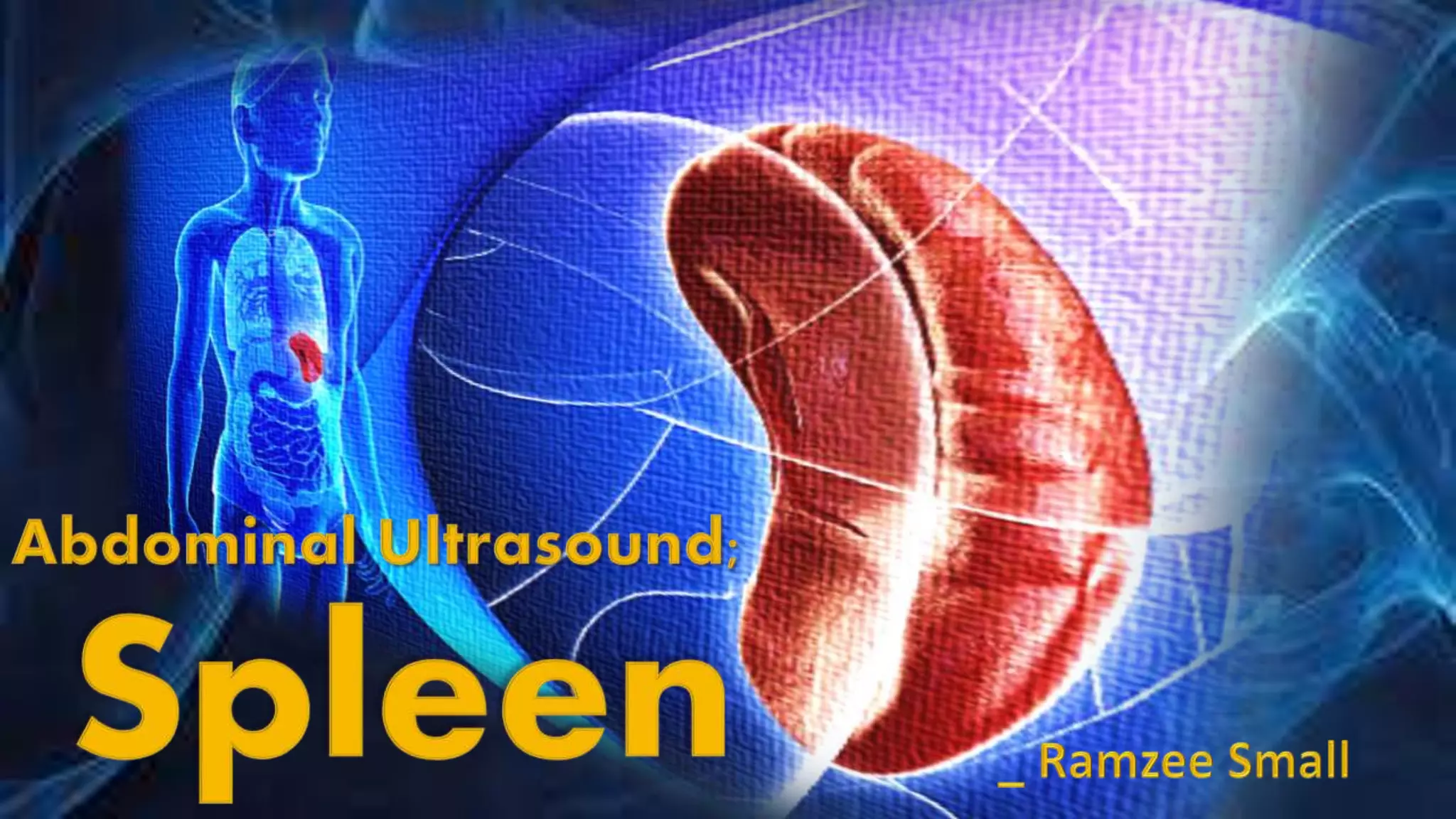
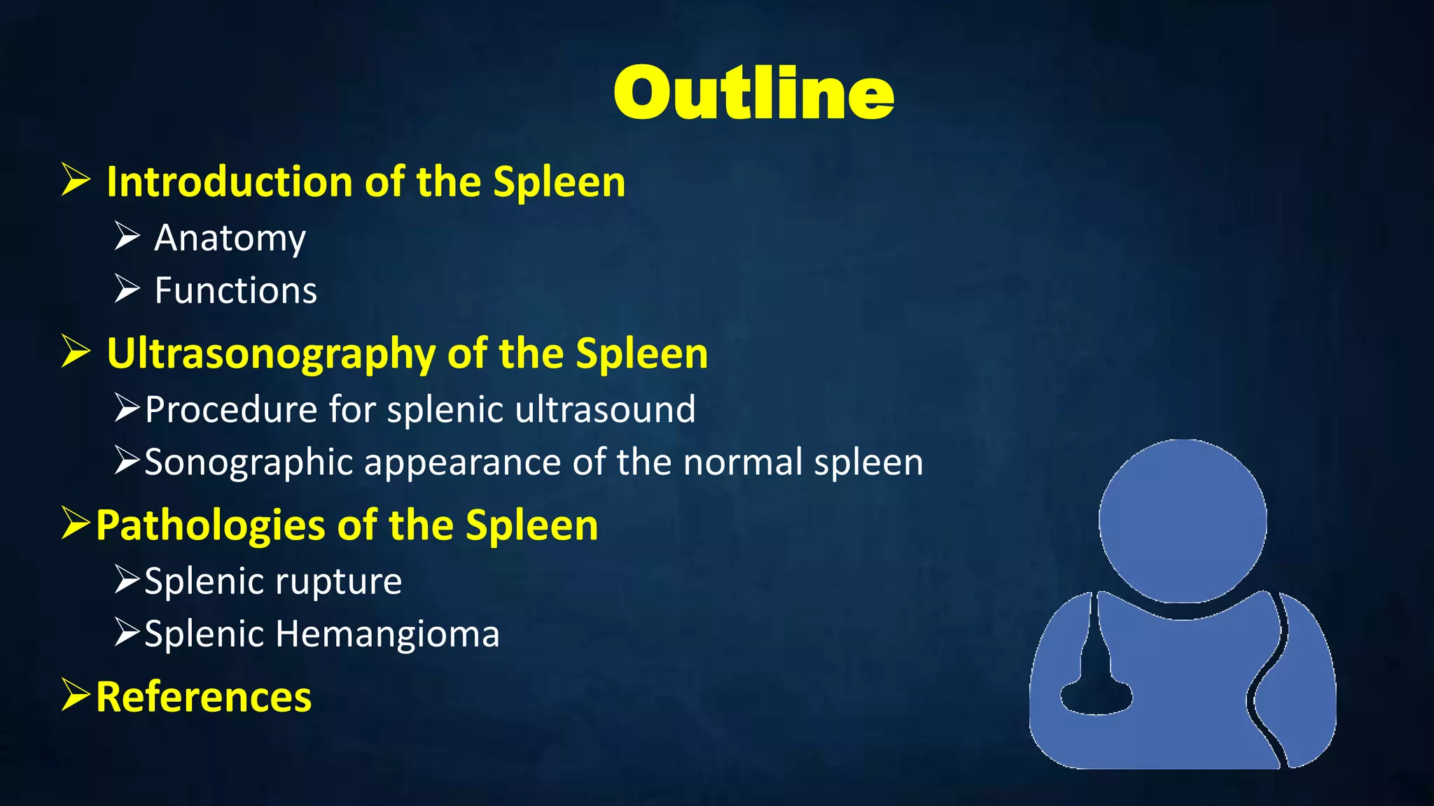


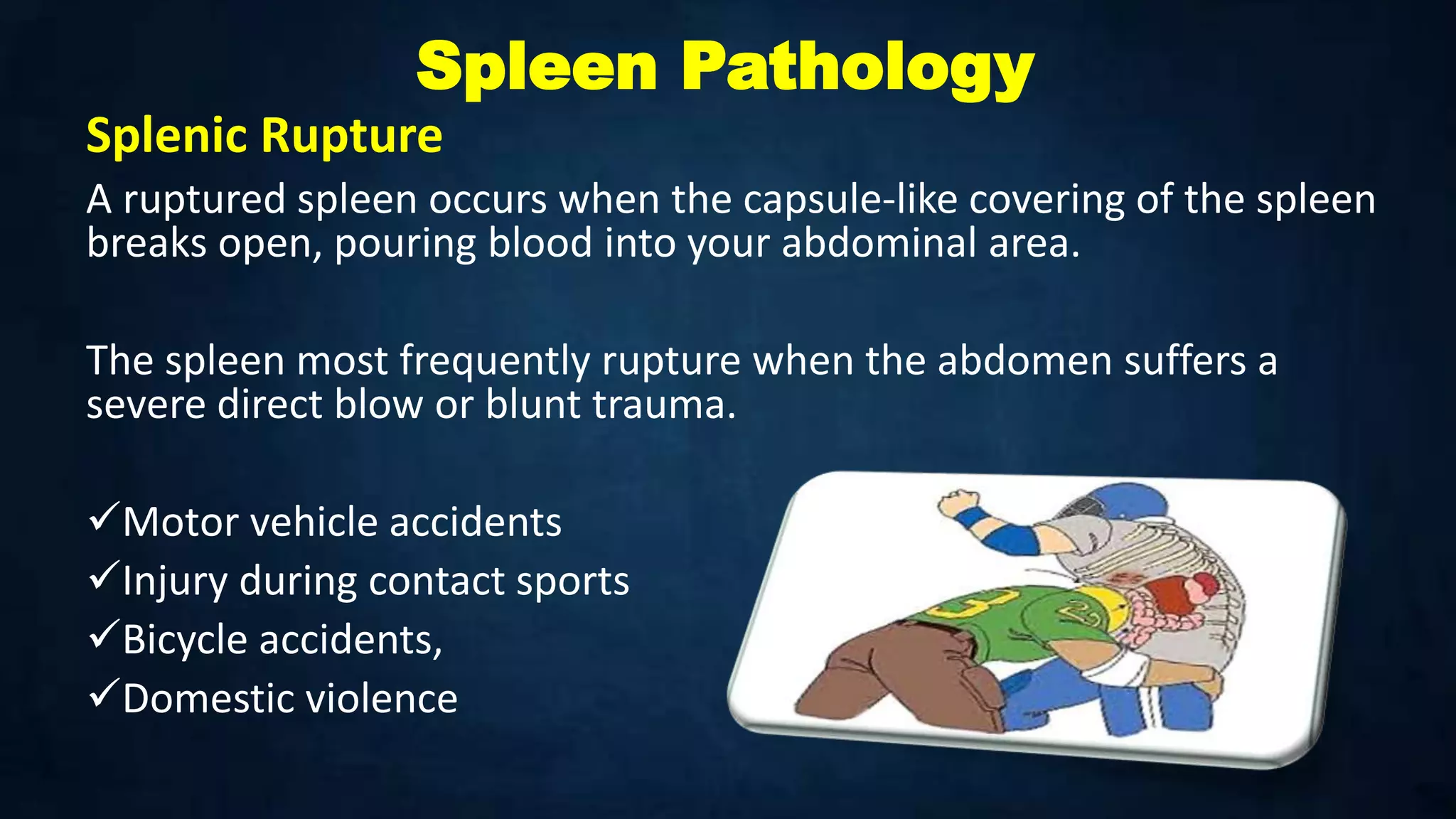


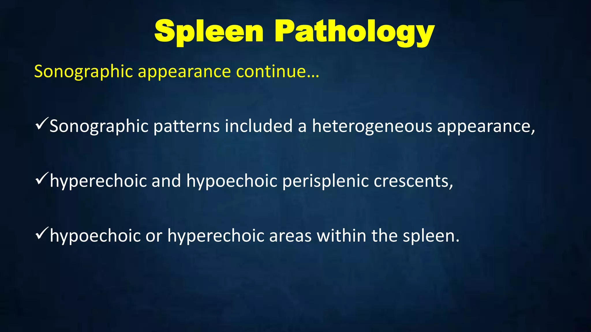


The document provides a comprehensive overview of the spleen, including its anatomy, functions, and the process of ultrasonography for diagnosis. It discusses various pathologies associated with the spleen, such as splenic rupture and hemangioma, along with their symptoms and sonographic appearances. Additionally, it includes references for further reading on the subject.


![Spleen[1]](https://cdn.slidesharecdn.com/ss_thumbnails/spleen1-171112094140-thumbnail.jpg?width=640&height=640&fit=bounds)


![Imaging ofsplenic diseases [Autosaved].pptx](https://cdn.slidesharecdn.com/ss_thumbnails/imagingodsplenicdiseasesautosaved-220822020559-3118a410-thumbnail.jpg?width=640&height=640&fit=bounds)


