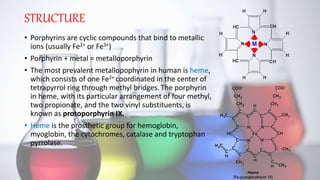
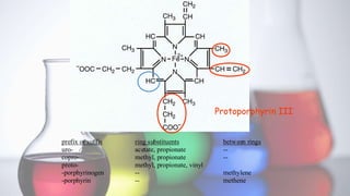
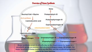
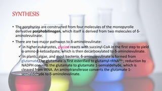
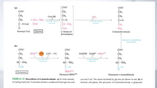
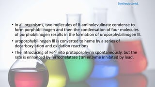
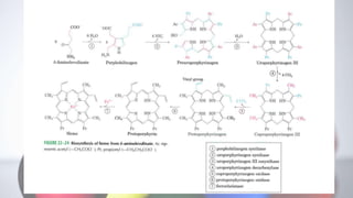
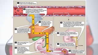
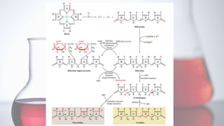
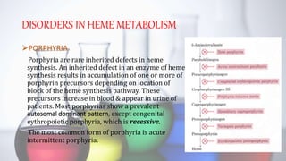
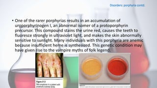
Content :-
What is porphyrin?
Biosynthesis of porphyrin (heme)
Site
Reactions
Regulation
Degradation of heme
Site
Reactions
Reference
What is Porphyrin?
Porphyrins are cyclic compound formed by the linkage of four pyrrole rings through methyne(=HC-)bridges.
Structure of hemoglobin & chlorophyll
Examples of some important humanand animal hemoproteins.
Biosynthesis of heme
Site of biosynthesis:-
-Liver (hepatocyte) & Bone Marrow
( erythryoid producing cells )
2) ALA dehydratase :-
The substrates are two molecules of ALA.
The product is porphobilinogen, the first pyrrole.
ALA dehydratase is a -SH containing enzyme.
It is very susceptible to inhibition by lead.
3) Uroporphyrinogen I synthase and uroporphyrinogen IIIcosynthase
Production of uroporphyrinogen III requires two enzymes.The substrates are four molecules of porphobilinogen.
4) Uroporphyrinogen decarboxylase:-
Decarboxylates the acetic acid groups, converting them to methyl groups.
5) Coproporphyrinogen III oxidase:-
Catalyzes the conversion of two propionic acid groups to vinyl groups
6) Protoporphyrinogen IX oxidase:-
Protoporphyrinogen IX oxidase converts the methylene bridges between the pyrrole rings to methenyl bridges.
7) Ferrochelatase:-
Ferrochelatase adds Fe++ to protoporphyrin IX, forming heme.
• The enzyme requires Fe++, ascorbic acid and cysteine (reducing agents).
• Ferrochelatase is inhibited by lead.
Regulation of heme synthesis
Feedback regulation:- heme is a feedback inhibitor of ALA synthase. ALA synthase occurs in both hepatic (ALAS 1) and erythroid (ALAS 2) forms.
Effects of drugs and steroids:- Certain drugs and steroids can increase heme synthesis via increased production of the ratelimiting enzyme, ALA synthase
Substrate availability:- Fe++ must be available for ferrochelatase.
Degradation of heme
Site of Degradation :-
- cells of the reticulo endothelial system in spleen, liver and bone marrow
Heme Degradation
Most of the heme which is degraded comes from hemoglobin in red blood cells, which have a life span of about 120 days.
There is thus a turnover of about 6 g/day of hemoglobin.
Normally , senescent red blood cells and heme from other sources are engulfed by cells of the reticuloendothelial system.
The globin is recycled or converted into amino acids, which in turn are recycled or catabolized as required.
Heme is oxidized.
Microsomal heme oxygenase system
Transport of bilirubin in Plasma
Bilirubin on release from macrophages circulates as unconjugated bilirubin in plasma tightly bound to albumin.
HARPER’S ILLUSTRATED BIOCHEMISTRY (28TH EDITION) by robert murray,david A.bender,peter j kennekky,victor w rodwell,p.antony weil.(page No-271)
Biochemistry Lippincott’s Illustrated Reviews by Richard Harvey& Denise Ferrier
The slides show the gastric and pancreatic function test along with the significance of these tests and the conditions in which the values of which increase.
cytochrome p450 is an super family of enzyme that contains a heme as co factor that function as monooxygenase. This enzyme has been identified in all kingdoms of life, like fungi, Protista, bacteria, and as well as in virus.
Heme Catabolism and Degradation Pathway #Bilirubin metabolismAHLAD T.O
This video is about heme catabolism or heme degradation. Heme is degraded into bilirubin in a heme degradation pathway. Heme degradation pathway or heme degradation biochemistry is important for understanding different types of bilirubin being elevated in jaundice. Types of jaundice can be classified based on the type of bilirubin being elevated in the blood. Heme catabolism or heme degradation is important concept
Heme Catabolism (Heme Degradation Pathway)
heme catabolism
Bilirubin metabolism ( Heme catabolism)
Heme Catabolism and Degradation Pathway - Biochemistry Lesson
Bilirubin metabolism
Heme catabolism
Heme catabolism || Bilirubin metabolism || #Biochemistry
steps
Introduction
Bilirubin formation pathway
Transport of bilirubin to liver
Uptake of bilirubin in liver
Conjugation of bilirubin
Excretion of bilirubin in bile canaliculi
Fate of conjugated bilirubin in intestine
Entero-hepatic circulation of urobilinogen
Final excretion of UBG and SBG
As the channel name suggests, our channel will be a perfect lounge for the malayali medicos..we wil be covering videos which will be like lecture classes related to the subjects biochemistry and microbiology in which we are specialised.. It will be a better learning experience for the students especially for those who are not able to understand and follow the normal classes in college..we assure the students that you will get a basic idea regarding the topic and extra reading can be done from the reference textbooks...
If you like my video
#like
#comment
#subscribe my channel
don't forget to subscribe my channel
Qualification
Ahlad.T.O
MSc MLT (Biochemistry)
Assistant Professor
Baby memorial college of allied Health science
Kozhikode
Our Partner Channel
Health & Voyage channel link - https://youtu.be/nzKqRVjlwc0
Heme catabolism
Heme degradation in macrophages
Bilirubin transport with albumin
Bilirubin metabolism
1. Uptake by hepatocytes by facilitated diffusion
2. Conjugation of unconjugated bilirubin
3. Secretion in to bile canaliculi by active transport through MRP 2
Congenital causes
UNCONJUGATED BILIRUBIN INCREASE
1. Crigler Najjar type 1
2. Crigler Najjar type 2
3. Gilbert syndrome
Conjugated bilirubin increase
1. Dubin Johnson syndrome
2. Rotor syndrome
#Mallu
#Microbiology
#Biochemistry
#MalluMedicosLounge
#HealthAndVoyage
#How to take online class
Introduction of metabolism, importance of metabolism, types of metabolism, carbohydrate metabolism, Glycolysis, electron transport & respiratory chain, TCA cycle, Abnormal metabolism, Protein metabolism, Urea cycle, Abnormal metabolism of proteins, Lipid metabolism, Beta-oxidation of fatty acids, Abnormal metabolism of fats. deficiency of enzymes causes, diseases, Interrelation metabolism
Model Attribute Check Company Auto PropertyCeline George
In Odoo, the multi-company feature allows you to manage multiple companies within a single Odoo database instance. Each company can have its own configurations while still sharing common resources such as products, customers, and suppliers.
Macroeconomics- Movie Location
This will be used as part of your Personal Professional Portfolio once graded.
Objective:
Prepare a presentation or a paper using research, basic comparative analysis, data organization and application of economic information. You will make an informed assessment of an economic climate outside of the United States to accomplish an entertainment industry objective.
Operation “Blue Star” is the only event in the history of Independent India where the state went into war with its own people. Even after about 40 years it is not clear if it was culmination of states anger over people of the region, a political game of power or start of dictatorial chapter in the democratic setup.
The people of Punjab felt alienated from main stream due to denial of their just demands during a long democratic struggle since independence. As it happen all over the word, it led to militant struggle with great loss of lives of military, police and civilian personnel. Killing of Indira Gandhi and massacre of innocent Sikhs in Delhi and other India cities was also associated with this movement.
How to Make a Field invisible in Odoo 17Celine George
It is possible to hide or invisible some fields in odoo. Commonly using “invisible” attribute in the field definition to invisible the fields. This slide will show how to make a field invisible in odoo 17.
Introduction to AI for Nonprofits with Tapp NetworkTechSoup
Dive into the world of AI! Experts Jon Hill and Tareq Monaur will guide you through AI's role in enhancing nonprofit websites and basic marketing strategies, making it easy to understand and apply.
Welcome to TechSoup New Member Orientation and Q&A (May 2024).pdfTechSoup
In this webinar you will learn how your organization can access TechSoup's wide variety of product discount and donation programs. From hardware to software, we'll give you a tour of the tools available to help your nonprofit with productivity, collaboration, financial management, donor tracking, security, and more.
2. STRUCTURE
• Porphyrins are cyclic compounds that bind to metallic
ions (usually Fe2+ or Fe3+)
• Porphyrin + metal = metalloporphyrin
• The most prevalent metallopophyrin in human is heme,
which consists of one Fe2+ coordinated in the center of
tetrapyrrol ring through methyl bridges. The porphyrin
in heme, with its particular arrangement of four methyl,
two propionate, and the two vinyl substituents, is
known as protoporphyrin IX.
• Heme is the prosthetic group for hemoglobin,
myoglobin, the cytochromes, catalase and tryptophan
pyrrolase.
3. Protoporphyrin III
prefix or suffix ring substituents between rings
uro- acetate, propionate --
copro- methyl, propionate --
proto- methyl, propionate, vinyl
-porphyrinogen -- methylene
-porphyrin -- methene
4. Uroporphyrinogen I Coproporphyrinogen I
Overviewof Heme Synthesis
Heme synthesis occurs in all cells due to the requirement for heme as a
prosthetic group on enzymes and electron transport chain. By weight, the
major locations of heme synthesis are the liver and the erythroid progenitor
cells of the bone marrow.
Succinyl CoA + Glycine
-aminolevulinic acid
Porphobilinogen Uroporphyrinogen III Coproporphyrinogen III
Coproporphyrinogen III
Protoporphyrinogen IX
Protoporphyrin IX
Heme
ALA synthase
cytoplasm
mitochondrial matrix
5. SYNTHESIS
• The porphyrins are constructed from four molecules of the monopyrolle
derivative porphobilinogen, which itself is derived from two molecules of δ-
aminolevulinate.
• There are two major pathways to δ-aminolevulinate:
in higher eukaryotes, glycine reacts with succinyl-CoA in the first step to yield
α-amino-β-ketoadipate, which is then decarboxylated to δ-aminolevulinate.
In plants, algae, and most bacteria, δ-aminolevulinate is formed from
glutamate.The glutamate is first esterified to glutamyl-tRNAGlu ; reduction by
NADPH converts the glutamate to glutamate 1-semialdehyde, which is
cleaved from tRNA. An aminotransferase converts the glutamate 1-
semialdehyde to δ-aminolevulinate.
6.
7. Synthesis contd.
• In all organisms, two molecules of δ-aminolevulinate condense to
form porphobilinogen and then the condensation of four molecules
of porphobilinogen results in the formation of uroporphybilinogen lll.
• uroporphybilinogen lll is converted to heme by a series of
decarboxylation and oxidation reactions
• The introducing of Fe+2 into protoporphyrin spontaneously, but the
rate is enhanced by ferrochelatase ( an enzyme inhibited by lead.
8.
9. REGULATION OF HEME BIOSYNTHESIS
• The two major sites of heme biosynthesis are erythroid cells, which synthesize
~85% of the body’s heme groups, and the liver, which synthesizes ~ 80% of the
remainder.
• An important function of heme in liver is as the prosthetic groups of the
cytochromes P450, a family of oxidative enzymes involved in detoxification,
whose members are required throughout a liver cell’s lifetime in amounts that
vary with conditions.
• In contrast, erythroid cells, in which heme is, of course, a hemoglobin
component, engage in heme synthesis only on differentiation, when they
synthesize hemoglobin in vast quantities.This is a onetime synthesis; the heme
must last the erythrocyte’s lifetime (normally 120 days) since heme and
hemoglobin synthesis stop on red cell maturation (protein synthesis stops on the
loss of nuclei and ribosomes).
• The different ways in which heme biosynthesis is regulated in liver and in
erythroid cells reflect these different demands: In liver, heme biosynthesis must
really be “controlled,” whereas in erythroid cells, the process is more like
breaking a dam.
10. Regulation contd.
IN LIVER :
• The main control target in heme biosynthesis is ALA synthase, the
enzyme catalyzing the pathway’s first committed step. Heme, or its
Fe(III) oxidation product hemin, controls this enzyme’s activity
through three mechanisms:
(1) feedback inhibition,
(2) inhibition of the transport of ALA synthase (ALAS) from its site of
synthesis in the cytosol to its reaction site in the mitochondrion, and
(3) repression of ALAS synthesis.
IN ERYTHROCYTES
• In erythroid cells, heme exerts quite a different effect on its
biosynthesis. Heme induces, rather than represses, protein synthesis
in reticulocytes (immature erythrocytes).
11. Regulation contd.
• Moreover, the rate-determining step of heme biosynthesis in
erythroid cells may not be the ALA synthase reaction.
• Experiments on various systems of differentiating erythroid cells
implicate ferrochelatase and porphobilinogen deaminase in the
control of heme biosynthesis in these cells. There are also indications
that cellular uptake of iron may be rate limiting.
• Iron is transported in the plasma complexed with the iron transport
protein transferrin. The rate at which the iron–transferrin complex
enters most cells, including those of liver, is controlled by receptor-
mediated endocytosis. However, lipid-soluble iron complexes that
diffuse directly into reticulocytes stimulate in vitro heme biosynthesis.
12. CATABOLISM OF HEME
• When hemoglobin is destroyed in the body, globin is degraded to its constituent
amino acids, which are reused, and the iron of heme enters the iron pool, also for
reuse. The iron-free porphyrin portion of heme is also degraded, mainly in the
reticuloendothelial cells of the liver, spleen, and bone marrow.
• The catabolism of heme from all of the heme proteins appears to be carried out
in the microsomal fractions of cells by a complex enzyme system called heme
oxygenase. By the time the heme derived from heme proteins reaches the
oxygenase system, the iron has usually been oxidized to the ferric form,
constituting hemin.
• the hemin is reduced to heme with NADPH, and, with the aid of more NADPH,
oxygen is added to the α-methyne bridge between pyrroles I and II of the
porphyrin. The ferrous iron is again oxidized to the ferric form. With the further
addition of oxygen, ferric ion is released, carbon monoxide is produced, and an
equimolar quantity of biliverdin results from the splitting of the tetrapyrrole ring.
13. Catabolism contd.
• In birds and amphibia, the green biliverdin IX is excreted; in
mammals, a soluble enzyme called biliverdin reductase reduces the
methylene bridge between pyrrole III and pyrrole IV to a methylene
group to produce bilirubin, a yellow pigment
• Bilirubin formed in peripheral tissues is transported to the liver by
plasma albumin. The further metabolism of bilirubin occurs primarily
in the liver. It can be divided into three processes:
(1) uptake of bilirubin by liver parenchymal cells,
(2) conjugation of bilirubin with glucuronate in the endoplasmic
reticulum, and
(3) secretion of conjugated bilirubin into the bile. Each of these
processes will be considered separately.
14. THE LIVER TAKES UP BILIRUBIN
• Bilirubin is only sparingly soluble in water, but its
solubility in plasma is increased by noncovalent
binding to albumin. Bilirubin in excess of this
quantity can be bound only loosely and thus can
easily be detached and diffuse into tissues.
• In the liver, the bilirubin is removed from albumin
and taken up at the sinusoidal surface of the
hepatocytes by a carrier-mediated saturable
system. This facilitated transport system has a very
large capacity, so that even under pathologic
conditions the system does not appear to be rate-
limiting in the metabolism of bilirubin.
Catabolism contd.
15. Conjugation of Bilirubin with Glucuronic Acid Occurs in the
Liver
• Bilirubin is nonpolar and would persist in cells (eg, bound to lipids) if
not rendered water-soluble. Hepatocytes convert bilirubin to a polar
form, which is readily excreted in the bile, by adding glucuronic acid
molecules to it. This process is called conjugation and can employ
polar molecules other than glucuronic acid (eg, sulfate).
• The conjugation of bilirubin is catalyzed by a specific
glucuronosyltransferase. The enzyme is mainly located in the
endoplasmic reticulum, uses UDP-glucuronic acid as the glucuronosyl
donor, and is referred to as bilirubin-UGT. Bilirubin monoglucuronide
is an intermediate and is subsequently converted to the diglucuronide
Catabolism contd.
16. Bilirubin Is Secreted into Bile
• Secretion of conjugated bilirubin into the bile occurs by an active transport
mechanism, which is probably rate-limiting for the entire process of
hepatic bilirubin metabolism.
• The protein involved is MRP-2 (multidrug-resistance-like protein 2), also
called multispecific organic anion transporter (MOAT). It is located in the
plasma membrane of the bile canalicular membrane and handles a number
of organic anions.
ConjugatedBilirubinIs Reduced to Urobilinogenby Intestinal Bacteria
• As the conjugated bilirubin reaches the terminal ileum and the large
intestine, the glucuronides are removed by specific bacterial enzymes (β-
glucuronidases), and the pigment is subsequently reduced by the fecal flora
to a group of colorless tetrapyrrolic compounds called urobilinogens. In the
terminal ileum and large intestine, a small fraction of the urobilinogens is
reabsorbed and reexcreted through the liver to constitute the
enterohepatic urobilinogen cycle.
Catabolism contd.
17.
18.
19. DISORDERS IN HEME METABOLISM
PORPHYRIA
Porphyria are rare inherited defects in heme
synthesis. An inherited defect in an enzyme of heme
synthesis results in accumulation of one or more of
porphyrin precursors depending on location of
block of the heme synthesis pathway. These
precursors increase in blood & appear in urine of
patients. Most porphyrias show a prevalent
autosomal dominant pattern, except congenital
eythropoietic porphyria, which is recessive.
The most common form of porphyria is acute
intermittent porphyria.
20. Disorders: porphyria contd.
• One of the rarer porphyrias results in an accumulation of
uroporphyrinogen I, an abnormal isomer of a protoporphyrin
precursor. This compound stains the urine red, causes the teeth to
fluoresce strongly in ultraviolet light, and makes the skin abnormally
sensitive to sunlight. Many individuals with this porphyria are anemic
because insufficient heme is synthesized. This genetic condition may
have given rise to the vampire myths of folk legend.
21.
22. JAUNDICE
• When bilirubin in the blood exceeds 1 mg/dL (17.1 μmol/L),
hyperbilirubinemia exists. Hyperbilirubinemia may be due to the
production of more bilirubin than the normal liver can excrete, or it
may result from the failure of a damaged liver to excrete bilirubin
produced in normal amounts. In the absence of hepatic damage,
obstruction of the excretory ducts of the liver—by preventing the
excretion of bilirubin—will also cause hyperbilirubinemia. In all these
situations, bilirubin accumulates in the blood, and when it reaches a
certain concentration (approximately 2–2.5 mg/dL), it diffuses into
the tissues, which then become yellow. That condition is called
jaundice or icterus.
Disorders contd.
23. Elevated Amounts of Unconjugated Bilirubin in Blood
Occur in a Number of Conditions
• Hemolytic anemias
• Neonatal “Physiologic Jaundice”
• Type I Crigler–Najjar syndrome
• Crigler–Najjar Syndrome, Type II.
• Gilbert Syndrome
• Toxic Hyperbilirubinemia
Disorders: jaundice contd.
24. Obstruction in the Biliary Tree Is the Most Common
Cause of Conjugated Hyperbilirubinemia
• Obstruction of the Biliary Tree
• Dubin–Johnson Syndrome
• Rotor Syndrome
Some Conjugated Bilirubin Can Bind Covalently to
Albumin
Disorders: jaundice contd.