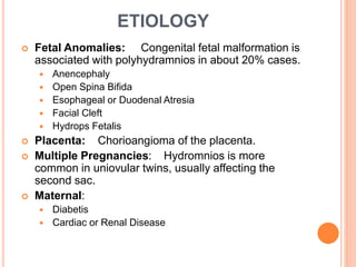
3. INTRODUCTION
Amniotic fluid normally increase in amount
throughout pregnancy from a few milliliters to a liter at
38th week. The fluid is not static ; the water and
solutes in it are change every few hours. There are
two chief abnormalities of amniotic fluid.
Polyhydramnios
Oligohydramnios
Polyhydramnios is a medical condition excess
of amniotic fluid in the amniotic sac. There are 2
clinical varieties of polyhydramnios.
Chronic
Acute
The opposite to Polyhydramnios is Oligohydramnios a
deficiency of amniotic fluid.
4. DEFINITION
Polyhydramnios is defined as a state where liquor
amnii exceeds 1500 to 2000 ml.
Clinical Definition: The excessive accumulation
of liquor amnii causing discomfort to the patient or when
a imaging help needed to substantiate the clinical
diagnosis of the lie and presentation of foetus.
The incidence varies from 1-2% of the cases. it is
more common of multipare than the premigravidae. it
occurs 1 in 1000 pregnancies.
INCIDENCE
5. ETIOLOGY
Fetal Anomalies: Congenital fetal malformation is
associated with polyhydramnios in about 20% cases.
Anencephaly
Open Spina Bifida
Esophageal or Duodenal Atresia
Facial Cleft
Hydrops Fetalis
Placenta: Chorioangioma of the placenta.
Multiple Pregnancies: Hydromnios is more
common in uniovular twins, usually affecting the
second sac.
Maternal:
Diabetis
Cardiac or Renal Disease
6. CLINICAL TYPES
Depending on the rapidly of onset, hydramnios may
be:
Chronic: It is the commonest onset in insidious
taking few weeks.
Acute: It is extremely rare acutely on pre-existing
chronic variety. The chronic variety is 10 times
commoner than the acute one.
7. CHRONIC POLYHYDRAMNIOS
Symptoms:
Dyspnoea.
Palpitation.
Oedema of legs.
Signs:
The patient may be dyspnoea state in the lying down
position.
Evidence of pre-eclampsia (edema, hypertension &
protenuria) may be present.
8. ABDOMINAL EXAMINATION
Inspection:
Abdominal marked enlarge.
The skin is tense shiny with large straiae.
Palpation:
Height of the uterus is more than the period of
amenorrhoea.
Girth of the abdomen round the umbilicus more than the
normal.
Foetal parts can’t be well defined: so also the
presentation or the position.
Auscultation:
Foetal heart sound is not heard easily.
9. INVESTIGATION
USG:
Amniotic fluid index is more than 25cm.
To note the lie and presentation of the foetus.
To diagnose any foetal congenital malformation.
10. COMPLICATION
Maternal:
During Pregnancy:
Pre eclampsia.
Malpresentation.
Pre rupture of the membrane.
Pre term labour.
During Labour:
Early rupture of the membrane.
Cord prolaps.
Increase operative delivery due to malpresentation.
Puerperium:
Sub involuation
Increase puerperial morbidity due to infection resulting from
increase operative interpherence and blood loss.
Foetal Complication:
Foetal death are mostly due to prematurity & congenital
abnormalities.
11. MANAGEMENT
Principles Of Management:
To relieve the symptoms.
To find out the causes.
To avoid and to deal with the complication.
Supportive Therapy:
Bed rest, treatment associate with condition like
preeclampsia.
Investigation are done to exclude congenital foetal
malformation.
Further management depends on
Response to treatment.
Period of gestation.
Presence of foetal malformation.
Associated complicating factors.
12. Response to treatment is good means pregnancy is
continued.
Unresponsive:
Pregnancy less than 37 weeks – Amniocentesis
Pregnancy more than 37 week – Induction of labour is done.
Amniocentesis, termination of pregnancy.
During Labour:
Usual management is followed.
If the uterine contration become sluggish, oxytocin
infusion may be started.
To prevent post partum haemorrhage.
IV administraion of methergin.