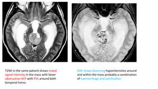
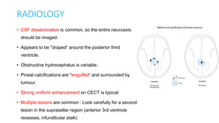
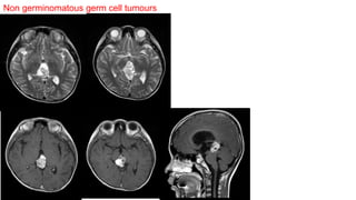
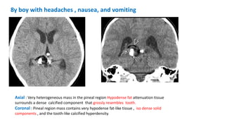
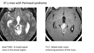
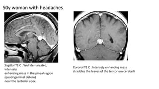
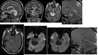
SlideShare a Scribd company logo
Pineal region tumours
Introduction,
clinical and radiological features.
By :Dr. Raj Kumar P
Moderator :Dr. Nishant Goyal
Associate prof.,
References :
Inderbir singh`s textbook of human histology.
Youman and winns neurological surgery.7th edition.
Rammurthy and Tandon`s Textbook of Neurosurgery.3rd edition.
Osborn brain 2nd edition.
Radiopedia.
Introduction
Overall clinical features
Individual tumours with specific clinical features and
radiology
Pineal gland
• The pineal gland is an encapsulated , extra axial structure.
• 1 to 3 % of all intracranial neoplasms.
• Avg measurements : 7.4 mm in (L), 6.9 mm in (W), and 2.5 mm in (H)
• Pineal masses originate infratentorially and expand into the posterior third
ventricle.
• Malignant tumors, particularly of glial origin, can invade into the midbrain
and thalamus.
Yamamoto Y, Kageyama N. Microsurgical anatomy of the pineal region. J Neurosurg 1980;53:205–221
NORMAL HISTOLOGY
• Made up of different types of cells , so diverse pathology.
• Histological features: Made up of
1. Capsule
2. Septa ( Trabeculae)
3. Parenchymal cells
• Pinealocytes
• Neuroglial cells
• Capsule : Derived from pia
matter : Thin collagenous
connective tissue.
• Septae: Arises from capsule
• Divides gland into incomplete
lobules.
• Blood vessels and sympathetic
nerve endings travel through
trabeculae and distributed
through out the gland.
Parenchymal cells
• Pinealocytes:
• 90 % of total cells.
• Larger than neuroglial cells.
• Large pale cells with prominent
nuclei.
• Neuroglial cells :
• Smaller than pinealocytes.
• Denser nuclei.
Corpora arenacea
• Characteristic of pineal
body.
• Calcified accretions in
intracellular spaces.
• AKA brain sand.
Tumours are grouped into 5 main categories
1. Germ cell tumors.
2. Papillary tumors of the pineal region.
3. Pineal parenchymal cell tumors.
4. Glial cell tumors.
5. Other miscellaneous tumors and cysts :- Meningioma, hemangioblastoma, choroid
plexus papilloma, metastatic tumour, chemodectoma, adenocarcinoma, and
lymphoma.
• Additionally, a variety of vascular lesions can occur, including cavernous malformations,
arteriovenous malformations, and vein of Galen malformations.
CLINICAL FEATURES
• one of three ways
1. Increased intracranial pressure from obstructive hydrocephalus.
2. Direct brainstem and cerebellar compression.
3. Endocrine dysfunction.
Raised ICP
• Headache : M.C initial symptom.
• Compression of aqueduct of sylvius  HCP.
• Further progression of HCP  Nausea , vomiting , cognitive
impairment , papilledema , ataxia.
• Rare : Pineal apoplexy  Hemorrhage in pineal tumour , acute
presentation.
Compression of midbrain
• Parinaud syndrome : Due to compression at the level of superior
colliculus.
• Paralysis of up gaze,
• Convergence or retraction nystagmus,
• Pupillary light-near dissociation.
• Sylvian aqueduct syndrome :
• Further compression leads to paralysis of downgaze or horizontal gaze.
• Dorsal midbrain compression or infiltration can lead to lid retraction
(Collier’s sign) or ptosis.
• IV nerve palsy with diplopia and head tilt.
• Interference with the cerebellar efferent pathways of the superior
cerebellar peduncles can cause ataxia and dysmetria.
• Hearing dysfunction : Probably caused by a disturbance in structures
associated with the inferior colliculi.
Endocrine dysfunction
• Secondary effects of hydrocephalus or from spread of tumor to the hypothalamic region.
• Diabetes insipidus :
• Germinoma spreading along the floor of the third ventricle.
• May develop early in the disease process, even before the tumor is radiographically apparent.
• Precocious puberty :
• AKA pseudoprecocious puberty , coz HPA is immature .
• Seen in choriocarcinoma or germinoma contains syncytiotrophoblastic cells producing ectopic β-
HCG.
Radiology of pineal region
tumours
Normal pineal gland radiology
• Avg measurements : 7.4 mm in (L), 6.9 mm in (W), and 2.5 mm in (H).
• Physiologic pineal gland calcification ("concretions") is common.
• Primary mineralization occurs in an organic matrix formed by pinealocytes.
• Pineal calcification increases with age.
• Reported prevalence is 1% < 6 y.
• 8% in < 10 y.
• 40% in < 30 y.
• >50% in all adults.
Pathology CT T1WI T2WI Contrast Others
Benign pineal cyst Hypodense Hypointense Hyperintense Rim
enhancment
Pinealocytoma Iso to hypodense.
Calcification
(peripheral)
ISO - Hypo Hyper Rim / nodular
pattern
enhancment
GRE : Blooming
Pinealoblastoma Large Hyperdense.
Heterogenous.
Calcification .
Iso - Hypo Iso – Hyper.
Cystic components
Heterogenous
enhancement
DWI – Restriction
GRE : Blooming
Germinoma Hyperdense .
Calcification
(engulfed).
Iso- Slightly
Hyerintense
Iso-hyper
Heterogenous.
Solid cystic.
Peri leisional
hyperintensity
Strong
homogenous
DWI-Restriction
GRE : Blooming
NGGCT Hetero. Iso to Hyper heterogenous Heterogenous
enhancment
Teratoma Heterogenous.
Hypodense – Fat.
Hyperdense – Tooth,
bone
Hetero
Fat- Hyper
Bone-Signal
void
Heterogenous
Solid + Cystic
Heterogenous
enhancement
Germ cell tumours
• Tumors of children and young adults; 80- 90% : < 20 y.
• Classified according to histology and immunohistochemistry.
• 8−15% of all primary CNS tumours in the paediatric age group in east asia(1).
• 3 to 4% in western countries(2).
• Divided into two basic groups.
1. Germinomas :- 2/3 of all GCT.
2. Nongerminomatous germ cell tumors : Rest of GCT.
1.Matsutani M, Sano K, Takakura K. Primary intracranial germ cell tumours: a clinical analysis of 153 histologicall verified cases. J Neurosurg
1997;86:146−455.
2. Schild SE, Scheithauer BW, Haddock MG, et al. Histologically confirmed pineal tumours and other germ cell tumours of the brain. Cancer.
1993;72:870−80.
• Teratoma
• Choreocarcinoma
• Embryonal cell carcinoma.
• Endodermal sinus tumour
• Other mixed variations.
• Normal mature pineal gland does not contain germ cells.
• Aetiology :
• Previously thought : “Aberrant migration" of cells from primordial germ layers.
• More recent hypotheses : Native stem cells of pluripotent or neural type, transformed as
neoplastically active germ cell elements.
• Germcell tumours:
• Preferentially effect midline structures.
• Majority around the third ventricle-pineal region followed by the suprasellar
compartment.
• Other sites include:
• Intraventricular.
• Basal ganglion.
• Germinomas : Predilection to arise from the suprasellar region.
• Diencephalon.
• Medulla.
• Sella.
Type of lesion Tumour marker
Pure germinoma -------
Choriocarcinoma HCG
Embryonal carcinoma PLAP
Yolk sac tumour AFP
Teratoma ------
Tumour markers
GERMINOMA
• Predilection for midline structures .
• 80-90% : “Hug" the midline, along the midline axis from the pineal gland ( ½ to
2/3 ) to the suprasellar region (1/4 to 1/3) .
• Size varies with location .
• 20% are multiple.
• The most frequent combination is a pineal plus a suprasellar ("bifocal" or "double
midline") germinoma.
• GROSS PATHOLOGY :
• Generally solid, friable, tan white masses that often infiltrate adjacent structures.
• Intratumoural cysts, small hemorrhagic foci, and CSF dissemination are common.
• CLINICAL FEATURES:
• Varies with location.
• Pineal germinomas : Headache and Parinaud syndrome.
• Suprasellar germinoma : M.C :- Central diabetes insipidus. Visual loss and
precocious puberty are other presentations.
• NATURAL HISTORY :
• CSF dissemination and
invasion are common.
• But pure germinomas 
Favorable response to
radiation therapy.
• The 5 yr survival for
treated patients with pure
germinoma > 90%.
• Mixed with
syncytiotrophoblastic giant
cells  Higher recurrence
rate and reduced long-
term survival.
CSF dissemination to the third,
lateral, and fourth
ventricles and
subarachnoid space.
Submento vertex view: basal
cisterns in the same case shows
diffuseCSF tumor
("carcinomatous meningitis")
filling the suprasellar cistern and
Coating of the brain.
19y man , NECT scan in a shows
hyperdense pineal mass "engulfing"
pineal gland calcifications.
Sagittal T1WI , well-defined pineal mass
compressing the tectal plate inferiorly,
causing severe obstructive hydrocephalus.
T2WI in the same patient shows mixed
signal intensity in the mass with Sever
obstructive HCP with PVL around both
temporal horns.
GRE shows blooming hypointensities around
and within the mass probably a combination
of haemorrhage and calcification
Sagittal T1 C : Shows that the mass
enhances intensely.
Tumour in the anterior recesses of the
third ventricle sand along the floor of the
fourth Ventricle.
Axial T1 C : Shows the enhancing mass and
sulcal-cisterna enhancement suggesting
CSF dissemination.
DWI show diffusion restriction. ADC map show moderate restriction
consistent with a high cellular mass.
24y male: Axial T2WI two masses:
one in the pineal gland Other in BG
and CC with multiple cysts, some
with haemorrhages.
The BG-CC mass contain multiple
small cyst and a large cyst in the
left frontal lobe. The pineal mass
extends anteriorly into the medial
thalami
T1 C: The pineal , thalamic mass
enhances intensely.
The cyst walls of the BG-CC frontal
lobe mass also enhance.
Enhancing tumour along the
ependymal surfaces of the left
frontal horn and third
ventricle/thalami. This
is a germinoma.
• Sagittal FLAIR : Shows a
pineal mass with Surrounding
hyperintensity extending into
the tectal plate and corpus
callosum splenium.
• Axial FLAIR : Pineal mass with
surrounding hyperintensity
extending into both thalami.
• Obstructive HCP with PVL.
• Axial T2WI: Pineal mass contains
multiple small cysts, surrounded by
hyperintensity extending into both
thalami.
• Coronal T2WI : Striking perilesional
hyperintensity that surrounds the
pineal mass.
16 y male with headaches, Parinaud syndrome
Axial T1 C : Pineal mass enhances strongly and appears well delineated.
Coronal T1 C show that the mass enhances ,while the perilesional hypo
intensity does not.
Initial stereotaxic biopsy disclosed only granulomatous inflammation.
Repeat biopsy confirmed germinoma
RADIOLOGY
• CSF dissemination is common, so the entire neuroaxis
should be imaged.
• Appears to be "draped“ around the posterior third
ventricle.
• Obstructive hydrocephalus is variable.
• Pineal calcifications are "engulfed“ and surrounded by
tumour.
• Strong uniform enhancement on CECT is typical.
• Multiple lesions are common : Look carefully for a second
lesion in the suprasellar region (anterior 3rd ventricle
recesses, infundibular stalk)
MRI
• T1- and T2WI :- Iso to slightly hyperintense to cortex.
• Intratumoural cysts are common, especially in larger and "ectopic" lesions.
• Hemorrhage is generally uncommon except in basal ganglionic
germinomas.
• T2* (GRE, SWI) may show "blooming" due to intratumoural calcification.
• Enhancement : Strong and usually homogeneous.
• Because of their high cellularity, germinomas may show restricted
diffusion.
• "Inflammatory" germinomas may show extensive, nonenhancing
peritumoral T2/FLAIR hyperintensity that extends into adjacent structures,
such as the midbrain and thalami.
• Biopsies , especially small stereotaxic sample may  only granulomatous
reaction  Mistaken for TB or neurosarcoidosis.
Nongerminomatous malignant germ cell tumors
• Adolescents : Peak incidence at 10-15 years of age.
• Prognosis : Poor , overall survival < 2 years.
• Tend to "hug" the midline.
• Differentiating intracranial germ cell neoplasms on the basis of imaging studies alone is difficult.
1. Yolk sac (endodermal sinus) tumour.
2. Embryonal carcinoma.
3. Choriocarcinoma.
4. Teratoma.
5. Mixed germ cell tumor.
• Yolk Sac Tumour :-
• 2% of all intracranial GCTs.
• Composed of primitive epithelial cells in a loose, variably cellular myxoid matrix.
• Peak incidence : Second decade.
• Imaging features are nonspecific.
• Embryonal Carcinoma :-
• Contains large, anaplastic epithelioid cells that are arranged in sheets, cords, and nests.
• Imaging findings are nonspecific and indistinguishable from germinoma.
Sagittal T1WI :
Shows a
Isointense
sellar/suprasellar
mass , pineal
lesion.
Coronal T2WI : The
intra/suprasellar
mass appears
moderately
hypointense.
T1 C : Both masses
enhance strongly but
heterogeneously .
This is embryonal
carcinoma.
22y male headaches, diabetes insipidus
• Choriocarcinoma :-
3 to 20 years of age , 4:1 male predominance.
Can be primary or metastatic, arising from an extracranial site such as the
retroperitoneum or mediastinum.
Primary is the rarest, most malignant of all the intracranial GCTs.
Dimorphic , contains cytotrophoblastic and syncytiotrophoblastic cells.
Precocious puberty : Most common presentation in males.
Intratumoural hemorrhages are common.
Markedly elevated serum / CSF β-hCG.
pineal and suprasellar regions.
• Imaging in Choriocarcinoma:
• Intratumoral hemorrhages with stripe-like or patchy hypointensities on
T2WI are common.
• Heterogeneous rim and nodular enhancement is seen in most cases.
• Extraneural/CSF metastases are common.
• Mixed Germ Cell Tumour :
• Composed any of the histologic subtypes.
• Often together with germinomatous elements.
• Mixed GCTs are more common than any pure germ cell lesion except for
germinoma.
• Imaging findings are nonspecific.
Non germinomatous germ cell tumours
Teratoma
• 2-4% of primary brain tumors in children.
• Almost half of all congenital (perinatal) brain tumors.
• 60% of prenatally detected parenchymal brain tumours.
• 2 peaks in age distribution , 10 % < 5 yr of age; nearly 50 % occur 5-15 yrs.
• Peri or antenatal presentation : Poor prognosis.
• Preferentially in the midline.
• Teratomas originate from "misenfolded" or displaced embryonic
stem cells.
• Cysts and hemorrhages are common features.
• 3 types
1. Mature
2. Immature
3. Teratoma with malignant differentiation.
•Mature Teratoma :
• Mitotic activity is low . WHO grade 1.
• Well-demarcated lobulated tumors, composed entirely of fully differentiated adult-type
elements from two or three embryonic germ layers.
• Ectodermal elements : Skin, hair, and dermal appendages (e.g., sebaceous glands) as
well as CNS tissue are common.
• Mesodermal elements: Cartilage, bony spicules, teeth, adipose tissue, and muscle may
be prominent features.
• Endodermal elements : Mucinous-appearing intratumoural cysts are common and are
often lined with respiratory or gastrointestinal epithelium
• The size varies , relatively small pineal lesions to huge holocranial lesions with
massive extracranial extension into the orbit, face, ears, and oral cavity.
• Intracranial component of these craniofacial teratomas may become so large that
there is virtually complete loss of normal intracranial architecture.
• In such cases, normal brain structures are basically unrecognizable.
• Imaging : Complex-appearing multiloculated lesion with fat, calcification,
numerous cysts, and other tissues .
• Hemorrhage is common.
• Enhancement is variable.
•Immature Teratoma:
• Giant immature teratomas  Seen in a fetus or newborn.
• Associated with stillbirth, perinatal death, or significant morbidity after attempted surgical
resection.
• The fetal ultrasound : Diagnosis relatively early in pregnancy (15-16 weeks).
• A rapidly growing heterogeneous mass with mixed hyper and hypoechogenic features.
• Macrocephaly, progressive hydrocephalus, and polyhydramnios are common.
• Contain a complex admixture of at least some fetal-type tissues from all three germ cell
layers in combination with more mature tissue elements.
• It is common to have cartilage, bone, intestinal mucous, and smooth muscle intermixed
with primitive neural ectodermal tissue.
• CT or MRI : Complete replacement of brain tissue by a complex mixed-density or signal
intensity mass.
• Hemorrhage and necrosis are common
•Teratoma With Malignant Transformation :
• Generally arise from immature teratomas.
• Contain somatic type cancers such as rhabdomyosarcoma or undifferentiated sarcoma.
Graphic showcases a pineal teratoma with the
typica heterogeneous tissue components (cysts,
solid tumour , calcifications, fat, etc.
Gross specimen of pineal teratoma shows a well
delineated heterogeneous mass with a lobulated
surface containing cysts ſt, fat , and connective tissue
8y boy with headaches , nausea, and vomiting
Axial : Very heterogeneous mass in the pineal region Hypodense fat attenuation tissue
surrounds a dense calcified component that grossly resembles tooth.
Coronal : Pineal region mass contains very hypodense fat-like tissue , iso dense solid
components , and the tooth-like calcified hyperdensity.
• Sagittal T1WI : Well-delineated,
lobulated pineal mass containing very
heterogeneous signa intensities. The
mass causing moderate obstructive
hydrocephalus.
• Sagittal T2WI : shows that the
heterogeneous appearing mass also
contains numerous cysts.
• Axial T1WI Shows T1 shortening
around the periphery of the mass
consistent with fat. The internal
signal void is caused by the densely
calcified component. A lobulated
mixed signal intensity component is
present in the posterior 3rd
ventricle.
• T1 C : Shows that the fat suppresses
and the lobulated component
enhances strongly.
New born with congenital teratoma presented with macrocephaly
Axial NECT : Shows a mixed-density midline
mass expanding and obstructing the lateral and
third ventricles.
More cephalad NECT shows a focus of
calcification in the mass.
• Axial T1WI : Lobulated mass , relatively
well demarcated and exhibit hypo, iso,
and hyperintense foci. The mass is very
• heterogeneous appearing on this
coronal T2WI.
• The mass fills most of the left lateral
ventricle, extends across the midline
into the right lateral ventricle , and
inferiorly through the foramen of
Monro into the third ventricle.
• Axial T1 C : Shows that part of the
mass enhances strongly but
heterogeneously. Intratumoural
cysts and tubular enhancement
from prominent neovascularity
• MR perfusion : in the same case
shows some intratumoural foci of
elevated rCBV.
Pineal parenchymal tumours
• Arises from pinealocytes or their precursors.
• < 0.2% of all brain tumours.
• 15 to 30% pineal gland tumours.
• 3 grades
1. Pinealocytoma (Grade : I)
2. Pineal parenchymal tumor of intermediate differentiation (PPTID)
(Grade : II/III)
3. Pineoblastoma(Grade : IV)
Pinealocytoma
• Slow-growing and well-differentiated PPT.
• Mitotic activity absent/low.
• Composed of mature cells that resemble normal pinealocytes.
• Pineocytomas are located behind the third ventricle.
• Rarely invade adjacent structures.
• Mostly < 3cm in size.
• Well circumscribed , round/lobular.
• Intratumoral cysts or hemorrhagic foci.
Imaging
• NCCT : Globular, well-delineated
• Mixed iso- to hypodense.
• Calcifications typically appear "exploded"
toward the periphery
• MRI :
• Iso- to hypointense on
T1WI.
• Hyperintense on T2WI
and FLAIR
• T2* GRE may show
"blooming" foci
secondary to
calcification or
haemorrhage.
• Pineocytomas typically
enhance avidly with
solid, rim, or even
nodular patterns
• T2WI : Cyst
surrounded by thin
rim of solid tissue.
• FLAIR : Cyst wall
enhancement , cyst
fluid didn’t
supress.
• Contrast: Cyst wall
enhancement.
58y woman with headaches and a normal neurologic examination
• Sagg T2WI : Mixed cystic and solid
pineal mas that compresses the tecta
plate. The cerebral aqueduct is patent.
• Axial T1WI : The cystic portion of the
mass is hyperintense compared with
CSF in the adjacent third ventricle.
• Axial FLAIR : Show that fluid in the
cyst does not suppress. There is no
evidence for obstructive
hydrocephalus.
• Axial T1 C : shows strong but
heterogeneous enhancement in the
solid portions of the mass.
• Pinealocytoma.
Pineal Parenchymal Tumor of Intermediate Differentiation
• Large, heterogeneous mass with peripheral calcification and variable cystic changes.
• More "aggressive" imaging appearance than pineocytoma.
• Extension into adjacent structures (e.g., the ventricles and thalami) is common.
• Size varies from less than 1 cm to large masses that are 4-6 cm in diameter
• CSF dissemination is uncommon but does occur, so imaging evaluation of the entire
neuroaxis should be performed.
57 y male with headache
Axial T1WI: Lobulated
mixed iso /hypointense
pineal mass causing
obstructive hydrocephalus.
Axial T2WI : Shows both solid
and cystic portions of the
mass.
Axial T1 C : Scan shows the solid
portions of the mass and cyst
enhance strongly.
This is PPTID, WHO grade II.
22 male with parinaud syndrome
Axial T1WI : Shows a large
heterogeneous mass causing
moderate obstructive
hydrocephalus.
Axial T2WI :Shows numerous
hyperintense cysts and solid
isointense nodules comprising
the mass.
Axial T1 C : Shows strong
enhancement of the solid
portions of the mass and
cyst walls.
Pathology was PPTID, WHO
grade III.
PINEALOBLASTOMA
• Soft, friable, diffusely
infiltrating tumor that invades
adjacent brain and obstructs
the cerebral aqueduct is
typical.
• Necrosis and intratumoral
hemorrhage are common, as
is CSF dissemination with
sheet-like coating of the brain
and spinal cord
NECT : Shows an ill-defined, slightly
hyperdense pineal region mass causing
Obstructive hydrocephalus. Some
calcifications are seen toward the periphery
of the mass.
DWI : Shows moderate diffusion restriction,
consistent
with high cellularity.
T2WI : In another patient with pineoblastoma
shows a large pineal mass that
causes severe obstructive
hydrocephalus.
T1 C+ : In the same patient shows that the
lesion enhances intensely, uniformly.
43 y , male with headache , vomiting from 11 days
Axial TWI : Hyperintense
pineal mass, acute
obstructive hydrocephalus
T1 C : Shows mass enhances
intensely, heterogeneously, no
evidence for CSF spread.
This is pineoblastoma, WHO
grade IV.
The same patient deteriorated
5 weeks later.
Repeat T1 C+ FS shows mass
has increased significantly,
and there is CSF
dissemination.
Imaging features
• Large, bulky, aggressive-looking pineal region masses that invade adjacent
brain and usually cause obstructive hydrocephalus.
• CSF dissemination is common, so the entire neuroaxis should be imaged
prior to surgical intervention.
• CT : A large, hyperdense, non homogeneous enhancing mass with
obstructive hydrocephalus is typical. If pineal calcifications are present,
they appear "exploded" toward the periphery of the tumor.
• MRI:
• Heterogeneous tumours that frequently demonstrate necrosis and
intratumoural hemorrhage.
• Mixed iso- to hypointense compared with brain on T1WI.
• Mixed iso- to hyperintense on T2WI.
• Enhance strongly but heterogeneously.
• Densely cellular tumors so , restriction on DWI is common.
Papillary tumours
• PTPRs tend to be large, relatively well
circumscribed, and often partially cystic.
• Strong but heterogeneous enhancement
is typical.
• No features that would distinguish these
tumors from pineal parenchymal tumors
of intermediate differentiation.
Benign pineal cysts
• Asymptomatic , incidentally found.
• Higher in females and increases with age.
• Histologically : Benign, normal variants of the pineal gland and consist of cystic
structures surrounded by normal pineal parenchymal tissue.
• They can be up to 2 cm in diameter, with a contrast-enhancing rim representing
compressed pineal gland tissue.
• Nearly always asymptomatic , do not require treatment unless they are causing
aqueductal obstruction.
• Sag T1WI : Hypointense lesion with
capsule.
• Sag T2WI : Hyperintense cystic
lesion.
• Axial T2WI : Hyperintense.
• Sag T1 C : Rim enhancement . Non
enhancing cyst.
Miscellaneous Pineal Neoplasms
• Rarely, tumors are composed of neoplastic elements other than parenchymal or
germ cells.
• Primary glial neoplasms such as astrocytoma (including glioblastoma) and
oligodendroglioma can occur within the pineal gland.
• Melanoma arising from pineal melanocytes.
• Metastases from extracranial sources also occasionally present as pineal masses.
• Imaging findings with intrinsic pineal gland masses are nonspecific.
• So biopsy is necessary to guide patient management.
• Masses in the pineal region can originate from non gland structures,
• The tectal plate.
• Third ventricle.
• Meninges of the tentorial apex.
• CSF spaces.
Pathology CT T1WI T2WI Contrast Others
Benign pineal cyst Hypodense Hypointense Hyperintense Rim
enhancment
Pinealocytoma Iso to hypodense.
Calcification
(peripheral)
ISO - Hypo Hyper Rim / nodular
pattern
enhancment
GRE : Blooming
Pinealoblastoma Large Hyperdense
Heterogenous
Calcification
Iso - Hypo Iso – Hyper
Cystic components
Heterogenous
enhancement
DWI – Restriction
GRE : Blooming
Germinoma Hyperdense
Calcification (engulfed)
Iso- Slightly
Hyerintense
Iso-hyper
Heterogenous
Solid cystic
Peri leisional
hyperintensity
Strong
homogenous
DWI-Restriction
GRE : Blooming
NGGCT Hetero Iso to Hyper heterogenous Heterogenous
enhancment
Teratoma Heterogenous
Hypodense – Fat
Hyperdense – Tooth,
bone
Hetero
Fat- Hyper
Bone-Signal
void
Heterogenous
Solid + Cystic
Heterogenous
enhancement
56y man with 4-week history of headache diplopia
Axial T2WI : Mixed iso and
hyperintense mass in the posterior
third ventricle/pineal region.
T1 C : Mass enhances strongly
Anaplastic astrocytoma, IDH wild type was found at
histopathology.
37 y man with Parinaud syndrome
Axial T2WI : A mixed signal
mass in the pineal region.
T1 C : Mixed solid, cystic
enhancing portions of the mass.
Oligodendroglioma was found at histopathology
50y woman with headaches
Sagittal T1 C : Well demarcated,
intensely
enhancing mass in the pineal region
(quadrigeminal cistern)
near the tentorial apex.
Coronal T1 C : Intensely enhancing mass
straddles the leaves of the tentorium cerebelli
Tentorial apex meningioma
Thank you

More Related Content

What's hot

Diagnostic Imaging of Brain Tumors
Diagnostic Imaging of Brain TumorsDiagnostic Imaging of Brain Tumors
Diagnostic Imaging of Brain Tumors
Mohamed M.A. Zaitoun
 
Oral cavity, pharynx radio-anatomy
Oral cavity, pharynx radio-anatomyOral cavity, pharynx radio-anatomy
Oral cavity, pharynx radio-anatomy
Dr. Mohit Goel
 
IMAGING OF INTRAVENTRICULAR TUMORS
IMAGING OF INTRAVENTRICULAR TUMORS IMAGING OF INTRAVENTRICULAR TUMORS
IMAGING OF INTRAVENTRICULAR TUMORS
Ameen Rageh
 
Skull base imaging
Skull base imagingSkull base imaging
Skull base imaging
Murali Chand Nallamothu
 
Larynx anatomy ct and mri
Larynx anatomy ct and mriLarynx anatomy ct and mri
Larynx anatomy ct and mri
Anish Choudhary
 
Anatomy of neck spaces
Anatomy of neck spacesAnatomy of neck spaces
Anatomy of neck spaces
Walid Rezk
 
Diagnostic Imaging of the Pituitary Gland
Diagnostic Imaging of the Pituitary GlandDiagnostic Imaging of the Pituitary Gland
Diagnostic Imaging of the Pituitary Gland
Mohamed M.A. Zaitoun
 
Presentation2.pptx. posterior fossa tumour
Presentation2.pptx. posterior fossa tumourPresentation2.pptx. posterior fossa tumour
Presentation2.pptx. posterior fossa tumourAbdellah Nazeer
 
PITUITARY TUMORS
PITUITARY TUMORSPITUITARY TUMORS
PITUITARY TUMORS
AvinashDahatre
 
radiological imaging of pancreatic malignancy - solid neoplasms radiological ...
radiological imaging of pancreatic malignancy - solid neoplasms radiological ...radiological imaging of pancreatic malignancy - solid neoplasms radiological ...
radiological imaging of pancreatic malignancy - solid neoplasms radiological ...
RagubharathiRavi
 
Presentation1.pptx sellar and para sellar masses
Presentation1.pptx sellar and para sellar massesPresentation1.pptx sellar and para sellar masses
Presentation1.pptx sellar and para sellar massesAbdellah Nazeer
 
Low Grade Gliomas
Low  Grade  GliomasLow  Grade  Gliomas
Low Grade Gliomas
Arnab Bose
 
WHO BRAIN TUMOR CLASSIFICATION 5th EDITION
WHO BRAIN TUMOR CLASSIFICATION 5th EDITIONWHO BRAIN TUMOR CLASSIFICATION 5th EDITION
WHO BRAIN TUMOR CLASSIFICATION 5th EDITION
Kanhu Charan
 
Presentation1.pptx, radiological imaging of spinal cord tumour.
Presentation1.pptx, radiological imaging of spinal cord tumour.Presentation1.pptx, radiological imaging of spinal cord tumour.
Presentation1.pptx, radiological imaging of spinal cord tumour.Abdellah Nazeer
 
Diagnostic Imaging of Pharynx & Larynx
Diagnostic Imaging of Pharynx & LarynxDiagnostic Imaging of Pharynx & Larynx
Diagnostic Imaging of Pharynx & Larynx
Mohamed M.A. Zaitoun
 
Presentation1.pptx, radiological anatomy of the brain.
Presentation1.pptx, radiological anatomy of the brain.Presentation1.pptx, radiological anatomy of the brain.
Presentation1.pptx, radiological anatomy of the brain.
Abdellah Nazeer
 
Presentation1.pptx, supratentorial brain tumour
Presentation1.pptx, supratentorial brain tumourPresentation1.pptx, supratentorial brain tumour
Presentation1.pptx, supratentorial brain tumourAbdellah Nazeer
 
Ultrasound of thyroid nodules
Ultrasound of thyroid nodulesUltrasound of thyroid nodules
Ultrasound of thyroid nodules
Samir Haffar
 
Brain tumours part 1
Brain tumours part 1Brain tumours part 1
Brain tumours part 1
Vrishit Saraswat
 

What's hot (20)

Medulloblastomas
MedulloblastomasMedulloblastomas
Medulloblastomas
 
Diagnostic Imaging of Brain Tumors
Diagnostic Imaging of Brain TumorsDiagnostic Imaging of Brain Tumors
Diagnostic Imaging of Brain Tumors
 
Oral cavity, pharynx radio-anatomy
Oral cavity, pharynx radio-anatomyOral cavity, pharynx radio-anatomy
Oral cavity, pharynx radio-anatomy
 
IMAGING OF INTRAVENTRICULAR TUMORS
IMAGING OF INTRAVENTRICULAR TUMORS IMAGING OF INTRAVENTRICULAR TUMORS
IMAGING OF INTRAVENTRICULAR TUMORS
 
Skull base imaging
Skull base imagingSkull base imaging
Skull base imaging
 
Larynx anatomy ct and mri
Larynx anatomy ct and mriLarynx anatomy ct and mri
Larynx anatomy ct and mri
 
Anatomy of neck spaces
Anatomy of neck spacesAnatomy of neck spaces
Anatomy of neck spaces
 
Diagnostic Imaging of the Pituitary Gland
Diagnostic Imaging of the Pituitary GlandDiagnostic Imaging of the Pituitary Gland
Diagnostic Imaging of the Pituitary Gland
 
Presentation2.pptx. posterior fossa tumour
Presentation2.pptx. posterior fossa tumourPresentation2.pptx. posterior fossa tumour
Presentation2.pptx. posterior fossa tumour
 
PITUITARY TUMORS
PITUITARY TUMORSPITUITARY TUMORS
PITUITARY TUMORS
 
radiological imaging of pancreatic malignancy - solid neoplasms radiological ...
radiological imaging of pancreatic malignancy - solid neoplasms radiological ...radiological imaging of pancreatic malignancy - solid neoplasms radiological ...
radiological imaging of pancreatic malignancy - solid neoplasms radiological ...
 
Presentation1.pptx sellar and para sellar masses
Presentation1.pptx sellar and para sellar massesPresentation1.pptx sellar and para sellar masses
Presentation1.pptx sellar and para sellar masses
 
Low Grade Gliomas
Low  Grade  GliomasLow  Grade  Gliomas
Low Grade Gliomas
 
WHO BRAIN TUMOR CLASSIFICATION 5th EDITION
WHO BRAIN TUMOR CLASSIFICATION 5th EDITIONWHO BRAIN TUMOR CLASSIFICATION 5th EDITION
WHO BRAIN TUMOR CLASSIFICATION 5th EDITION
 
Presentation1.pptx, radiological imaging of spinal cord tumour.
Presentation1.pptx, radiological imaging of spinal cord tumour.Presentation1.pptx, radiological imaging of spinal cord tumour.
Presentation1.pptx, radiological imaging of spinal cord tumour.
 
Diagnostic Imaging of Pharynx & Larynx
Diagnostic Imaging of Pharynx & LarynxDiagnostic Imaging of Pharynx & Larynx
Diagnostic Imaging of Pharynx & Larynx
 
Presentation1.pptx, radiological anatomy of the brain.
Presentation1.pptx, radiological anatomy of the brain.Presentation1.pptx, radiological anatomy of the brain.
Presentation1.pptx, radiological anatomy of the brain.
 
Presentation1.pptx, supratentorial brain tumour
Presentation1.pptx, supratentorial brain tumourPresentation1.pptx, supratentorial brain tumour
Presentation1.pptx, supratentorial brain tumour
 
Ultrasound of thyroid nodules
Ultrasound of thyroid nodulesUltrasound of thyroid nodules
Ultrasound of thyroid nodules
 
Brain tumours part 1
Brain tumours part 1Brain tumours part 1
Brain tumours part 1
 

Similar to Pineal region tumours seminar

Pediatric GIT Imaging
Pediatric GIT ImagingPediatric GIT Imaging
Pediatric GIT Imaging
Ali Aboelsouad
 
Craniopharyngioma and vestibular schwanoma kiran
Craniopharyngioma and vestibular schwanoma kiranCraniopharyngioma and vestibular schwanoma kiran
Craniopharyngioma and vestibular schwanoma kiran
Kiran Ramakrishna
 
Imaging in Pediatric Retroperitoneal Masses
Imaging in Pediatric Retroperitoneal MassesImaging in Pediatric Retroperitoneal Masses
Imaging in Pediatric Retroperitoneal Masses
Dr.Suhas Basavaiah
 
Ca Papillary(Thyroid Gland)
Ca Papillary(Thyroid Gland)Ca Papillary(Thyroid Gland)
Ca Papillary(Thyroid Gland)
DRASIMSHAHZAD1
 
Basic and advanced imaging in adult gliomas
Basic and advanced imaging in adult gliomasBasic and advanced imaging in adult gliomas
Basic and advanced imaging in adult gliomas
DR. KANIKA BHARGAVA
 
Brain tumours part 3
Brain tumours part 3Brain tumours part 3
Brain tumours part 3
Vrishit Saraswat
 
Thyroid Malignancies
Thyroid MalignanciesThyroid Malignancies
Thyroid Malignancies
NoshirwanGazder
 
IMAGING OF INFRATENTORIAL BRAIN TUMORS.pptx
IMAGING OF INFRATENTORIAL BRAIN TUMORS.pptxIMAGING OF INFRATENTORIAL BRAIN TUMORS.pptx
IMAGING OF INFRATENTORIAL BRAIN TUMORS.pptx
Arya Anish
 
Pediatric brain tumors Dr. Muhammad Bin Zulfiqar
Pediatric brain tumors Dr. Muhammad Bin Zulfiqar Pediatric brain tumors Dr. Muhammad Bin Zulfiqar
Pediatric brain tumors Dr. Muhammad Bin Zulfiqar
Dr. Muhammad Bin Zulfiqar
 
CA HYPOPHARYNX.pptx
CA HYPOPHARYNX.pptxCA HYPOPHARYNX.pptx
CA HYPOPHARYNX.pptx
Dr Monica P
 
Brain tumours part2
Brain tumours part2Brain tumours part2
Brain tumours part2
Vrishit Saraswat
 
NEOPLASMS OF THYROID slide share.pptx
NEOPLASMS OF THYROID slide share.pptxNEOPLASMS OF THYROID slide share.pptx
NEOPLASMS OF THYROID slide share.pptx
madhurikakarnati
 
testicular tumors
testicular tumorstesticular tumors
testicular tumors
DrAyush Garg
 
Common neck swellings
Common neck swellings Common neck swellings
Common neck swellings
OmarAlaidaroos3
 
Ependymoma
EpendymomaEpendymoma
Ependymoma
Inga Nalivaiko
 
Case of the week : Thyroglossal cyst
Case of the week : Thyroglossal cystCase of the week : Thyroglossal cyst
Case of the week : Thyroglossal cyst
Dr Abdalla M. Gamal
 
Pharyngeal tumor
Pharyngeal tumorPharyngeal tumor
Pharyngeal tumor
ahmadtalalqa
 
Pharyngeal tumor
Pharyngeal tumorPharyngeal tumor
Pharyngeal tumor
ahmadtalalqa
 
Testicular tumor
Testicular tumorTesticular tumor
Testicular tumor
Faryal Tebani
 
Pineal region tumors
Pineal region tumorsPineal region tumors
Pineal region tumors
Govind Gaikwad
 

Similar to Pineal region tumours seminar (20)

Pediatric GIT Imaging
Pediatric GIT ImagingPediatric GIT Imaging
Pediatric GIT Imaging
 
Craniopharyngioma and vestibular schwanoma kiran
Craniopharyngioma and vestibular schwanoma kiranCraniopharyngioma and vestibular schwanoma kiran
Craniopharyngioma and vestibular schwanoma kiran
 
Imaging in Pediatric Retroperitoneal Masses
Imaging in Pediatric Retroperitoneal MassesImaging in Pediatric Retroperitoneal Masses
Imaging in Pediatric Retroperitoneal Masses
 
Ca Papillary(Thyroid Gland)
Ca Papillary(Thyroid Gland)Ca Papillary(Thyroid Gland)
Ca Papillary(Thyroid Gland)
 
Basic and advanced imaging in adult gliomas
Basic and advanced imaging in adult gliomasBasic and advanced imaging in adult gliomas
Basic and advanced imaging in adult gliomas
 
Brain tumours part 3
Brain tumours part 3Brain tumours part 3
Brain tumours part 3
 
Thyroid Malignancies
Thyroid MalignanciesThyroid Malignancies
Thyroid Malignancies
 
IMAGING OF INFRATENTORIAL BRAIN TUMORS.pptx
IMAGING OF INFRATENTORIAL BRAIN TUMORS.pptxIMAGING OF INFRATENTORIAL BRAIN TUMORS.pptx
IMAGING OF INFRATENTORIAL BRAIN TUMORS.pptx
 
Pediatric brain tumors Dr. Muhammad Bin Zulfiqar
Pediatric brain tumors Dr. Muhammad Bin Zulfiqar Pediatric brain tumors Dr. Muhammad Bin Zulfiqar
Pediatric brain tumors Dr. Muhammad Bin Zulfiqar
 
CA HYPOPHARYNX.pptx
CA HYPOPHARYNX.pptxCA HYPOPHARYNX.pptx
CA HYPOPHARYNX.pptx
 
Brain tumours part2
Brain tumours part2Brain tumours part2
Brain tumours part2
 
NEOPLASMS OF THYROID slide share.pptx
NEOPLASMS OF THYROID slide share.pptxNEOPLASMS OF THYROID slide share.pptx
NEOPLASMS OF THYROID slide share.pptx
 
testicular tumors
testicular tumorstesticular tumors
testicular tumors
 
Common neck swellings
Common neck swellings Common neck swellings
Common neck swellings
 
Ependymoma
EpendymomaEpendymoma
Ependymoma
 
Case of the week : Thyroglossal cyst
Case of the week : Thyroglossal cystCase of the week : Thyroglossal cyst
Case of the week : Thyroglossal cyst
 
Pharyngeal tumor
Pharyngeal tumorPharyngeal tumor
Pharyngeal tumor
 
Pharyngeal tumor
Pharyngeal tumorPharyngeal tumor
Pharyngeal tumor
 
Testicular tumor
Testicular tumorTesticular tumor
Testicular tumor
 
Pineal region tumors
Pineal region tumorsPineal region tumors
Pineal region tumors
 

Recently uploaded

Ophthalmology Clinical Tests for OSCE exam
Ophthalmology Clinical Tests for OSCE examOphthalmology Clinical Tests for OSCE exam
Ophthalmology Clinical Tests for OSCE exam
KafrELShiekh University
 
Evaluation of antidepressant activity of clitoris ternatea in animals
Evaluation of antidepressant activity of clitoris ternatea in animalsEvaluation of antidepressant activity of clitoris ternatea in animals
Evaluation of antidepressant activity of clitoris ternatea in animals
Shweta
 
24 Upakrama.pptx class ppt useful in all
24 Upakrama.pptx class ppt useful in all24 Upakrama.pptx class ppt useful in all
24 Upakrama.pptx class ppt useful in all
DrSathishMS1
 
Surgical Site Infections, pathophysiology, and prevention.pptx
Surgical Site Infections, pathophysiology, and prevention.pptxSurgical Site Infections, pathophysiology, and prevention.pptx
Surgical Site Infections, pathophysiology, and prevention.pptx
jval Landero
 
Surat @ℂall @Girls ꧁❤8527049040❤꧂@ℂall @Girls Service Vip Top Model Safe
Surat @ℂall @Girls ꧁❤8527049040❤꧂@ℂall @Girls Service Vip Top Model SafeSurat @ℂall @Girls ꧁❤8527049040❤꧂@ℂall @Girls Service Vip Top Model Safe
Surat @ℂall @Girls ꧁❤8527049040❤꧂@ℂall @Girls Service Vip Top Model Safe
Savita Shen $i11
 
Triangles of Neck and Clinical Correlation by Dr. RIG.pptx
Triangles of Neck and Clinical Correlation by Dr. RIG.pptxTriangles of Neck and Clinical Correlation by Dr. RIG.pptx
Triangles of Neck and Clinical Correlation by Dr. RIG.pptx
Dr. Rabia Inam Gandapore
 
Tom Selleck Health: A Comprehensive Look at the Iconic Actor’s Wellness Journey
Tom Selleck Health: A Comprehensive Look at the Iconic Actor’s Wellness JourneyTom Selleck Health: A Comprehensive Look at the Iconic Actor’s Wellness Journey
Tom Selleck Health: A Comprehensive Look at the Iconic Actor’s Wellness Journey
greendigital
 
TEST BANK for Operations Management, 14th Edition by William J. Stevenson, Ve...
TEST BANK for Operations Management, 14th Edition by William J. Stevenson, Ve...TEST BANK for Operations Management, 14th Edition by William J. Stevenson, Ve...
TEST BANK for Operations Management, 14th Edition by William J. Stevenson, Ve...
kevinkariuki227
 
New Directions in Targeted Therapeutic Approaches for Older Adults With Mantl...
New Directions in Targeted Therapeutic Approaches for Older Adults With Mantl...New Directions in Targeted Therapeutic Approaches for Older Adults With Mantl...
New Directions in Targeted Therapeutic Approaches for Older Adults With Mantl...
i3 Health
 
ANATOMY AND PHYSIOLOGY OF URINARY SYSTEM.pptx
ANATOMY AND PHYSIOLOGY OF URINARY SYSTEM.pptxANATOMY AND PHYSIOLOGY OF URINARY SYSTEM.pptx
ANATOMY AND PHYSIOLOGY OF URINARY SYSTEM.pptx
Swetaba Besh
 
ARTHROLOGY PPT NCISM SYLLABUS AYURVEDA STUDENTS
ARTHROLOGY PPT NCISM SYLLABUS AYURVEDA STUDENTSARTHROLOGY PPT NCISM SYLLABUS AYURVEDA STUDENTS
ARTHROLOGY PPT NCISM SYLLABUS AYURVEDA STUDENTS
Dr. Vinay Pareek
 
Physiology of Chemical Sensation of smell.pdf
Physiology of Chemical Sensation of smell.pdfPhysiology of Chemical Sensation of smell.pdf
Physiology of Chemical Sensation of smell.pdf
MedicoseAcademics
 
basicmodesofventilation2022-220313203758.pdf
basicmodesofventilation2022-220313203758.pdfbasicmodesofventilation2022-220313203758.pdf
basicmodesofventilation2022-220313203758.pdf
aljamhori teaching hospital
 
For Better Surat #ℂall #Girl Service ❤85270-49040❤ Surat #ℂall #Girls
For Better Surat #ℂall #Girl Service ❤85270-49040❤ Surat #ℂall #GirlsFor Better Surat #ℂall #Girl Service ❤85270-49040❤ Surat #ℂall #Girls
For Better Surat #ℂall #Girl Service ❤85270-49040❤ Surat #ℂall #Girls
Savita Shen $i11
 
The hemodynamic and autonomic determinants of elevated blood pressure in obes...
The hemodynamic and autonomic determinants of elevated blood pressure in obes...The hemodynamic and autonomic determinants of elevated blood pressure in obes...
The hemodynamic and autonomic determinants of elevated blood pressure in obes...
Catherine Liao
 
THOA 2.ppt Human Organ Transplantation Act
THOA 2.ppt Human Organ Transplantation ActTHOA 2.ppt Human Organ Transplantation Act
THOA 2.ppt Human Organ Transplantation Act
DrSathishMS1
 
POST OPERATIVE OLIGURIA and its management
POST OPERATIVE OLIGURIA and its managementPOST OPERATIVE OLIGURIA and its management
POST OPERATIVE OLIGURIA and its management
touseefaziz1
 
Hemodialysis: Chapter 3, Dialysis Water Unit - Dr.Gawad
Hemodialysis: Chapter 3, Dialysis Water Unit - Dr.GawadHemodialysis: Chapter 3, Dialysis Water Unit - Dr.Gawad
Hemodialysis: Chapter 3, Dialysis Water Unit - Dr.Gawad
NephroTube - Dr.Gawad
 
micro teaching on communication m.sc nursing.pdf
micro teaching on communication m.sc nursing.pdfmicro teaching on communication m.sc nursing.pdf
micro teaching on communication m.sc nursing.pdf
Anurag Sharma
 
ARTIFICIAL INTELLIGENCE IN HEALTHCARE.pdf
ARTIFICIAL INTELLIGENCE IN  HEALTHCARE.pdfARTIFICIAL INTELLIGENCE IN  HEALTHCARE.pdf
ARTIFICIAL INTELLIGENCE IN HEALTHCARE.pdf
Anujkumaranit
 

Recently uploaded (20)

Ophthalmology Clinical Tests for OSCE exam
Ophthalmology Clinical Tests for OSCE examOphthalmology Clinical Tests for OSCE exam
Ophthalmology Clinical Tests for OSCE exam
 
Evaluation of antidepressant activity of clitoris ternatea in animals
Evaluation of antidepressant activity of clitoris ternatea in animalsEvaluation of antidepressant activity of clitoris ternatea in animals
Evaluation of antidepressant activity of clitoris ternatea in animals
 
24 Upakrama.pptx class ppt useful in all
24 Upakrama.pptx class ppt useful in all24 Upakrama.pptx class ppt useful in all
24 Upakrama.pptx class ppt useful in all
 
Surgical Site Infections, pathophysiology, and prevention.pptx
Surgical Site Infections, pathophysiology, and prevention.pptxSurgical Site Infections, pathophysiology, and prevention.pptx
Surgical Site Infections, pathophysiology, and prevention.pptx
 
Surat @ℂall @Girls ꧁❤8527049040❤꧂@ℂall @Girls Service Vip Top Model Safe
Surat @ℂall @Girls ꧁❤8527049040❤꧂@ℂall @Girls Service Vip Top Model SafeSurat @ℂall @Girls ꧁❤8527049040❤꧂@ℂall @Girls Service Vip Top Model Safe
Surat @ℂall @Girls ꧁❤8527049040❤꧂@ℂall @Girls Service Vip Top Model Safe
 
Triangles of Neck and Clinical Correlation by Dr. RIG.pptx
Triangles of Neck and Clinical Correlation by Dr. RIG.pptxTriangles of Neck and Clinical Correlation by Dr. RIG.pptx
Triangles of Neck and Clinical Correlation by Dr. RIG.pptx
 
Tom Selleck Health: A Comprehensive Look at the Iconic Actor’s Wellness Journey
Tom Selleck Health: A Comprehensive Look at the Iconic Actor’s Wellness JourneyTom Selleck Health: A Comprehensive Look at the Iconic Actor’s Wellness Journey
Tom Selleck Health: A Comprehensive Look at the Iconic Actor’s Wellness Journey
 
TEST BANK for Operations Management, 14th Edition by William J. Stevenson, Ve...
TEST BANK for Operations Management, 14th Edition by William J. Stevenson, Ve...TEST BANK for Operations Management, 14th Edition by William J. Stevenson, Ve...
TEST BANK for Operations Management, 14th Edition by William J. Stevenson, Ve...
 
New Directions in Targeted Therapeutic Approaches for Older Adults With Mantl...
New Directions in Targeted Therapeutic Approaches for Older Adults With Mantl...New Directions in Targeted Therapeutic Approaches for Older Adults With Mantl...
New Directions in Targeted Therapeutic Approaches for Older Adults With Mantl...
 
ANATOMY AND PHYSIOLOGY OF URINARY SYSTEM.pptx
ANATOMY AND PHYSIOLOGY OF URINARY SYSTEM.pptxANATOMY AND PHYSIOLOGY OF URINARY SYSTEM.pptx
ANATOMY AND PHYSIOLOGY OF URINARY SYSTEM.pptx
 
ARTHROLOGY PPT NCISM SYLLABUS AYURVEDA STUDENTS
ARTHROLOGY PPT NCISM SYLLABUS AYURVEDA STUDENTSARTHROLOGY PPT NCISM SYLLABUS AYURVEDA STUDENTS
ARTHROLOGY PPT NCISM SYLLABUS AYURVEDA STUDENTS
 
Physiology of Chemical Sensation of smell.pdf
Physiology of Chemical Sensation of smell.pdfPhysiology of Chemical Sensation of smell.pdf
Physiology of Chemical Sensation of smell.pdf
 
basicmodesofventilation2022-220313203758.pdf
basicmodesofventilation2022-220313203758.pdfbasicmodesofventilation2022-220313203758.pdf
basicmodesofventilation2022-220313203758.pdf
 
For Better Surat #ℂall #Girl Service ❤85270-49040❤ Surat #ℂall #Girls
For Better Surat #ℂall #Girl Service ❤85270-49040❤ Surat #ℂall #GirlsFor Better Surat #ℂall #Girl Service ❤85270-49040❤ Surat #ℂall #Girls
For Better Surat #ℂall #Girl Service ❤85270-49040❤ Surat #ℂall #Girls
 
The hemodynamic and autonomic determinants of elevated blood pressure in obes...
The hemodynamic and autonomic determinants of elevated blood pressure in obes...The hemodynamic and autonomic determinants of elevated blood pressure in obes...
The hemodynamic and autonomic determinants of elevated blood pressure in obes...
 
THOA 2.ppt Human Organ Transplantation Act
THOA 2.ppt Human Organ Transplantation ActTHOA 2.ppt Human Organ Transplantation Act
THOA 2.ppt Human Organ Transplantation Act
 
POST OPERATIVE OLIGURIA and its management
POST OPERATIVE OLIGURIA and its managementPOST OPERATIVE OLIGURIA and its management
POST OPERATIVE OLIGURIA and its management
 
Hemodialysis: Chapter 3, Dialysis Water Unit - Dr.Gawad
Hemodialysis: Chapter 3, Dialysis Water Unit - Dr.GawadHemodialysis: Chapter 3, Dialysis Water Unit - Dr.Gawad
Hemodialysis: Chapter 3, Dialysis Water Unit - Dr.Gawad
 
micro teaching on communication m.sc nursing.pdf
micro teaching on communication m.sc nursing.pdfmicro teaching on communication m.sc nursing.pdf
micro teaching on communication m.sc nursing.pdf
 
ARTIFICIAL INTELLIGENCE IN HEALTHCARE.pdf
ARTIFICIAL INTELLIGENCE IN  HEALTHCARE.pdfARTIFICIAL INTELLIGENCE IN  HEALTHCARE.pdf
ARTIFICIAL INTELLIGENCE IN HEALTHCARE.pdf
 

Pineal region tumours seminar

  • 1. Pineal region tumours Introduction, clinical and radiological features. By :Dr. Raj Kumar P Moderator :Dr. Nishant Goyal Associate prof., References : Inderbir singh`s textbook of human histology. Youman and winns neurological surgery.7th edition. Rammurthy and Tandon`s Textbook of Neurosurgery.3rd edition. Osborn brain 2nd edition. Radiopedia.
  • 2. Introduction Overall clinical features Individual tumours with specific clinical features and radiology
  • 3. Pineal gland • The pineal gland is an encapsulated , extra axial structure. • 1 to 3 % of all intracranial neoplasms. • Avg measurements : 7.4 mm in (L), 6.9 mm in (W), and 2.5 mm in (H) • Pineal masses originate infratentorially and expand into the posterior third ventricle. • Malignant tumors, particularly of glial origin, can invade into the midbrain and thalamus. Yamamoto Y, Kageyama N. Microsurgical anatomy of the pineal region. J Neurosurg 1980;53:205–221
  • 4. NORMAL HISTOLOGY • Made up of different types of cells , so diverse pathology. • Histological features: Made up of 1. Capsule 2. Septa ( Trabeculae) 3. Parenchymal cells • Pinealocytes • Neuroglial cells
  • 5. • Capsule : Derived from pia matter : Thin collagenous connective tissue. • Septae: Arises from capsule • Divides gland into incomplete lobules. • Blood vessels and sympathetic nerve endings travel through trabeculae and distributed through out the gland.
  • 6. Parenchymal cells • Pinealocytes: • 90 % of total cells. • Larger than neuroglial cells. • Large pale cells with prominent nuclei. • Neuroglial cells : • Smaller than pinealocytes. • Denser nuclei.
  • 7. Corpora arenacea • Characteristic of pineal body. • Calcified accretions in intracellular spaces. • AKA brain sand.
  • 8. Tumours are grouped into 5 main categories 1. Germ cell tumors. 2. Papillary tumors of the pineal region. 3. Pineal parenchymal cell tumors. 4. Glial cell tumors. 5. Other miscellaneous tumors and cysts :- Meningioma, hemangioblastoma, choroid plexus papilloma, metastatic tumour, chemodectoma, adenocarcinoma, and lymphoma. • Additionally, a variety of vascular lesions can occur, including cavernous malformations, arteriovenous malformations, and vein of Galen malformations.
  • 9. CLINICAL FEATURES • one of three ways 1. Increased intracranial pressure from obstructive hydrocephalus. 2. Direct brainstem and cerebellar compression. 3. Endocrine dysfunction.
  • 10. Raised ICP • Headache : M.C initial symptom. • Compression of aqueduct of sylvius  HCP. • Further progression of HCP  Nausea , vomiting , cognitive impairment , papilledema , ataxia. • Rare : Pineal apoplexy  Hemorrhage in pineal tumour , acute presentation.
  • 11. Compression of midbrain • Parinaud syndrome : Due to compression at the level of superior colliculus. • Paralysis of up gaze, • Convergence or retraction nystagmus, • Pupillary light-near dissociation. • Sylvian aqueduct syndrome : • Further compression leads to paralysis of downgaze or horizontal gaze.
  • 12. • Dorsal midbrain compression or infiltration can lead to lid retraction (Collier’s sign) or ptosis. • IV nerve palsy with diplopia and head tilt. • Interference with the cerebellar efferent pathways of the superior cerebellar peduncles can cause ataxia and dysmetria. • Hearing dysfunction : Probably caused by a disturbance in structures associated with the inferior colliculi.
  • 13. Endocrine dysfunction • Secondary effects of hydrocephalus or from spread of tumor to the hypothalamic region. • Diabetes insipidus : • Germinoma spreading along the floor of the third ventricle. • May develop early in the disease process, even before the tumor is radiographically apparent. • Precocious puberty : • AKA pseudoprecocious puberty , coz HPA is immature . • Seen in choriocarcinoma or germinoma contains syncytiotrophoblastic cells producing ectopic β- HCG.
  • 14. Radiology of pineal region tumours
  • 15. Normal pineal gland radiology • Avg measurements : 7.4 mm in (L), 6.9 mm in (W), and 2.5 mm in (H). • Physiologic pineal gland calcification ("concretions") is common. • Primary mineralization occurs in an organic matrix formed by pinealocytes. • Pineal calcification increases with age. • Reported prevalence is 1% < 6 y. • 8% in < 10 y. • 40% in < 30 y. • >50% in all adults.
  • 16. Pathology CT T1WI T2WI Contrast Others Benign pineal cyst Hypodense Hypointense Hyperintense Rim enhancment Pinealocytoma Iso to hypodense. Calcification (peripheral) ISO - Hypo Hyper Rim / nodular pattern enhancment GRE : Blooming Pinealoblastoma Large Hyperdense. Heterogenous. Calcification . Iso - Hypo Iso – Hyper. Cystic components Heterogenous enhancement DWI – Restriction GRE : Blooming Germinoma Hyperdense . Calcification (engulfed). Iso- Slightly Hyerintense Iso-hyper Heterogenous. Solid cystic. Peri leisional hyperintensity Strong homogenous DWI-Restriction GRE : Blooming NGGCT Hetero. Iso to Hyper heterogenous Heterogenous enhancment Teratoma Heterogenous. Hypodense – Fat. Hyperdense – Tooth, bone Hetero Fat- Hyper Bone-Signal void Heterogenous Solid + Cystic Heterogenous enhancement
  • 17.
  • 18. Germ cell tumours • Tumors of children and young adults; 80- 90% : < 20 y. • Classified according to histology and immunohistochemistry. • 8−15% of all primary CNS tumours in the paediatric age group in east asia(1). • 3 to 4% in western countries(2). • Divided into two basic groups. 1. Germinomas :- 2/3 of all GCT. 2. Nongerminomatous germ cell tumors : Rest of GCT. 1.Matsutani M, Sano K, Takakura K. Primary intracranial germ cell tumours: a clinical analysis of 153 histologicall verified cases. J Neurosurg 1997;86:146−455. 2. Schild SE, Scheithauer BW, Haddock MG, et al. Histologically confirmed pineal tumours and other germ cell tumours of the brain. Cancer. 1993;72:870−80.
  • 19. • Teratoma • Choreocarcinoma • Embryonal cell carcinoma. • Endodermal sinus tumour • Other mixed variations. • Normal mature pineal gland does not contain germ cells. • Aetiology : • Previously thought : “Aberrant migration" of cells from primordial germ layers. • More recent hypotheses : Native stem cells of pluripotent or neural type, transformed as neoplastically active germ cell elements.
  • 20. • Germcell tumours: • Preferentially effect midline structures. • Majority around the third ventricle-pineal region followed by the suprasellar compartment. • Other sites include: • Intraventricular. • Basal ganglion. • Germinomas : Predilection to arise from the suprasellar region. • Diencephalon. • Medulla. • Sella.
  • 21. Type of lesion Tumour marker Pure germinoma ------- Choriocarcinoma HCG Embryonal carcinoma PLAP Yolk sac tumour AFP Teratoma ------ Tumour markers
  • 22. GERMINOMA • Predilection for midline structures . • 80-90% : “Hug" the midline, along the midline axis from the pineal gland ( ½ to 2/3 ) to the suprasellar region (1/4 to 1/3) . • Size varies with location . • 20% are multiple. • The most frequent combination is a pineal plus a suprasellar ("bifocal" or "double midline") germinoma.
  • 23. • GROSS PATHOLOGY : • Generally solid, friable, tan white masses that often infiltrate adjacent structures. • Intratumoural cysts, small hemorrhagic foci, and CSF dissemination are common. • CLINICAL FEATURES: • Varies with location. • Pineal germinomas : Headache and Parinaud syndrome. • Suprasellar germinoma : M.C :- Central diabetes insipidus. Visual loss and precocious puberty are other presentations.
  • 24. • NATURAL HISTORY : • CSF dissemination and invasion are common. • But pure germinomas  Favorable response to radiation therapy. • The 5 yr survival for treated patients with pure germinoma > 90%. • Mixed with syncytiotrophoblastic giant cells  Higher recurrence rate and reduced long- term survival. CSF dissemination to the third, lateral, and fourth ventricles and subarachnoid space. Submento vertex view: basal cisterns in the same case shows diffuseCSF tumor ("carcinomatous meningitis") filling the suprasellar cistern and Coating of the brain.
  • 25. 19y man , NECT scan in a shows hyperdense pineal mass "engulfing" pineal gland calcifications. Sagittal T1WI , well-defined pineal mass compressing the tectal plate inferiorly, causing severe obstructive hydrocephalus.
  • 26. T2WI in the same patient shows mixed signal intensity in the mass with Sever obstructive HCP with PVL around both temporal horns. GRE shows blooming hypointensities around and within the mass probably a combination of haemorrhage and calcification
  • 27. Sagittal T1 C : Shows that the mass enhances intensely. Tumour in the anterior recesses of the third ventricle sand along the floor of the fourth Ventricle. Axial T1 C : Shows the enhancing mass and sulcal-cisterna enhancement suggesting CSF dissemination.
  • 28. DWI show diffusion restriction. ADC map show moderate restriction consistent with a high cellular mass.
  • 29. 24y male: Axial T2WI two masses: one in the pineal gland Other in BG and CC with multiple cysts, some with haemorrhages. The BG-CC mass contain multiple small cyst and a large cyst in the left frontal lobe. The pineal mass extends anteriorly into the medial thalami T1 C: The pineal , thalamic mass enhances intensely. The cyst walls of the BG-CC frontal lobe mass also enhance. Enhancing tumour along the ependymal surfaces of the left frontal horn and third ventricle/thalami. This is a germinoma.
  • 30. • Sagittal FLAIR : Shows a pineal mass with Surrounding hyperintensity extending into the tectal plate and corpus callosum splenium. • Axial FLAIR : Pineal mass with surrounding hyperintensity extending into both thalami. • Obstructive HCP with PVL. • Axial T2WI: Pineal mass contains multiple small cysts, surrounded by hyperintensity extending into both thalami. • Coronal T2WI : Striking perilesional hyperintensity that surrounds the pineal mass. 16 y male with headaches, Parinaud syndrome
  • 31. Axial T1 C : Pineal mass enhances strongly and appears well delineated. Coronal T1 C show that the mass enhances ,while the perilesional hypo intensity does not. Initial stereotaxic biopsy disclosed only granulomatous inflammation. Repeat biopsy confirmed germinoma
  • 32. RADIOLOGY • CSF dissemination is common, so the entire neuroaxis should be imaged. • Appears to be "draped“ around the posterior third ventricle. • Obstructive hydrocephalus is variable. • Pineal calcifications are "engulfed“ and surrounded by tumour. • Strong uniform enhancement on CECT is typical. • Multiple lesions are common : Look carefully for a second lesion in the suprasellar region (anterior 3rd ventricle recesses, infundibular stalk)
  • 33. MRI • T1- and T2WI :- Iso to slightly hyperintense to cortex. • Intratumoural cysts are common, especially in larger and "ectopic" lesions. • Hemorrhage is generally uncommon except in basal ganglionic germinomas. • T2* (GRE, SWI) may show "blooming" due to intratumoural calcification. • Enhancement : Strong and usually homogeneous. • Because of their high cellularity, germinomas may show restricted diffusion. • "Inflammatory" germinomas may show extensive, nonenhancing peritumoral T2/FLAIR hyperintensity that extends into adjacent structures, such as the midbrain and thalami. • Biopsies , especially small stereotaxic sample may  only granulomatous reaction  Mistaken for TB or neurosarcoidosis.
  • 34. Nongerminomatous malignant germ cell tumors • Adolescents : Peak incidence at 10-15 years of age. • Prognosis : Poor , overall survival < 2 years. • Tend to "hug" the midline. • Differentiating intracranial germ cell neoplasms on the basis of imaging studies alone is difficult. 1. Yolk sac (endodermal sinus) tumour. 2. Embryonal carcinoma. 3. Choriocarcinoma. 4. Teratoma. 5. Mixed germ cell tumor.
  • 35. • Yolk Sac Tumour :- • 2% of all intracranial GCTs. • Composed of primitive epithelial cells in a loose, variably cellular myxoid matrix. • Peak incidence : Second decade. • Imaging features are nonspecific. • Embryonal Carcinoma :- • Contains large, anaplastic epithelioid cells that are arranged in sheets, cords, and nests. • Imaging findings are nonspecific and indistinguishable from germinoma.
  • 36. Sagittal T1WI : Shows a Isointense sellar/suprasellar mass , pineal lesion. Coronal T2WI : The intra/suprasellar mass appears moderately hypointense. T1 C : Both masses enhance strongly but heterogeneously . This is embryonal carcinoma. 22y male headaches, diabetes insipidus
  • 37. • Choriocarcinoma :- 3 to 20 years of age , 4:1 male predominance. Can be primary or metastatic, arising from an extracranial site such as the retroperitoneum or mediastinum. Primary is the rarest, most malignant of all the intracranial GCTs. Dimorphic , contains cytotrophoblastic and syncytiotrophoblastic cells. Precocious puberty : Most common presentation in males. Intratumoural hemorrhages are common. Markedly elevated serum / CSF β-hCG. pineal and suprasellar regions.
  • 38. • Imaging in Choriocarcinoma: • Intratumoral hemorrhages with stripe-like or patchy hypointensities on T2WI are common. • Heterogeneous rim and nodular enhancement is seen in most cases. • Extraneural/CSF metastases are common. • Mixed Germ Cell Tumour : • Composed any of the histologic subtypes. • Often together with germinomatous elements. • Mixed GCTs are more common than any pure germ cell lesion except for germinoma. • Imaging findings are nonspecific.
  • 39. Non germinomatous germ cell tumours
  • 40. Teratoma • 2-4% of primary brain tumors in children. • Almost half of all congenital (perinatal) brain tumors. • 60% of prenatally detected parenchymal brain tumours. • 2 peaks in age distribution , 10 % < 5 yr of age; nearly 50 % occur 5-15 yrs. • Peri or antenatal presentation : Poor prognosis.
  • 41. • Preferentially in the midline. • Teratomas originate from "misenfolded" or displaced embryonic stem cells. • Cysts and hemorrhages are common features. • 3 types 1. Mature 2. Immature 3. Teratoma with malignant differentiation.
  • 42. •Mature Teratoma : • Mitotic activity is low . WHO grade 1. • Well-demarcated lobulated tumors, composed entirely of fully differentiated adult-type elements from two or three embryonic germ layers. • Ectodermal elements : Skin, hair, and dermal appendages (e.g., sebaceous glands) as well as CNS tissue are common. • Mesodermal elements: Cartilage, bony spicules, teeth, adipose tissue, and muscle may be prominent features. • Endodermal elements : Mucinous-appearing intratumoural cysts are common and are often lined with respiratory or gastrointestinal epithelium
  • 43. • The size varies , relatively small pineal lesions to huge holocranial lesions with massive extracranial extension into the orbit, face, ears, and oral cavity. • Intracranial component of these craniofacial teratomas may become so large that there is virtually complete loss of normal intracranial architecture. • In such cases, normal brain structures are basically unrecognizable. • Imaging : Complex-appearing multiloculated lesion with fat, calcification, numerous cysts, and other tissues . • Hemorrhage is common. • Enhancement is variable.
  • 44. •Immature Teratoma: • Giant immature teratomas  Seen in a fetus or newborn. • Associated with stillbirth, perinatal death, or significant morbidity after attempted surgical resection. • The fetal ultrasound : Diagnosis relatively early in pregnancy (15-16 weeks). • A rapidly growing heterogeneous mass with mixed hyper and hypoechogenic features. • Macrocephaly, progressive hydrocephalus, and polyhydramnios are common. • Contain a complex admixture of at least some fetal-type tissues from all three germ cell layers in combination with more mature tissue elements.
  • 45. • It is common to have cartilage, bone, intestinal mucous, and smooth muscle intermixed with primitive neural ectodermal tissue. • CT or MRI : Complete replacement of brain tissue by a complex mixed-density or signal intensity mass. • Hemorrhage and necrosis are common •Teratoma With Malignant Transformation : • Generally arise from immature teratomas. • Contain somatic type cancers such as rhabdomyosarcoma or undifferentiated sarcoma.
  • 46. Graphic showcases a pineal teratoma with the typica heterogeneous tissue components (cysts, solid tumour , calcifications, fat, etc. Gross specimen of pineal teratoma shows a well delineated heterogeneous mass with a lobulated surface containing cysts ſt, fat , and connective tissue
  • 47. 8y boy with headaches , nausea, and vomiting Axial : Very heterogeneous mass in the pineal region Hypodense fat attenuation tissue surrounds a dense calcified component that grossly resembles tooth. Coronal : Pineal region mass contains very hypodense fat-like tissue , iso dense solid components , and the tooth-like calcified hyperdensity.
  • 48. • Sagittal T1WI : Well-delineated, lobulated pineal mass containing very heterogeneous signa intensities. The mass causing moderate obstructive hydrocephalus. • Sagittal T2WI : shows that the heterogeneous appearing mass also contains numerous cysts. • Axial T1WI Shows T1 shortening around the periphery of the mass consistent with fat. The internal signal void is caused by the densely calcified component. A lobulated mixed signal intensity component is present in the posterior 3rd ventricle. • T1 C : Shows that the fat suppresses and the lobulated component enhances strongly.
  • 49. New born with congenital teratoma presented with macrocephaly Axial NECT : Shows a mixed-density midline mass expanding and obstructing the lateral and third ventricles. More cephalad NECT shows a focus of calcification in the mass.
  • 50. • Axial T1WI : Lobulated mass , relatively well demarcated and exhibit hypo, iso, and hyperintense foci. The mass is very • heterogeneous appearing on this coronal T2WI. • The mass fills most of the left lateral ventricle, extends across the midline into the right lateral ventricle , and inferiorly through the foramen of Monro into the third ventricle. • Axial T1 C : Shows that part of the mass enhances strongly but heterogeneously. Intratumoural cysts and tubular enhancement from prominent neovascularity • MR perfusion : in the same case shows some intratumoural foci of elevated rCBV.
  • 51. Pineal parenchymal tumours • Arises from pinealocytes or their precursors. • < 0.2% of all brain tumours. • 15 to 30% pineal gland tumours. • 3 grades 1. Pinealocytoma (Grade : I) 2. Pineal parenchymal tumor of intermediate differentiation (PPTID) (Grade : II/III) 3. Pineoblastoma(Grade : IV)
  • 52. Pinealocytoma • Slow-growing and well-differentiated PPT. • Mitotic activity absent/low. • Composed of mature cells that resemble normal pinealocytes. • Pineocytomas are located behind the third ventricle. • Rarely invade adjacent structures. • Mostly < 3cm in size. • Well circumscribed , round/lobular. • Intratumoral cysts or hemorrhagic foci.
  • 53. Imaging • NCCT : Globular, well-delineated • Mixed iso- to hypodense. • Calcifications typically appear "exploded" toward the periphery
  • 54. • MRI : • Iso- to hypointense on T1WI. • Hyperintense on T2WI and FLAIR • T2* GRE may show "blooming" foci secondary to calcification or haemorrhage. • Pineocytomas typically enhance avidly with solid, rim, or even nodular patterns
  • 55. • T2WI : Cyst surrounded by thin rim of solid tissue. • FLAIR : Cyst wall enhancement , cyst fluid didn’t supress. • Contrast: Cyst wall enhancement.
  • 56. 58y woman with headaches and a normal neurologic examination • Sagg T2WI : Mixed cystic and solid pineal mas that compresses the tecta plate. The cerebral aqueduct is patent. • Axial T1WI : The cystic portion of the mass is hyperintense compared with CSF in the adjacent third ventricle. • Axial FLAIR : Show that fluid in the cyst does not suppress. There is no evidence for obstructive hydrocephalus. • Axial T1 C : shows strong but heterogeneous enhancement in the solid portions of the mass. • Pinealocytoma.
  • 57. Pineal Parenchymal Tumor of Intermediate Differentiation • Large, heterogeneous mass with peripheral calcification and variable cystic changes. • More "aggressive" imaging appearance than pineocytoma. • Extension into adjacent structures (e.g., the ventricles and thalami) is common. • Size varies from less than 1 cm to large masses that are 4-6 cm in diameter • CSF dissemination is uncommon but does occur, so imaging evaluation of the entire neuroaxis should be performed.
  • 58. 57 y male with headache Axial T1WI: Lobulated mixed iso /hypointense pineal mass causing obstructive hydrocephalus. Axial T2WI : Shows both solid and cystic portions of the mass. Axial T1 C : Scan shows the solid portions of the mass and cyst enhance strongly. This is PPTID, WHO grade II.
  • 59. 22 male with parinaud syndrome Axial T1WI : Shows a large heterogeneous mass causing moderate obstructive hydrocephalus. Axial T2WI :Shows numerous hyperintense cysts and solid isointense nodules comprising the mass. Axial T1 C : Shows strong enhancement of the solid portions of the mass and cyst walls. Pathology was PPTID, WHO grade III.
  • 60. PINEALOBLASTOMA • Soft, friable, diffusely infiltrating tumor that invades adjacent brain and obstructs the cerebral aqueduct is typical. • Necrosis and intratumoral hemorrhage are common, as is CSF dissemination with sheet-like coating of the brain and spinal cord
  • 61. NECT : Shows an ill-defined, slightly hyperdense pineal region mass causing Obstructive hydrocephalus. Some calcifications are seen toward the periphery of the mass. DWI : Shows moderate diffusion restriction, consistent with high cellularity. T2WI : In another patient with pineoblastoma shows a large pineal mass that causes severe obstructive hydrocephalus. T1 C+ : In the same patient shows that the lesion enhances intensely, uniformly.
  • 62. 43 y , male with headache , vomiting from 11 days Axial TWI : Hyperintense pineal mass, acute obstructive hydrocephalus T1 C : Shows mass enhances intensely, heterogeneously, no evidence for CSF spread. This is pineoblastoma, WHO grade IV. The same patient deteriorated 5 weeks later. Repeat T1 C+ FS shows mass has increased significantly, and there is CSF dissemination.
  • 63. Imaging features • Large, bulky, aggressive-looking pineal region masses that invade adjacent brain and usually cause obstructive hydrocephalus. • CSF dissemination is common, so the entire neuroaxis should be imaged prior to surgical intervention. • CT : A large, hyperdense, non homogeneous enhancing mass with obstructive hydrocephalus is typical. If pineal calcifications are present, they appear "exploded" toward the periphery of the tumor.
  • 64. • MRI: • Heterogeneous tumours that frequently demonstrate necrosis and intratumoural hemorrhage. • Mixed iso- to hypointense compared with brain on T1WI. • Mixed iso- to hyperintense on T2WI. • Enhance strongly but heterogeneously. • Densely cellular tumors so , restriction on DWI is common.
  • 65. Papillary tumours • PTPRs tend to be large, relatively well circumscribed, and often partially cystic. • Strong but heterogeneous enhancement is typical. • No features that would distinguish these tumors from pineal parenchymal tumors of intermediate differentiation.
  • 66. Benign pineal cysts • Asymptomatic , incidentally found. • Higher in females and increases with age. • Histologically : Benign, normal variants of the pineal gland and consist of cystic structures surrounded by normal pineal parenchymal tissue. • They can be up to 2 cm in diameter, with a contrast-enhancing rim representing compressed pineal gland tissue. • Nearly always asymptomatic , do not require treatment unless they are causing aqueductal obstruction.
  • 67. • Sag T1WI : Hypointense lesion with capsule. • Sag T2WI : Hyperintense cystic lesion. • Axial T2WI : Hyperintense. • Sag T1 C : Rim enhancement . Non enhancing cyst.
  • 68. Miscellaneous Pineal Neoplasms • Rarely, tumors are composed of neoplastic elements other than parenchymal or germ cells. • Primary glial neoplasms such as astrocytoma (including glioblastoma) and oligodendroglioma can occur within the pineal gland. • Melanoma arising from pineal melanocytes. • Metastases from extracranial sources also occasionally present as pineal masses. • Imaging findings with intrinsic pineal gland masses are nonspecific. • So biopsy is necessary to guide patient management. • Masses in the pineal region can originate from non gland structures, • The tectal plate. • Third ventricle. • Meninges of the tentorial apex. • CSF spaces.
  • 69. Pathology CT T1WI T2WI Contrast Others Benign pineal cyst Hypodense Hypointense Hyperintense Rim enhancment Pinealocytoma Iso to hypodense. Calcification (peripheral) ISO - Hypo Hyper Rim / nodular pattern enhancment GRE : Blooming Pinealoblastoma Large Hyperdense Heterogenous Calcification Iso - Hypo Iso – Hyper Cystic components Heterogenous enhancement DWI – Restriction GRE : Blooming Germinoma Hyperdense Calcification (engulfed) Iso- Slightly Hyerintense Iso-hyper Heterogenous Solid cystic Peri leisional hyperintensity Strong homogenous DWI-Restriction GRE : Blooming NGGCT Hetero Iso to Hyper heterogenous Heterogenous enhancment Teratoma Heterogenous Hypodense – Fat Hyperdense – Tooth, bone Hetero Fat- Hyper Bone-Signal void Heterogenous Solid + Cystic Heterogenous enhancement
  • 70. 56y man with 4-week history of headache diplopia Axial T2WI : Mixed iso and hyperintense mass in the posterior third ventricle/pineal region. T1 C : Mass enhances strongly
  • 71. Anaplastic astrocytoma, IDH wild type was found at histopathology.
  • 72. 37 y man with Parinaud syndrome Axial T2WI : A mixed signal mass in the pineal region. T1 C : Mixed solid, cystic enhancing portions of the mass.
  • 73. Oligodendroglioma was found at histopathology
  • 74. 50y woman with headaches Sagittal T1 C : Well demarcated, intensely enhancing mass in the pineal region (quadrigeminal cistern) near the tentorial apex. Coronal T1 C : Intensely enhancing mass straddles the leaves of the tentorium cerebelli
  • 76.