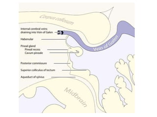
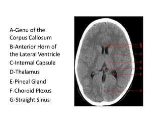
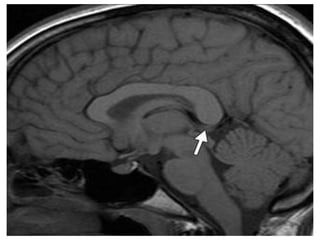
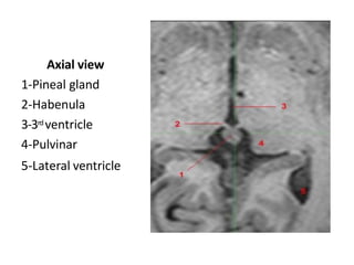
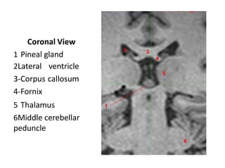
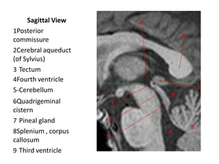
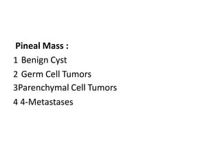
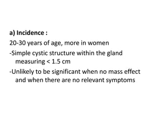
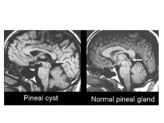
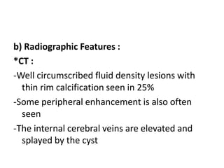
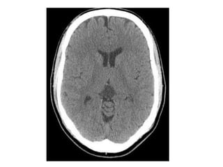
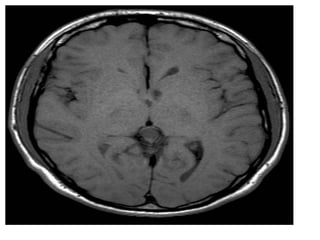
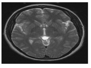
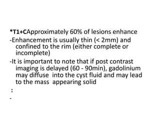
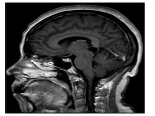
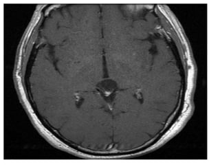
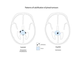
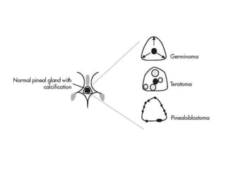
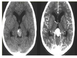
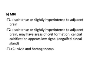
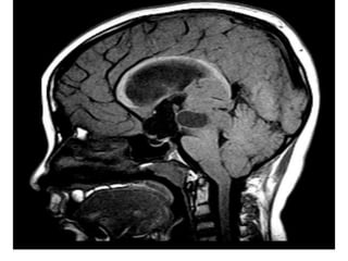
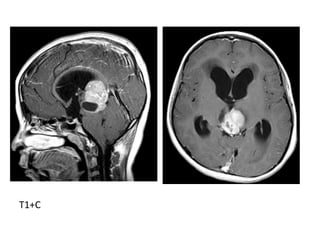
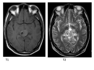
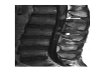
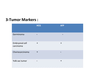
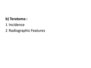
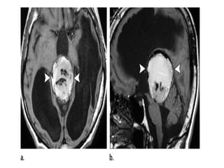
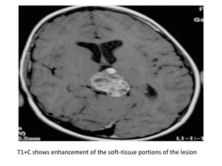
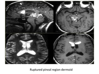
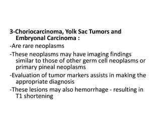
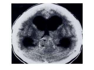
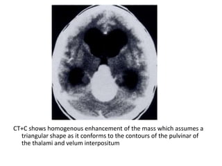
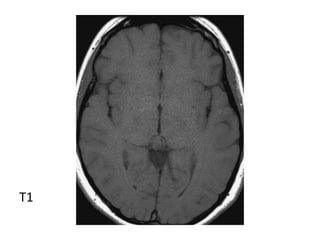
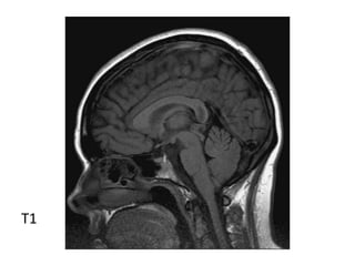
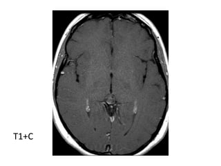
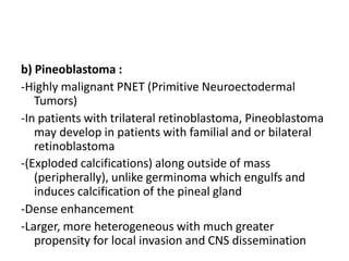
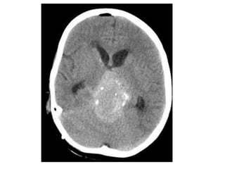
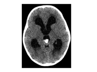
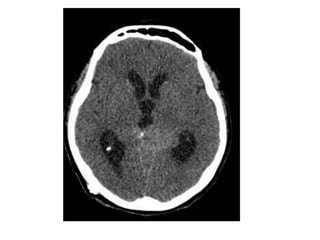
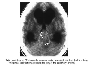
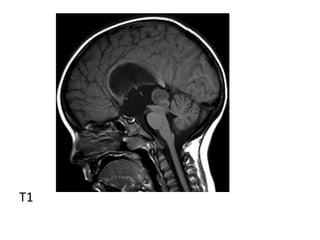
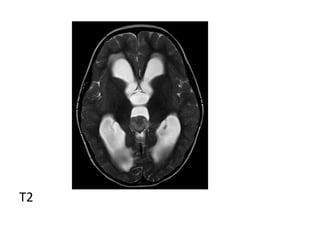
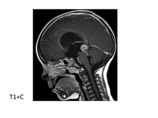
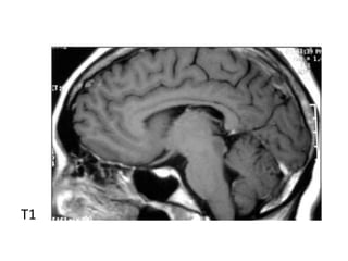
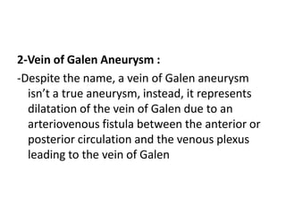
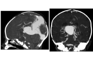
Pineal Region Masses
A-Genu of the
Corpus Callosum
B-Anterior Horn of
the Lateral Ventricle
C-Internal Capsule
D-Thalamus
E-Pineal Gland
F-Choroid Plexus
G-Straight Sinus
Axial view
1-Pineal gland
2-Habenula
3-3rd ventricle
4-Pulvinar
5-Lateral ventricle
Coronal View
1 Pineal gland
2Lateral ventricle
3-Corpus callosum
4-Fornix
5 Thalamus
6Middle cerebellar
peduncle
Sagittal View
1Posterior
commissure
2Cerebral aqueduct
(of Sylvius)
3 Tectum
4Fourth ventricle
5-Cerebellum
6Quadrigeminal
cistern
7 Pineal gland
8Splenium , corpus
callosum
9 Third ventricle
Pineal Mass :
1 Benign Cyst
2 Germ Cell Tumors
3Parenchymal Cell Tumors
4 4-Metastases
1-Benign Cyst :
a) Incidence
b) Radiographic Features
c) Differential Diagnosis
a) Incidence :
20-30 years of age, more in women
-Simple cystic structure within the gland
measuring < 1.5 cm
-Unlikely to be significant when no mass effect
and when there are no relevant symptoms
b) Radiographic Features :
*CT :
-Well circumscribed fluid density lesions with
thin rim calcification seen in 25%
-Some peripheral enhancement is also often
seen
-The internal cerebral veins are elevated and
splayed by the cyst
*MRI :
-Slightly higher signal than CSF on all sequences
-A common incidental finding on MRI studies
*T1 :
-Typically iso to low signal compared to brain
parenchyma
-55 to 60% are somewhat hyperintense when
compared to CSF
-Generally homogenous signal
*T1+CApproximately 60% of lesions enhance
-Enhancement is usually thin (< 2mm) and
confined to the rim (either complete or
incomplete)
-It is important to note that if post contrast
imaging is delayed (60 - 90min), gadolinium
may diffuse into the cyst fluid and may lead
to the mass appearing solid
:
-
c) Differential Diagnosis :
-A differential consideration is Pineocytoma
which would shows internal enhancement and
may have cystic component, however a truly
cystic Pineocytoma is considered very rare
2-Germ Cell Tumors :
a) Germinoma (most common)
b) Teratoma
c) Embryonal Cell Carcinoma
d) Choriocarcinoma
a) Germinoma :
1 Incidence
2Radiographic Features
3-Tumor Markers
1-Incidence :
-Most common pineal germ cell tumor (equivalent to
seminoma in testes and dysgerminoma in ovary)
-Males predominate (10:1), age 10 to 30 years
-In females, more commonly located in suprasellar location
-Serum markers (alpha-fetoprotein) may also be
positive
2-Radiographic Features :
a) CT
-Well defined slightly hyperdense mass that
engulfs a prominent calcified pineal gland
-Homogeneous intense enhancement
-Central calcification due to pineal engulfment
(rare)
b) MRI
-T1 : isointense or slightly hyperintense to adjacent
brain
-T2 : isointense or slightly hyperintense to adjacent
brain, may have areas of cyst formation, central
calcification appears low signal (engulfed pineal
gland)
-T1+C : vivid and homogeneous
T1
T1+C
T1 T2
3-Tumor Markers :
HCG AFP
Germinoma - -
Embryonal cell
carcinoma
+ +
Choriocarcinoma + -
Yolk sac tumor - +
b) Teratoma :
1 Incidence
2 Radiographic Features
1-Incidence :
-Second most common pineal germ cell tumor
-Almost exclusively in male children
-The most common congenital intracranial
tumor and are usually diagnosed prenatally
-Presence of fat and calcification is
diagnostically helpful with little to no
enhancement
2-Radiographic Features :
*CT :
-Demonstrates at least some fat and some
calcification which is usually solid / clump like
-They usually have cystic and solid components,
contributing to an irregular outline, solid
components demonstrate variable
enhancement
CT+C , there is relatively homogeneous enhancement of the non-calcified
solid portions of the tumor , the cystic region doesn’t enhance
*MRI :
-T1 :
-Hyperintense components due to fat and
proteinaceous / lipid rich fluid
-Intermediate components of soft tissue
-Hypointense components due to calcification and
blood products
-T2 :
-Mixed signal from differing components
-T1+C :
-Little or no enhancement
-Solid soft tissue components show enhancement
T1+C shows enhancement of the soft-tissue portions of the lesion
Ruptured pineal region dermoid
3-Choriocarcinoma, Yolk Sac Tumors and
Embryonal Carcinoma :
-Are rare neoplasms
-These neoplasms may have imaging findings
similar to those of other germ cell neoplasms or
primary pineal neoplasms
-Evaluation of tumor markers assists in making the
appropriate diagnosis
-These lesions may also hemorrhage - resulting in
T1 shortening
a)Sagittal T1 weighted MR
image shows an
heterogeneous pineal region
mass with foci of T1
shortening due to
hemorrhage , note the
associated hydrocephalus
b)Axial postcontrast T1-
weighted MR image shows
that the pineal region mass
also has an heterogeneous
enhancement with foci of
necrosis/cyst , involvement of
the tectal plate and both
thalami are also present (not
shown) , evaluation of serum
oncoproteins demonstrated
high level of b-hCG , biopsy of
the lesion revealed that it
corresponds to
Choriocarcinoma
3-Parenchymal Cell Tumors :
a) Pineocytoma
b) Pineoblastoma
a) Pineocytoma :
-No male predilection
-Older age group, mean age 35 years
-Slow growing, dissemination is uncommon
-No helpful imaging features, cannot be
distinguished by imaging features from a
Pineoblastoma
CT+C shows homogenous enhancement of the mass which assumes a
triangular shape as it conforms to the contours of the pulvinar of
the thalami and velum interpositum
T1
T1
T1+C
b) Pineoblastoma :
-Highly malignant PNET (Primitive Neuroectodermal
Tumors)
-In patients with trilateral retinoblastoma, Pineoblastoma
may develop in patients with familial and or bilateral
retinoblastoma
-(Exploded calcifications) along outside of mass
(peripherally), unlike germinoma which engulfs and
induces calcification of the pineal gland
-Dense enhancement
-Larger, more heterogeneous with much greater
propensity for local invasion and CNS dissemination
Axial nonenhanced CT shows a large pineal region mass with resultant hydrocephalus ,
the pineal calcifications are exploded toward the periphery (arrows)
T1
T2
T1+C
b) Extrinsic Pineal Mass :
1 Gliomas
2Vein of Galen Aneurysm
3-Meningioma
4-Quadrigeminal Plate Lipoma
1-Gliomas :
-Gliomas (most commonly astrocytomas) of
varying grade may occur in adjacent intra-axial
structures such as the tectum, midbrain or
splenium of the corpus callosum
Tectal Glioma :
a) Incidence
b) Radiographic Features
a) Incidence :
-Tectal plate gliomas are encountered in
children and adolescents
-Usually low grade tectal tumor causing
aqueduct stenosis
-Typically low grade astrocytoma with good
prognosis
b) Radiographic Features :
-CT :
-Typical CT finding is homogenous expansion of
tectal plate, isodense to grey matter with
minimal enhancement in postcontrast image
CT shows non-communicating hydrocephalus , note the low density lesion of
the tectal plate
-MRI :
-Typically the tumors demonstrate expansion of the
tectal plate by a solid nodule of tissue
*T1 :
-Iso to slightly hypointense to grey matter
*T2 :
-Hyperintense to grey matter
*T1+C :
-Usually no enhancement
-Higher grade tumors tend to be larger and tend to
enhance more vividly
T1
T2 FLAIR shows a tectal mass leading to obliteration of the
cerebral aqueduct
T1+C shows a slightly hypointense tectal mass which does not
enhance , the mass obstructs the aqueduct and is stable over
8 years
2-Vein of Galen Aneurysm :
-Despite the name, a vein of Galen aneurysm
isn’t a true aneurysm, instead, it represents
dilatation of the vein of Galen due to an
arteriovenous fistula between the anterior or
posterior circulation and the venous plexus
leading to the vein of Galen
3-Meningioma :
-The tentorial apex, adjacent to the pineal gland,
is a characteristic location for meningioma
-The tentorial meningioma tends to depress the
internal cerebral veins, in contrast to a pineal-
based mass which typically elevates the
internal cerebral veins
4-Quadrigeminal Plate Lipoma :
-At CT, lipomas have low attenuation, consistent
with fat
-At MR imaging, they have the same signal
characteristics as fat (hyperintense on T1 with
saturation on fat-saturated images)
-No enhancement is seen on postcontrast
images
Quadrigeminal plate lipoma , (a) axial T1 , (b) sagittal T1 show a lobulated ,
hyperintense mass of the quadrigeminal plate , note the associated
thinning of the posterior body (arrow) and splenium of the corpus
callosum
pineal gland 28.pptx

pineal gland 28.pptx

  • 1.
  • 3.
    A-Genu of the CorpusCallosum B-Anterior Horn of the Lateral Ventricle C-Internal Capsule D-Thalamus E-Pineal Gland F-Choroid Plexus G-Straight Sinus
  • 5.
    Axial view 1-Pineal gland 2-Habenula 3-3rdventricle 4-Pulvinar 5-Lateral ventricle
  • 6.
    Coronal View 1 Pinealgland 2Lateral ventricle 3-Corpus callosum 4-Fornix 5 Thalamus 6Middle cerebellar peduncle
  • 7.
    Sagittal View 1Posterior commissure 2Cerebral aqueduct (ofSylvius) 3 Tectum 4Fourth ventricle 5-Cerebellum 6Quadrigeminal cistern 7 Pineal gland 8Splenium , corpus callosum 9 Third ventricle
  • 8.
    Pineal Mass : 1Benign Cyst 2 Germ Cell Tumors 3Parenchymal Cell Tumors 4 4-Metastases
  • 9.
    1-Benign Cyst : a)Incidence b) Radiographic Features c) Differential Diagnosis
  • 10.
    a) Incidence : 20-30years of age, more in women -Simple cystic structure within the gland measuring < 1.5 cm -Unlikely to be significant when no mass effect and when there are no relevant symptoms
  • 12.
    b) Radiographic Features: *CT : -Well circumscribed fluid density lesions with thin rim calcification seen in 25% -Some peripheral enhancement is also often seen -The internal cerebral veins are elevated and splayed by the cyst
  • 14.
    *MRI : -Slightly highersignal than CSF on all sequences -A common incidental finding on MRI studies *T1 : -Typically iso to low signal compared to brain parenchyma -55 to 60% are somewhat hyperintense when compared to CSF -Generally homogenous signal
  • 19.
    *T1+CApproximately 60% oflesions enhance -Enhancement is usually thin (< 2mm) and confined to the rim (either complete or incomplete) -It is important to note that if post contrast imaging is delayed (60 - 90min), gadolinium may diffuse into the cyst fluid and may lead to the mass appearing solid : -
  • 22.
    c) Differential Diagnosis: -A differential consideration is Pineocytoma which would shows internal enhancement and may have cystic component, however a truly cystic Pineocytoma is considered very rare
  • 23.
    2-Germ Cell Tumors: a) Germinoma (most common) b) Teratoma c) Embryonal Cell Carcinoma d) Choriocarcinoma
  • 24.
    a) Germinoma : 1Incidence 2Radiographic Features 3-Tumor Markers
  • 25.
    1-Incidence : -Most commonpineal germ cell tumor (equivalent to seminoma in testes and dysgerminoma in ovary) -Males predominate (10:1), age 10 to 30 years -In females, more commonly located in suprasellar location -Serum markers (alpha-fetoprotein) may also be positive
  • 26.
    2-Radiographic Features : a)CT -Well defined slightly hyperdense mass that engulfs a prominent calcified pineal gland -Homogeneous intense enhancement -Central calcification due to pineal engulfment (rare)
  • 31.
    b) MRI -T1 :isointense or slightly hyperintense to adjacent brain -T2 : isointense or slightly hyperintense to adjacent brain, may have areas of cyst formation, central calcification appears low signal (engulfed pineal gland) -T1+C : vivid and homogeneous
  • 32.
  • 33.
  • 34.
  • 36.
    3-Tumor Markers : HCGAFP Germinoma - - Embryonal cell carcinoma + + Choriocarcinoma + - Yolk sac tumor - +
  • 37.
    b) Teratoma : 1Incidence 2 Radiographic Features
  • 38.
    1-Incidence : -Second mostcommon pineal germ cell tumor -Almost exclusively in male children -The most common congenital intracranial tumor and are usually diagnosed prenatally -Presence of fat and calcification is diagnostically helpful with little to no enhancement
  • 39.
    2-Radiographic Features : *CT: -Demonstrates at least some fat and some calcification which is usually solid / clump like -They usually have cystic and solid components, contributing to an irregular outline, solid components demonstrate variable enhancement
  • 41.
    CT+C , thereis relatively homogeneous enhancement of the non-calcified solid portions of the tumor , the cystic region doesn’t enhance
  • 42.
    *MRI : -T1 : -Hyperintensecomponents due to fat and proteinaceous / lipid rich fluid -Intermediate components of soft tissue -Hypointense components due to calcification and blood products -T2 : -Mixed signal from differing components -T1+C : -Little or no enhancement -Solid soft tissue components show enhancement
  • 44.
    T1+C shows enhancementof the soft-tissue portions of the lesion
  • 45.
  • 46.
    3-Choriocarcinoma, Yolk SacTumors and Embryonal Carcinoma : -Are rare neoplasms -These neoplasms may have imaging findings similar to those of other germ cell neoplasms or primary pineal neoplasms -Evaluation of tumor markers assists in making the appropriate diagnosis -These lesions may also hemorrhage - resulting in T1 shortening
  • 47.
    a)Sagittal T1 weightedMR image shows an heterogeneous pineal region mass with foci of T1 shortening due to hemorrhage , note the associated hydrocephalus b)Axial postcontrast T1- weighted MR image shows that the pineal region mass also has an heterogeneous enhancement with foci of necrosis/cyst , involvement of the tectal plate and both thalami are also present (not shown) , evaluation of serum oncoproteins demonstrated high level of b-hCG , biopsy of the lesion revealed that it corresponds to Choriocarcinoma
  • 48.
    3-Parenchymal Cell Tumors: a) Pineocytoma b) Pineoblastoma
  • 50.
    a) Pineocytoma : -Nomale predilection -Older age group, mean age 35 years -Slow growing, dissemination is uncommon -No helpful imaging features, cannot be distinguished by imaging features from a Pineoblastoma
  • 52.
    CT+C shows homogenousenhancement of the mass which assumes a triangular shape as it conforms to the contours of the pulvinar of the thalami and velum interpositum
  • 53.
  • 54.
  • 55.
  • 56.
    b) Pineoblastoma : -Highlymalignant PNET (Primitive Neuroectodermal Tumors) -In patients with trilateral retinoblastoma, Pineoblastoma may develop in patients with familial and or bilateral retinoblastoma -(Exploded calcifications) along outside of mass (peripherally), unlike germinoma which engulfs and induces calcification of the pineal gland -Dense enhancement -Larger, more heterogeneous with much greater propensity for local invasion and CNS dissemination
  • 60.
    Axial nonenhanced CTshows a large pineal region mass with resultant hydrocephalus , the pineal calcifications are exploded toward the periphery (arrows)
  • 61.
  • 62.
  • 63.
  • 64.
    b) Extrinsic PinealMass : 1 Gliomas 2Vein of Galen Aneurysm 3-Meningioma 4-Quadrigeminal Plate Lipoma
  • 65.
    1-Gliomas : -Gliomas (mostcommonly astrocytomas) of varying grade may occur in adjacent intra-axial structures such as the tectum, midbrain or splenium of the corpus callosum Tectal Glioma : a) Incidence b) Radiographic Features
  • 67.
    a) Incidence : -Tectalplate gliomas are encountered in children and adolescents -Usually low grade tectal tumor causing aqueduct stenosis -Typically low grade astrocytoma with good prognosis
  • 68.
    b) Radiographic Features: -CT : -Typical CT finding is homogenous expansion of tectal plate, isodense to grey matter with minimal enhancement in postcontrast image
  • 69.
    CT shows non-communicatinghydrocephalus , note the low density lesion of the tectal plate
  • 71.
    -MRI : -Typically thetumors demonstrate expansion of the tectal plate by a solid nodule of tissue *T1 : -Iso to slightly hypointense to grey matter *T2 : -Hyperintense to grey matter *T1+C : -Usually no enhancement -Higher grade tumors tend to be larger and tend to enhance more vividly
  • 72.
  • 73.
    T2 FLAIR showsa tectal mass leading to obliteration of the cerebral aqueduct
  • 74.
    T1+C shows aslightly hypointense tectal mass which does not enhance , the mass obstructs the aqueduct and is stable over 8 years
  • 75.
    2-Vein of GalenAneurysm : -Despite the name, a vein of Galen aneurysm isn’t a true aneurysm, instead, it represents dilatation of the vein of Galen due to an arteriovenous fistula between the anterior or posterior circulation and the venous plexus leading to the vein of Galen
  • 77.
    3-Meningioma : -The tentorialapex, adjacent to the pineal gland, is a characteristic location for meningioma -The tentorial meningioma tends to depress the internal cerebral veins, in contrast to a pineal- based mass which typically elevates the internal cerebral veins
  • 79.
    4-Quadrigeminal Plate Lipoma: -At CT, lipomas have low attenuation, consistent with fat -At MR imaging, they have the same signal characteristics as fat (hyperintense on T1 with saturation on fat-saturated images) -No enhancement is seen on postcontrast images
  • 80.
    Quadrigeminal plate lipoma, (a) axial T1 , (b) sagittal T1 show a lobulated , hyperintense mass of the quadrigeminal plate , note the associated thinning of the posterior body (arrow) and splenium of the corpus callosum