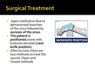
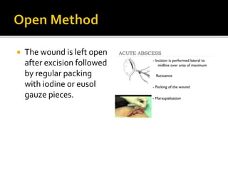
- DrAnil Chaudhary
 Definition: Infection of the skin and
subcutaneous tissue at or near the upper part
of the natal cleft of the buttocks.
 The term pilonidal is derived from the
Latin pilus (hair) and nidus (nest)
 Jeep-bottom because it was very common
in jeep drivers.
SITES
 Midline over the coccyx
 Umbilicus
 lnterdigital in barbers
RISK FACTORS
 Male gender (3rd decade)
 Overweight
 Occupational: prolonged
sitting
 Excessive body hair (Coarse
or stiff hair)
 Previous injury in the area
 Hairs broken off by
vibration and friction tend
to accumulate in the nates.
Thus, it accumulates in the
gluteal cleft and enters the
opening of the
sudoriferous glands.
 Pointed end of the dead
hair is inside (blind end of
the sinus)
 The hair follicle is never
demonstrated in the wall
of the pilonidal sinus but
hair is the content of
pilonidal sinus.
 Low grade fever
 Swelling of cyst
 Pain during sitting or standing
 Soreness and reddish skin around the
depression
 Foul smell in the pocket
 Hair protruding from the affected area
 External opening of the sinus
seen just above the anal
verge in the midline over the
coccyx.
 These cavities will
often discharge serous
fluid and can periodically
become acutely infected to
form a pilonidal abscess
(when infected it can leak pus
blood and have a foul odour)
 A secondary opening may be
present on either side of the
midline often far out on the
buttocks or in the perineum.
 Inject methylene blue to
demonstrate branches
of the sinus followed by
excision of the sinus.
 The patient is
positioned prone with
buttocks elevated (Jack
knife position).
 After excision there are
two methods to treat the
wound -Open and
Closed methods
 The wound is left open
after excision followed
by regular packing
with iodine or eusol
gauze pieces.
 The wound is closed by
'z' plasty.This method
carries 10-20% chances
of recurrence.
 V-Y Advancement Flap
 Rhomboid flap
(Limberg flap) can be
raised to close the
defect also.
 Karydakis believed that hair insertion was the cause for
pilonidal sinus
 Low recurrence rates due to wound placed away from mid-
line
 Resulting new natal cleft was shallower
PROBLEMS:
 Sutures taken over the pre-sacral fascia cause pain
 Use of shallow cleft
 Causes less pain as
presacral fascia not
included
 Patients with acute pilonidal sinus – I & D
 Chronic pilonidal sinus – an excision of the
sinus and all tracts
 A primary closure is associated with faster
wound healing – however, a delayed closure
is associated with less recurrence
 Patients undergoing primary wound closure –
off midline closure recommended
 Recurrent pilonidal sinuses.
i. Diverticulum of main channel has been
overlooked at primary operation
ii. New hair may enter through the scar
iii. Tearing of scar resulting in the formed
crevice to get infected
iv. Very, very rarely carcinoma can arise in a
chronic pilonidal sinus.
PILONIDAL SINUS2.pptx

PILONIDAL SINUS2.pptx

  • 1.
  • 2.
     Definition: Infectionof the skin and subcutaneous tissue at or near the upper part of the natal cleft of the buttocks.  The term pilonidal is derived from the Latin pilus (hair) and nidus (nest)  Jeep-bottom because it was very common in jeep drivers.
  • 3.
    SITES  Midline overthe coccyx  Umbilicus  lnterdigital in barbers RISK FACTORS  Male gender (3rd decade)  Overweight  Occupational: prolonged sitting  Excessive body hair (Coarse or stiff hair)  Previous injury in the area
  • 5.
     Hairs brokenoff by vibration and friction tend to accumulate in the nates. Thus, it accumulates in the gluteal cleft and enters the opening of the sudoriferous glands.  Pointed end of the dead hair is inside (blind end of the sinus)  The hair follicle is never demonstrated in the wall of the pilonidal sinus but hair is the content of pilonidal sinus.
  • 6.
     Low gradefever  Swelling of cyst  Pain during sitting or standing  Soreness and reddish skin around the depression  Foul smell in the pocket  Hair protruding from the affected area
  • 7.
     External openingof the sinus seen just above the anal verge in the midline over the coccyx.  These cavities will often discharge serous fluid and can periodically become acutely infected to form a pilonidal abscess (when infected it can leak pus blood and have a foul odour)  A secondary opening may be present on either side of the midline often far out on the buttocks or in the perineum.
  • 8.
     Inject methyleneblue to demonstrate branches of the sinus followed by excision of the sinus.  The patient is positioned prone with buttocks elevated (Jack knife position).  After excision there are two methods to treat the wound -Open and Closed methods
  • 9.
     The woundis left open after excision followed by regular packing with iodine or eusol gauze pieces.
  • 10.
     The woundis closed by 'z' plasty.This method carries 10-20% chances of recurrence.  V-Y Advancement Flap  Rhomboid flap (Limberg flap) can be raised to close the defect also.
  • 11.
     Karydakis believedthat hair insertion was the cause for pilonidal sinus  Low recurrence rates due to wound placed away from mid- line  Resulting new natal cleft was shallower PROBLEMS:  Sutures taken over the pre-sacral fascia cause pain
  • 12.
     Use ofshallow cleft  Causes less pain as presacral fascia not included
  • 13.
     Patients withacute pilonidal sinus – I & D  Chronic pilonidal sinus – an excision of the sinus and all tracts  A primary closure is associated with faster wound healing – however, a delayed closure is associated with less recurrence  Patients undergoing primary wound closure – off midline closure recommended
  • 14.
     Recurrent pilonidalsinuses. i. Diverticulum of main channel has been overlooked at primary operation ii. New hair may enter through the scar iii. Tearing of scar resulting in the formed crevice to get infected iv. Very, very rarely carcinoma can arise in a chronic pilonidal sinus.