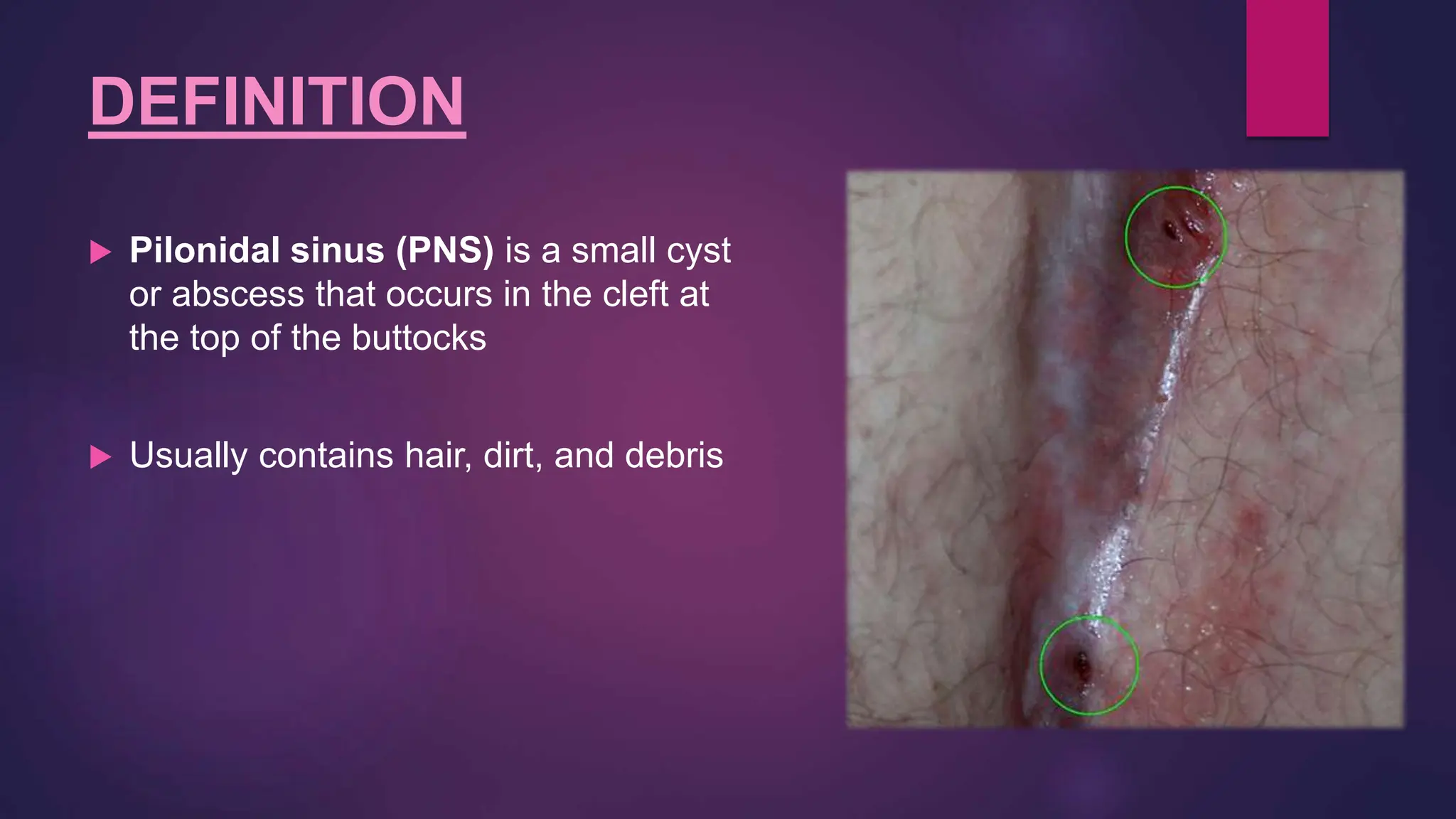
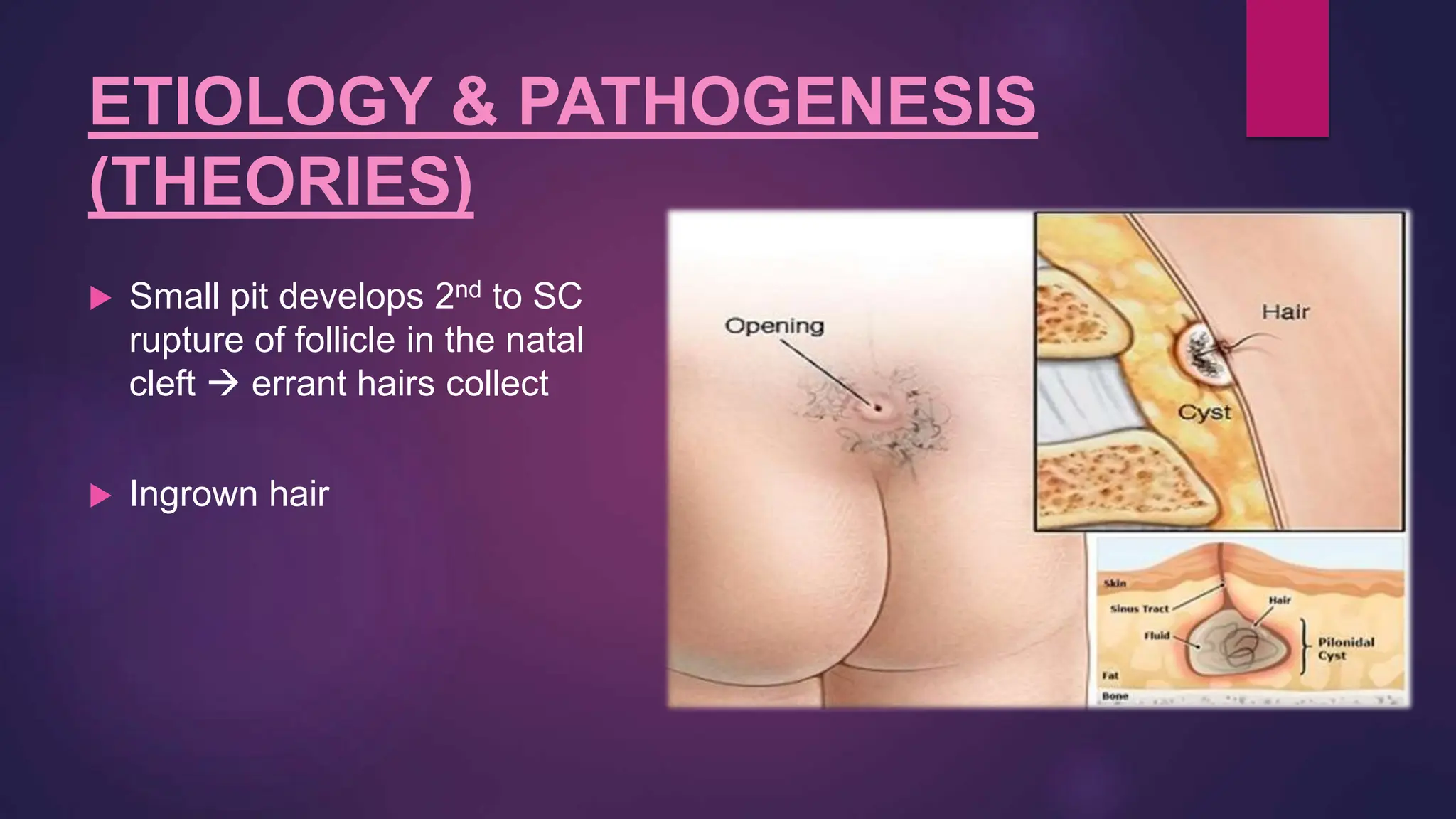
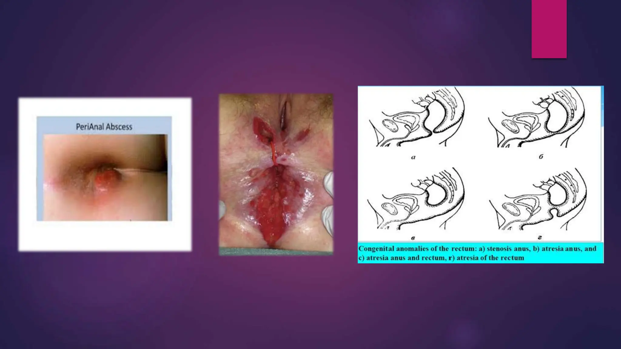
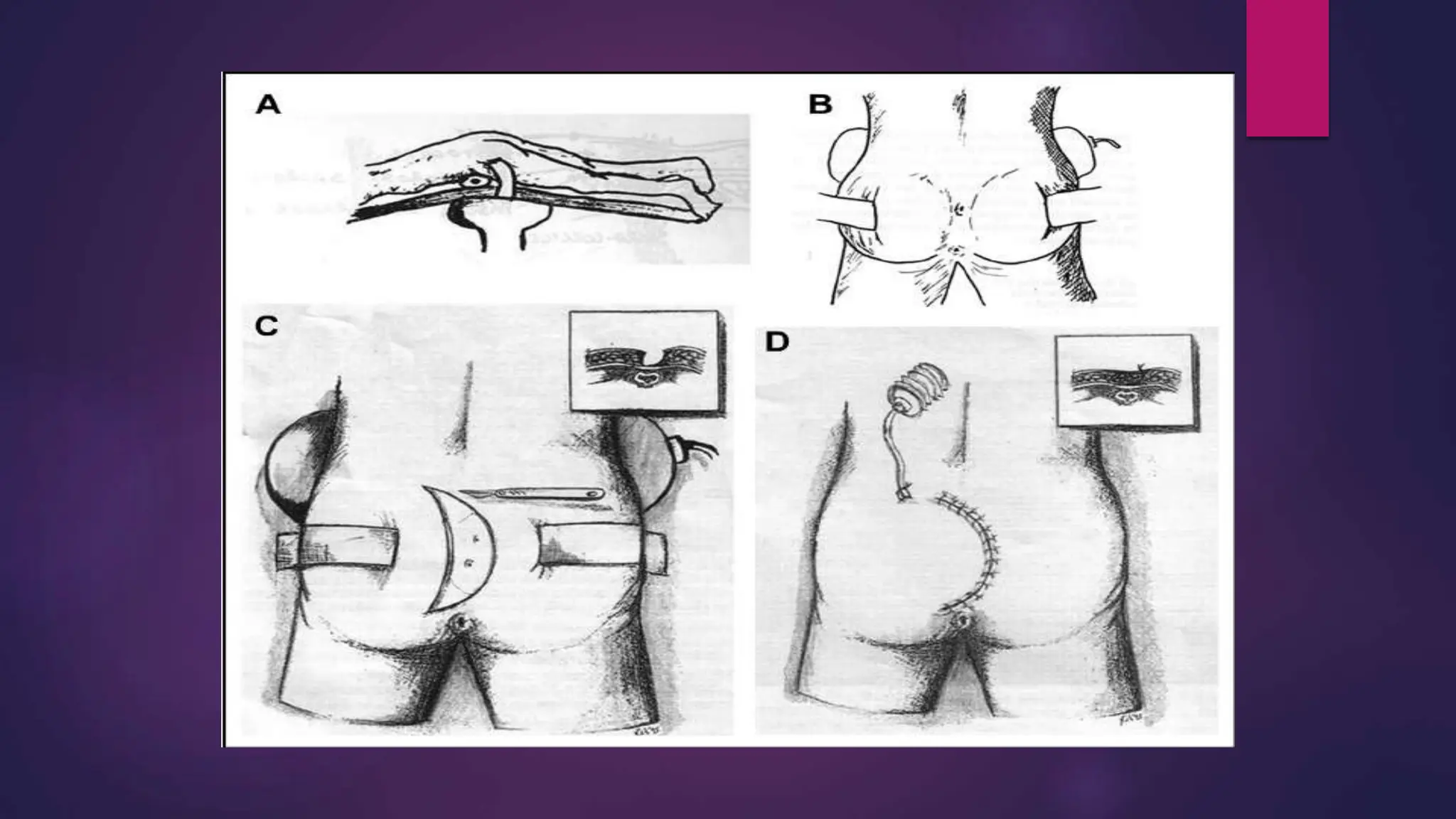
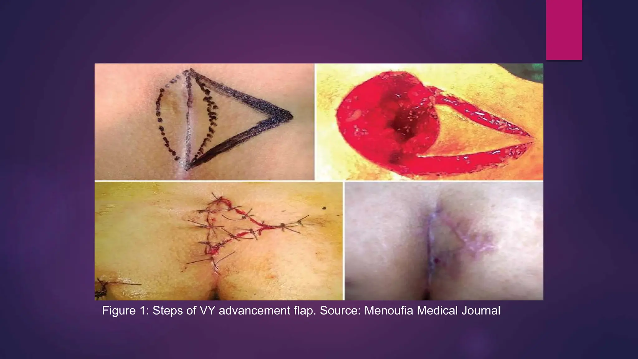
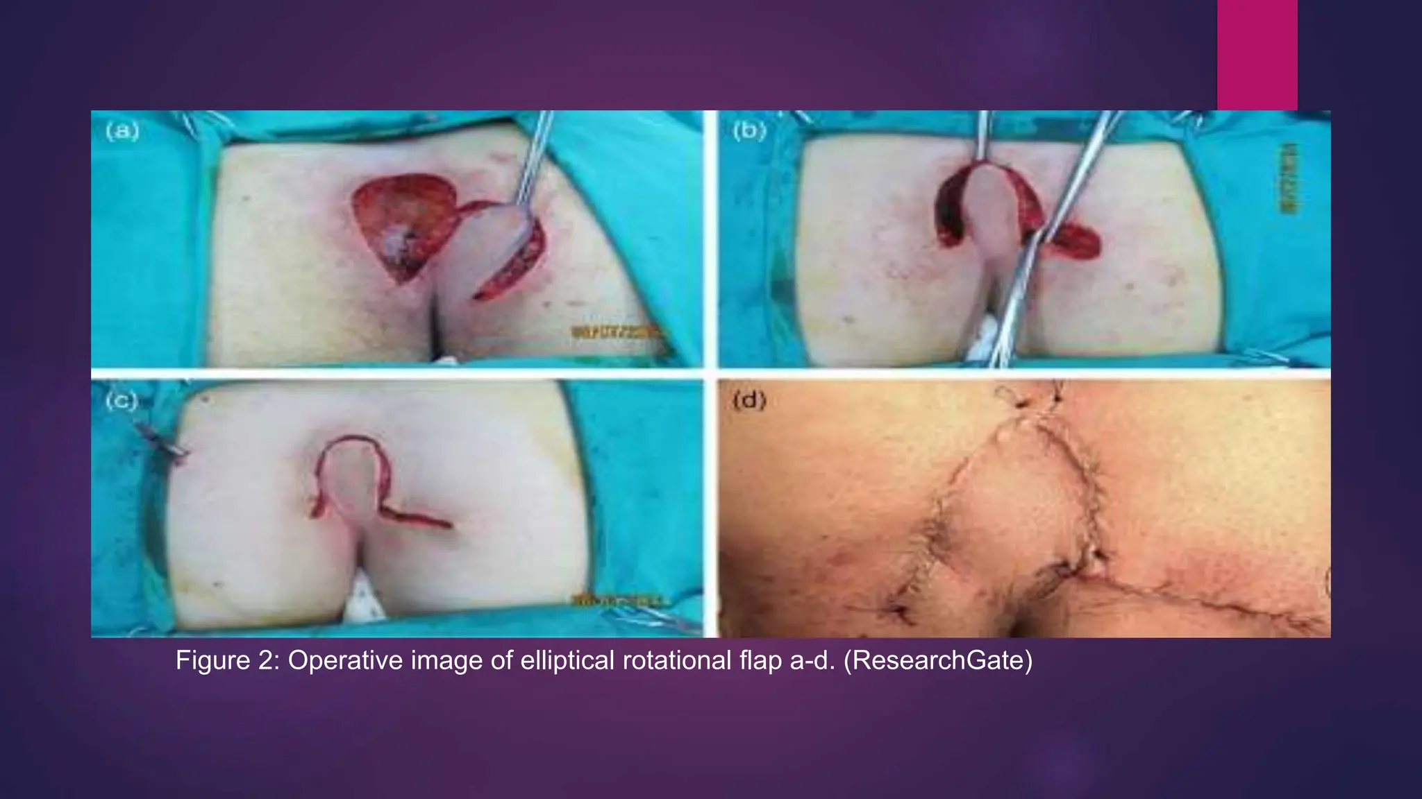
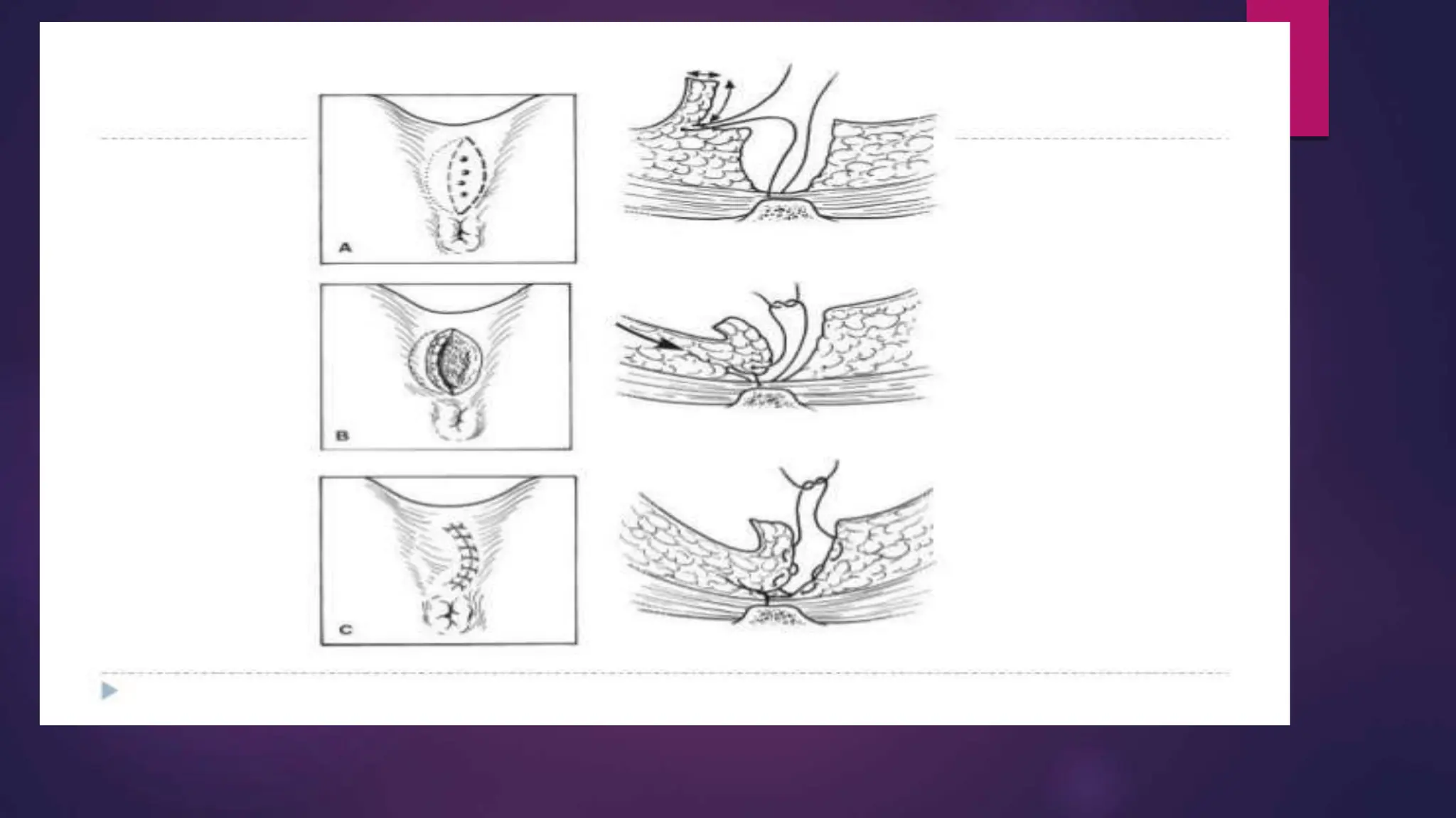
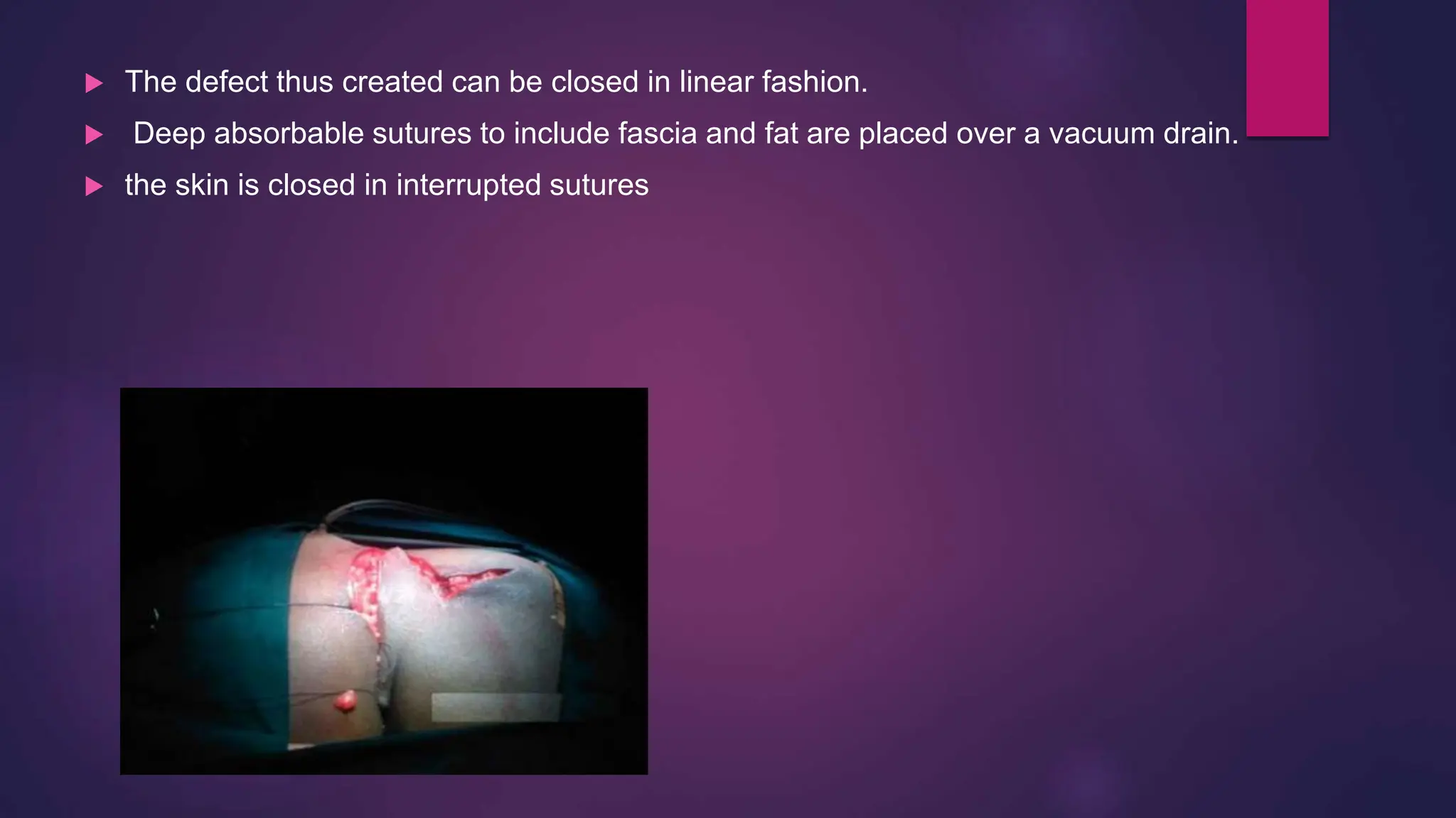
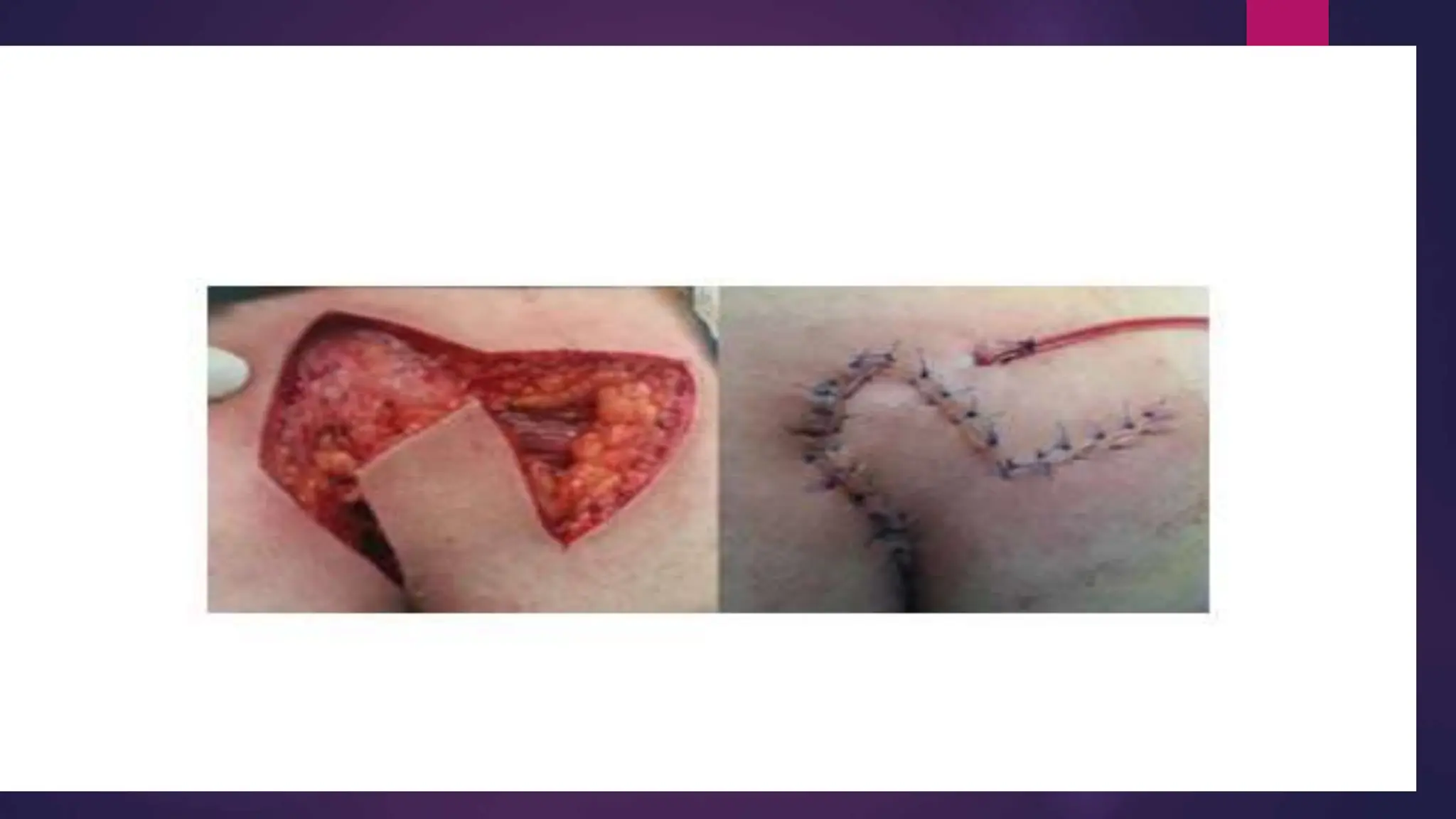
PILONIDA
L
SINUS
ID : 2, 3, 4, 5, 6, 7
DEFINITION
 Pilonidal sinus (PNS) is a small cyst
or abscess that occurs in the cleft at
the top of the buttocks
 Usually contains hair, dirt, and debris
ETIOLOGY & PATHOGENESIS
(THEORIES)
 Small pit develops 2nd to SC
rupture of follicle in the natal
cleft  errant hairs collect
 Ingrown hair
RISK FACTORS
 Male
 Hirsutism
 Chronic trauma
 Spending long time sitting
 Excessive sweating
 Deep natal cleft
CLINICAL
PICTURE
ID : 8, 9, 10, 11, 13, 14
CLINICAL PICTURES
1. Usually occur in natal cleft
2. Also seen in inter-digital clefts & axilla
3. Young adults
4. Male : Female 4 : 1
 ACUTE
I. Acute abscess
II. Cellulitis
III. Sweilling & fluctuation
 CHRONIC
I. Painless
II. Recurr
III. Multiple opening in the midline
DIFFERENTIAL
DIAGNOSES
Diseases Differential criteria
Anal fistula Palpable tract leading to 2ry opening
Hidradenitis suprativa >30 years old, comorbidities, folliculitis
and local friction, other sites of
affection (sweat glands)
Syphilis Manifestations of syphilis
Congenital abnormalities May be continous with spinal cord-
CSF leak
Perirectal abscess Other sites
Pyodermal gangrenosum >40 years with comorbities, ulcerative
lesion
TB Manifestations of TB
• Anal Fistulas and Fissures
• Hidradenitis Suppurativa
Several surgical studies mention the
difficulty in differentiating pilonidal disease
from anal fistula and hidradenitis
suppurativa. Pilonidal disease may result in
sinuses that reach the perianal region and
simulate an anal fistula.
Hidradenitis suppurativa is a chronic
inflammatory disease of the apocrine sweat
glands in which folliculitis and local friction
also play a role, in patients aged 30 years
or older, especially with comorbidities such
as diabetes and obesity. This disease often
affects the groin, axillary, perianal, perineal,
and inframammary regions. These patients
need surgical referral because this condition
is likely to be a long-term concern.
 Perirectal Abscess
 Pyoderma gangrenosum
 Syphilis
 Tuberculosis
Location of the lesion is the best
means to differentiate this entity from
pilonidal disease. Perirectal
abscesses frequently require surgical
consultation in the ED for formal
drainage in the operating room.
Pyoderma gangrenosum is an
ulcerative lesion also generally seen
in the fourth decade of life with other
comorbidities.
Management of
Pilonidal Sinus
ID : 15, 16, 17, 18, 19, 20
Diagnosis of Pilonidal Sinus
BY AHMAD NAZREEN BIN DIN
11-6-15
Diagnosis of PNS
History -Recurrent swelling in sacrococcygeal region
-History of trauma at the area
- intermittent swelling and drainage, including
purulent, mucoid, or bloody fluid from the area.
- -Chronic pilonidal disease often manifests as
recurrent or persistent drainage and pain
Physical Examination A tender, swollen lesion in the sacrococcygeal
region about 4-5 cm posterior to the anal orifice.
Treatment of PNS
Acute/Pilonidal abscess
-Incision & Drainage + Antibiotic
Chronic Pilonidal sinus
-Sinus excision
-Lay-open
-Open method
-Marsupilization
-Closed method
Complex/Recurrent PNS
-Z-plasty
-D-shaped eccentric excision
-VY advancement flap
-Elliptical rotational flap
-Rhomboid flap
Myocutaneous flap
Treatment of Chronic Pilonidal Sinus
AHMAD NAUFAL BIN SOKRI 11-6-16
AHMAD HARIZ IZZUDDIN BIN ABDUL AZIZ 11-6-17
A. Lay open / marsupialization
B. Closed method
 Once the sinus has been excised through an elliptical incision,
the relatively small midline wound will be closed primarily with
minimal tension
 Indication for closed method;
• Short strictly midline tract
• Far from anal orifice
• Pliable tissues
• Shallow tract with no deep pockets in a non hairy person
without active acute infection
Post operative care
 Change the dressing twice daily at least
 Use tube or sitz bath to clean the area
 Return for weekly examination
 Each visits, careful removal of all local hair is crucial to reduce the
recurrence
Treatment of Complex or Recurrent
Disease
AHYAD BIN MD DESA 11-6-18
ADIB ‘AFIFI BIN ABDUL RAHIM 11-6-19
IZDIHAR BIN ZAMRI 11-6-20
A. Z-plasty
 It eliminates the deep natal cleft by bringing healthy, lateral skin and SC
tissue into the midline
 Mean postoperative stay was 3.5 days ( 2-5 days) and return to work was
between 7 – 18 days (Mean 12.5 days).
Z-plasty (cont.)
B. D-shaped Eccentric Excision
 The sinus is excised enblock up to the sacral fascia through a D shaped
incision. The flaps are undermined and approximated without any tension.
C. V-Y advancement flap
Technique:
 The diseased tissue is excised and then the defect is closed with a V-
shaped full-thickness flap extending down to the gluteal fascia, which is
completely mobilized from the gluteus maximus muscle to prevent tension.
 The suture line is in the shape of a Y; hence, the name is V-Y
advancement flap.
Figure 1: Steps of VY advancement flap. Source: Menoufia Medical Journal
Advantages:
 offers tension-free, recurrence-free, and reliable skin coverage while
flattening the natal cleft that predisposes to recurrences.
 Reliable flap closure reduces hospital stay, costs, as well as disability and
time spent off work
D. Elliptical rotational flap
Technique:
 The sinus tracts are all excised & an elliptical flap is designed and rotated
to cover the defect.
 This method offers patient comfort, shorter hospital stay and no recurrence
rate.
Figure 2: Operative image of elliptical rotational flap a-d. (ResearchGate)
E. Karydakis Procedure
 Operations were performed under spinal or general anesthesia
 Patient in the prone position and strapped apart
 The surgical site was shaved before the operation, and the skin was
prepared with povidone-iodine solution
 Methylene Blue is inserted to outline track
 IV antibiotic
 Undercut the flap
 The sinus tract was removed down to the sacrococcygeal fascia by a semilateral elliptic
incision
 flap was placed on the contralateral side with cutaneous-subcutaneous
fatty tissue
 surgical drain was placed in the cavity
 The skin was sutured using 3–0 polypropylene suture
F. Rhomboid Flap
 The patient was put in prone position, under SA with buttocks strapped apart.
 A rhombic area of skin is marked over pilonidal sinus involving all midline pits and lateral
extension if any
 Marking with letter
 Excision till deep fascia
 Raising of flap and rotating over the defect.
 The defect thus created can be closed in linear fashion.
 Deep absorbable sutures to include fascia and fat are placed over a vacuum drain.
 the skin is closed in interrupted sutures
COMPLICATION,
POST-OP
COMPLICATION &
PROGNOSIS
ID : 21, 23, 26, 28, 30, 31
Complication
 Abscess formation
 Recurrence of PNS
 Systemic infection
 SCC (rare)
Post-OP Complication
 Bleeding / hematoma formation
 Infection
 Wound dehiscence
 Recurrence  use and
instead to reduce recurrence.
 Depends on severity of disease ( )
 Usually good if the patient keep a or
 For healing duration and recurrence it depends on
done
1. Incision wound closed with stitches  take 4 weeks to heal (
)
2. Incision wound is left open, it take from a few weeks to several
months to heal. ( )
Prognosis

Pilonidal sinus diagnosis and management (colorectal surgery)

  • 1.
  • 2.
    DEFINITION  Pilonidal sinus(PNS) is a small cyst or abscess that occurs in the cleft at the top of the buttocks  Usually contains hair, dirt, and debris
  • 3.
    ETIOLOGY & PATHOGENESIS (THEORIES) Small pit develops 2nd to SC rupture of follicle in the natal cleft  errant hairs collect  Ingrown hair
  • 4.
    RISK FACTORS  Male Hirsutism  Chronic trauma  Spending long time sitting  Excessive sweating  Deep natal cleft
  • 5.
    CLINICAL PICTURE ID : 8,9, 10, 11, 13, 14
  • 6.
    CLINICAL PICTURES 1. Usuallyoccur in natal cleft 2. Also seen in inter-digital clefts & axilla 3. Young adults 4. Male : Female 4 : 1
  • 7.
     ACUTE I. Acuteabscess II. Cellulitis III. Sweilling & fluctuation  CHRONIC I. Painless II. Recurr III. Multiple opening in the midline
  • 8.
  • 9.
    Diseases Differential criteria Analfistula Palpable tract leading to 2ry opening Hidradenitis suprativa >30 years old, comorbidities, folliculitis and local friction, other sites of affection (sweat glands) Syphilis Manifestations of syphilis Congenital abnormalities May be continous with spinal cord- CSF leak Perirectal abscess Other sites Pyodermal gangrenosum >40 years with comorbities, ulcerative lesion TB Manifestations of TB
  • 11.
    Diseases Differential criteria Analfistula Palpable tract leading to 2ry opening Hidradenitis suprativa >30 years old, comorbidities, folliculitis and local friction, other sites of affection (sweat glands) Syphilis Manifestations of syphilis Congenital abnormalities May be continous with spinal cord- CSF leak Perirectal abscess Other sites Pyodermal gangrenosum >40 years with comorbities, ulcerative lesion TB Manifestations of TB
  • 13.
    • Anal Fistulasand Fissures • Hidradenitis Suppurativa Several surgical studies mention the difficulty in differentiating pilonidal disease from anal fistula and hidradenitis suppurativa. Pilonidal disease may result in sinuses that reach the perianal region and simulate an anal fistula. Hidradenitis suppurativa is a chronic inflammatory disease of the apocrine sweat glands in which folliculitis and local friction also play a role, in patients aged 30 years or older, especially with comorbidities such as diabetes and obesity. This disease often affects the groin, axillary, perianal, perineal, and inframammary regions. These patients need surgical referral because this condition is likely to be a long-term concern.
  • 14.
     Perirectal Abscess Pyoderma gangrenosum  Syphilis  Tuberculosis Location of the lesion is the best means to differentiate this entity from pilonidal disease. Perirectal abscesses frequently require surgical consultation in the ED for formal drainage in the operating room. Pyoderma gangrenosum is an ulcerative lesion also generally seen in the fourth decade of life with other comorbidities.
  • 15.
    Management of Pilonidal Sinus ID: 15, 16, 17, 18, 19, 20
  • 16.
    Diagnosis of PilonidalSinus BY AHMAD NAZREEN BIN DIN 11-6-15
  • 17.
    Diagnosis of PNS History-Recurrent swelling in sacrococcygeal region -History of trauma at the area - intermittent swelling and drainage, including purulent, mucoid, or bloody fluid from the area. - -Chronic pilonidal disease often manifests as recurrent or persistent drainage and pain Physical Examination A tender, swollen lesion in the sacrococcygeal region about 4-5 cm posterior to the anal orifice.
  • 18.
    Treatment of PNS Acute/Pilonidalabscess -Incision & Drainage + Antibiotic Chronic Pilonidal sinus -Sinus excision -Lay-open -Open method -Marsupilization -Closed method Complex/Recurrent PNS -Z-plasty -D-shaped eccentric excision -VY advancement flap -Elliptical rotational flap -Rhomboid flap Myocutaneous flap
  • 19.
    Treatment of ChronicPilonidal Sinus AHMAD NAUFAL BIN SOKRI 11-6-16 AHMAD HARIZ IZZUDDIN BIN ABDUL AZIZ 11-6-17
  • 20.
    A. Lay open/ marsupialization
  • 21.
    B. Closed method Once the sinus has been excised through an elliptical incision, the relatively small midline wound will be closed primarily with minimal tension  Indication for closed method; • Short strictly midline tract • Far from anal orifice • Pliable tissues • Shallow tract with no deep pockets in a non hairy person without active acute infection
  • 22.
    Post operative care Change the dressing twice daily at least  Use tube or sitz bath to clean the area  Return for weekly examination  Each visits, careful removal of all local hair is crucial to reduce the recurrence
  • 23.
    Treatment of Complexor Recurrent Disease AHYAD BIN MD DESA 11-6-18 ADIB ‘AFIFI BIN ABDUL RAHIM 11-6-19 IZDIHAR BIN ZAMRI 11-6-20
  • 24.
    A. Z-plasty  Iteliminates the deep natal cleft by bringing healthy, lateral skin and SC tissue into the midline  Mean postoperative stay was 3.5 days ( 2-5 days) and return to work was between 7 – 18 days (Mean 12.5 days).
  • 25.
  • 26.
    B. D-shaped EccentricExcision  The sinus is excised enblock up to the sacral fascia through a D shaped incision. The flaps are undermined and approximated without any tension.
  • 28.
    C. V-Y advancementflap Technique:  The diseased tissue is excised and then the defect is closed with a V- shaped full-thickness flap extending down to the gluteal fascia, which is completely mobilized from the gluteus maximus muscle to prevent tension.  The suture line is in the shape of a Y; hence, the name is V-Y advancement flap.
  • 29.
    Figure 1: Stepsof VY advancement flap. Source: Menoufia Medical Journal
  • 30.
    Advantages:  offers tension-free,recurrence-free, and reliable skin coverage while flattening the natal cleft that predisposes to recurrences.  Reliable flap closure reduces hospital stay, costs, as well as disability and time spent off work
  • 31.
    D. Elliptical rotationalflap Technique:  The sinus tracts are all excised & an elliptical flap is designed and rotated to cover the defect.  This method offers patient comfort, shorter hospital stay and no recurrence rate.
  • 32.
    Figure 2: Operativeimage of elliptical rotational flap a-d. (ResearchGate)
  • 33.
    E. Karydakis Procedure Operations were performed under spinal or general anesthesia  Patient in the prone position and strapped apart  The surgical site was shaved before the operation, and the skin was prepared with povidone-iodine solution  Methylene Blue is inserted to outline track  IV antibiotic
  • 34.
     Undercut theflap  The sinus tract was removed down to the sacrococcygeal fascia by a semilateral elliptic incision
  • 35.
     flap wasplaced on the contralateral side with cutaneous-subcutaneous fatty tissue  surgical drain was placed in the cavity  The skin was sutured using 3–0 polypropylene suture
  • 37.
    F. Rhomboid Flap The patient was put in prone position, under SA with buttocks strapped apart.  A rhombic area of skin is marked over pilonidal sinus involving all midline pits and lateral extension if any
  • 38.
     Marking withletter  Excision till deep fascia
  • 39.
     Raising offlap and rotating over the defect.
  • 40.
     The defectthus created can be closed in linear fashion.  Deep absorbable sutures to include fascia and fat are placed over a vacuum drain.  the skin is closed in interrupted sutures
  • 42.
  • 43.
    Complication  Abscess formation Recurrence of PNS  Systemic infection  SCC (rare)
  • 44.
    Post-OP Complication  Bleeding/ hematoma formation  Infection  Wound dehiscence  Recurrence  use and instead to reduce recurrence.
  • 45.
     Depends onseverity of disease ( )  Usually good if the patient keep a or  For healing duration and recurrence it depends on done 1. Incision wound closed with stitches  take 4 weeks to heal ( ) 2. Incision wound is left open, it take from a few weeks to several months to heal. ( ) Prognosis