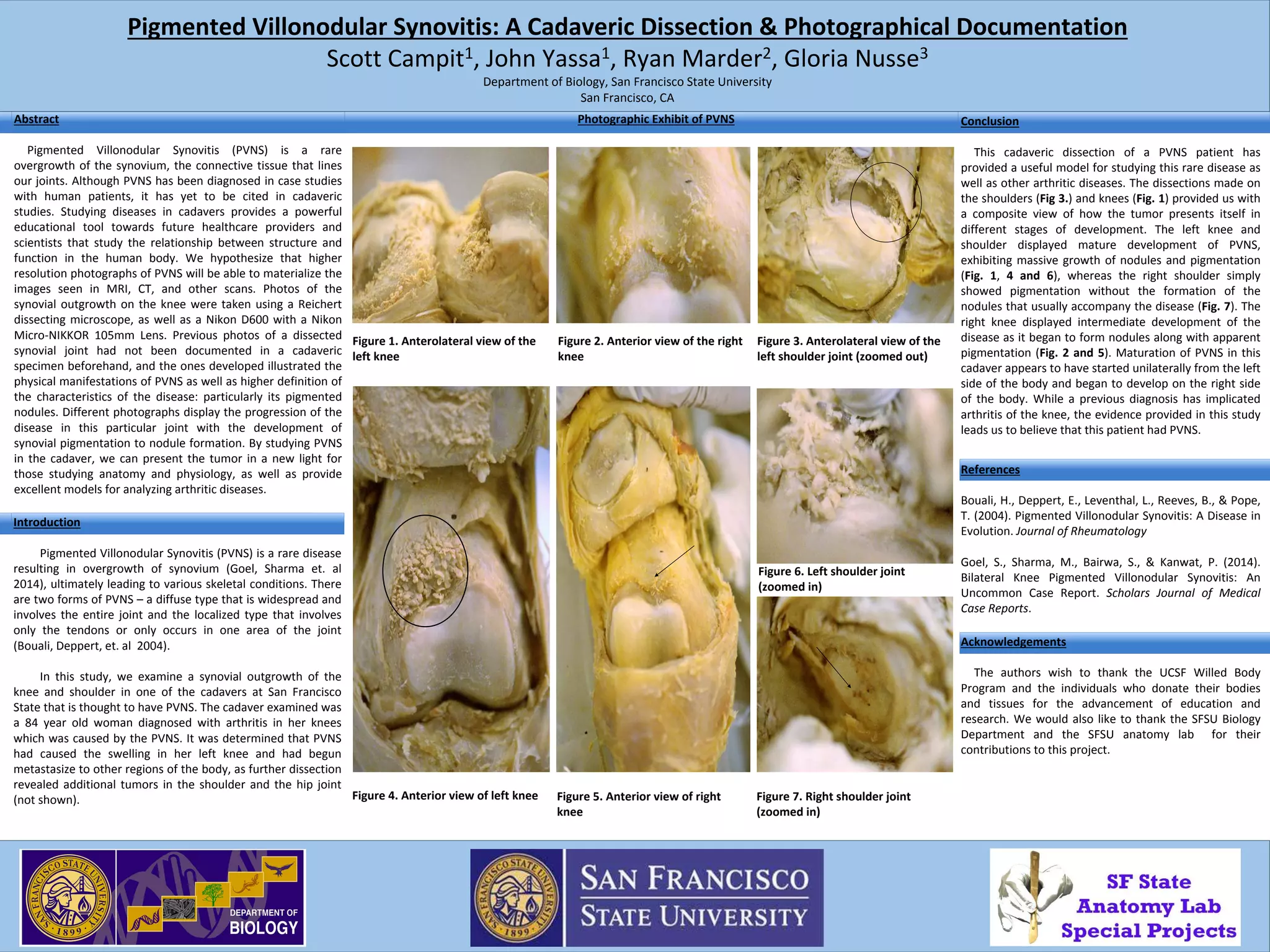
SlideShare a Scribd company logo
1 of 1
Abstract
Pigmented Villonodular Synovitis (PVNS) is a rare
overgrowth of the synovium, the connective tissue that lines
our joints. Although PVNS has been diagnosed in case studies
with human patients, it has yet to be cited in cadaveric
studies. Studying diseases in cadavers provides a powerful
educational tool towards future healthcare providers and
scientists that study the relationship between structure and
function in the human body. We hypothesize that higher
resolution photographs of PVNS will be able to materialize the
images seen in MRI, CT, and other scans. Photos of the
synovial outgrowth on the knee were taken using a Reichert
dissecting microscope, as well as a Nikon D600 with a Nikon
Micro-NIKKOR 105mm Lens. Previous photos of a dissected
synovial joint had not been documented in a cadaveric
specimen beforehand, and the ones developed illustrated the
physical manifestations of PVNS as well as higher definition of
the characteristics of the disease: particularly its pigmented
nodules. Different photographs display the progression of the
disease in this particular joint with the development of
synovial pigmentation to nodule formation. By studying PVNS
in the cadaver, we can present the tumor in a new light for
those studying anatomy and physiology, as well as provide
excellent models for analyzing arthritic diseases.
Pigmented Villonodular Synovitis: A Cadaveric Dissection & Photographical Documentation
Scott Campit1, John Yassa1, Ryan Marder2, Gloria Nusse3
Department of Biology, San Francisco State University
San Francisco, CA
Photographic Exhibit of PVNS Conclusion
This cadaveric dissection of a PVNS patient has
provided a useful model for studying this rare disease as
well as other arthritic diseases. The dissections made on
the shoulders (Fig 3.) and knees (Fig. 1) provided us with
a composite view of how the tumor presents itself in
different stages of development. The left knee and
shoulder displayed mature development of PVNS,
exhibiting massive growth of nodules and pigmentation
(Fig. 1, 4 and 6), whereas the right shoulder simply
showed pigmentation without the formation of the
nodules that usually accompany the disease (Fig. 7). The
right knee displayed intermediate development of the
disease as it began to form nodules along with apparent
pigmentation (Fig. 2 and 5). Maturation of PVNS in this
cadaver appears to have started unilaterally from the left
side of the body and began to develop on the right side
of the body. While a previous diagnosis has implicated
arthritis of the knee, the evidence provided in this study
leads us to believe that this patient had PVNS.
Acknowledgements
The authors wish to thank the UCSF Willed Body
Program and the individuals who donate their bodies
and tissues for the advancement of education and
research. We would also like to thank the SFSU Biology
Department and the SFSU anatomy lab for their
contributions to this project.
References
Bouali, H., Deppert, E., Leventhal, L., Reeves, B., & Pope,
T. (2004). Pigmented Villonodular Synovitis: A Disease in
Evolution. Journal of Rheumatology
Goel, S., Sharma, M., Bairwa, S., & Kanwat, P. (2014).
Bilateral Knee Pigmented Villonodular Synovitis: An
Uncommon Case Report. Scholars Journal of Medical
Case Reports.
Figure 2. Anterior view of the right
knee
Figure 1. Anterolateral view of the
left knee
Figure 4. Anterior view of left knee
Figure 3. Anterolateral view of the
left shoulder joint (zoomed out)
Figure 5. Anterior view of right
knee
Figure 6. Left shoulder joint
(zoomed in)
Figure 7. Right shoulder joint
(zoomed in)
Introduction
Pigmented Villonodular Synovitis (PVNS) is a rare disease
resulting in overgrowth of synovium (Goel, Sharma et. al
2014), ultimately leading to various skeletal conditions. There
are two forms of PVNS – a diffuse type that is widespread and
involves the entire joint and the localized type that involves
only the tendons or only occurs in one area of the joint
(Bouali, Deppert, et. al 2004).
In this study, we examine a synovial outgrowth of the
knee and shoulder in one of the cadavers at San Francisco
State that is thought to have PVNS. The cadaver examined was
a 84 year old woman diagnosed with arthritis in her knees
which was caused by the PVNS. It was determined that PVNS
had caused the swelling in her left knee and had begun
metastasize to other regions of the body, as further dissection
revealed additional tumors in the shoulder and the hip joint
(not shown).

More Related Content

What's hot

What's hot (20)

Case record...Spinal metastasis
Case record...Spinal metastasisCase record...Spinal metastasis
Case record...Spinal metastasis
 
Analysis of focal lucent bony lesion
Analysis of focal lucent bony lesionAnalysis of focal lucent bony lesion
Analysis of focal lucent bony lesion
 
An Unusual Case of Frequent Knee Locking _Crimson Publishers
An Unusual Case of Frequent Knee Locking _Crimson PublishersAn Unusual Case of Frequent Knee Locking _Crimson Publishers
An Unusual Case of Frequent Knee Locking _Crimson Publishers
 
Alberto Bazzocchi , today multiparametric imaging of bone and soft tissue tum...
Alberto Bazzocchi , today multiparametric imaging of bone and soft tissue tum...Alberto Bazzocchi , today multiparametric imaging of bone and soft tissue tum...
Alberto Bazzocchi , today multiparametric imaging of bone and soft tissue tum...
 
International Journal of Orthopedics: Research & Therapy
International Journal of Orthopedics: Research & TherapyInternational Journal of Orthopedics: Research & Therapy
International Journal of Orthopedics: Research & Therapy
 
Heel pain
Heel pain Heel pain
Heel pain
 
Diagnostic Imaging of Endocrine bone disease
Diagnostic Imaging of Endocrine bone diseaseDiagnostic Imaging of Endocrine bone disease
Diagnostic Imaging of Endocrine bone disease
 
Imaging of inflammatory arthritis ppt
Imaging of inflammatory arthritis pptImaging of inflammatory arthritis ppt
Imaging of inflammatory arthritis ppt
 
Presentation1.pptx, radiological imaging of osteoarthritis.
Presentation1.pptx, radiological imaging of osteoarthritis.Presentation1.pptx, radiological imaging of osteoarthritis.
Presentation1.pptx, radiological imaging of osteoarthritis.
 
Case report on metachronous Osteosarcoma
Case report on metachronous OsteosarcomaCase report on metachronous Osteosarcoma
Case report on metachronous Osteosarcoma
 
Diagnosing osteoarthritis
Diagnosing osteoarthritisDiagnosing osteoarthritis
Diagnosing osteoarthritis
 
Radiological evaluation of Arthritis
Radiological evaluation of  ArthritisRadiological evaluation of  Arthritis
Radiological evaluation of Arthritis
 
Benign bone tumors
Benign bone tumorsBenign bone tumors
Benign bone tumors
 
Studying relation between sitting position and knee osteoarthriti
Studying relation between sitting position and knee osteoarthritiStudying relation between sitting position and knee osteoarthriti
Studying relation between sitting position and knee osteoarthriti
 
Injerto oseo
Injerto oseoInjerto oseo
Injerto oseo
 
Diagnostic Imaging of Bone Dysplasia
Diagnostic Imaging of Bone DysplasiaDiagnostic Imaging of Bone Dysplasia
Diagnostic Imaging of Bone Dysplasia
 
Diagnostic Imaging of Paget's Disease
Diagnostic Imaging of Paget's DiseaseDiagnostic Imaging of Paget's Disease
Diagnostic Imaging of Paget's Disease
 
Diagnostic Imaging of Mandible & Maxilla
Diagnostic Imaging of Mandible & MaxillaDiagnostic Imaging of Mandible & Maxilla
Diagnostic Imaging of Mandible & Maxilla
 
Surgery in myeloma
Surgery in myelomaSurgery in myeloma
Surgery in myeloma
 
Inflammatory Arthritis
Inflammatory ArthritisInflammatory Arthritis
Inflammatory Arthritis
 

Similar to COSE2015

Ankylosing spondylitis clinical feature and diagnosis
Ankylosing spondylitis clinical feature and diagnosisAnkylosing spondylitis clinical feature and diagnosis
Ankylosing spondylitis clinical feature and diagnosis
dattasrisaila
 
Study on Clinical observation in Patients Presenting With Right Iliac Fossa P...
Study on Clinical observation in Patients Presenting With Right Iliac Fossa P...Study on Clinical observation in Patients Presenting With Right Iliac Fossa P...
Study on Clinical observation in Patients Presenting With Right Iliac Fossa P...
QUESTJOURNAL
 

Similar to COSE2015 (20)

Pvns dr.sameer
Pvns dr.sameerPvns dr.sameer
Pvns dr.sameer
 
Distal_Radioulnar_Joint_Replacement_with.pdf
Distal_Radioulnar_Joint_Replacement_with.pdfDistal_Radioulnar_Joint_Replacement_with.pdf
Distal_Radioulnar_Joint_Replacement_with.pdf
 
Arthroscopic Resection of Localized Pigmented Villonodular Synovitis of the Knee
Arthroscopic Resection of Localized Pigmented Villonodular Synovitis of the KneeArthroscopic Resection of Localized Pigmented Villonodular Synovitis of the Knee
Arthroscopic Resection of Localized Pigmented Villonodular Synovitis of the Knee
 
Benefits of Mechanical Manipulation of the Sacroiliac Joint: A Transient Syno...
Benefits of Mechanical Manipulation of the Sacroiliac Joint: A Transient Syno...Benefits of Mechanical Manipulation of the Sacroiliac Joint: A Transient Syno...
Benefits of Mechanical Manipulation of the Sacroiliac Joint: A Transient Syno...
 
Ankylosing spondylitis clinical feature and diagnosis
Ankylosing spondylitis clinical feature and diagnosisAnkylosing spondylitis clinical feature and diagnosis
Ankylosing spondylitis clinical feature and diagnosis
 
Scientific Journal of Research in Dentistry
Scientific Journal of Research in DentistryScientific Journal of Research in Dentistry
Scientific Journal of Research in Dentistry
 
Primary Pelvic Hydatid Cyst.pdf
Primary Pelvic Hydatid Cyst.pdfPrimary Pelvic Hydatid Cyst.pdf
Primary Pelvic Hydatid Cyst.pdf
 
Tuberculous dactylitis pseudotumor of an adult thumb.pdf
Tuberculous dactylitis pseudotumor of an adult thumb.pdfTuberculous dactylitis pseudotumor of an adult thumb.pdf
Tuberculous dactylitis pseudotumor of an adult thumb.pdf
 
International Journal of Orthopedics: Research & Therapy
International Journal of Orthopedics: Research & TherapyInternational Journal of Orthopedics: Research & Therapy
International Journal of Orthopedics: Research & Therapy
 
Case 1
Case 1Case 1
Case 1
 
PDF Disfunción sacroiliaca evaluación y manejo
PDF Disfunción sacroiliaca evaluación y manejoPDF Disfunción sacroiliaca evaluación y manejo
PDF Disfunción sacroiliaca evaluación y manejo
 
Extraskeletal Osteochondroma of the Heel: A Case Report
Extraskeletal Osteochondroma of the Heel: A Case ReportExtraskeletal Osteochondroma of the Heel: A Case Report
Extraskeletal Osteochondroma of the Heel: A Case Report
 
Banal vs tb infection of spine team viii
Banal vs tb infection of spine team viiiBanal vs tb infection of spine team viii
Banal vs tb infection of spine team viii
 
Study on Clinical observation in Patients Presenting With Right Iliac Fossa P...
Study on Clinical observation in Patients Presenting With Right Iliac Fossa P...Study on Clinical observation in Patients Presenting With Right Iliac Fossa P...
Study on Clinical observation in Patients Presenting With Right Iliac Fossa P...
 
Subperiosteal resection of mid-clavicle in sprengel's.pdf
Subperiosteal resection of mid-clavicle in sprengel's.pdfSubperiosteal resection of mid-clavicle in sprengel's.pdf
Subperiosteal resection of mid-clavicle in sprengel's.pdf
 
Spinal tuberculosis
Spinal tuberculosisSpinal tuberculosis
Spinal tuberculosis
 
PVNS,SYNOVIAL CHONDROMATOSIS & LOOSE BODIES
PVNS,SYNOVIAL CHONDROMATOSIS & LOOSE BODIESPVNS,SYNOVIAL CHONDROMATOSIS & LOOSE BODIES
PVNS,SYNOVIAL CHONDROMATOSIS & LOOSE BODIES
 
MANAJEMEN SEPTIK ARTHRITIS pada lututmanusia
MANAJEMEN SEPTIK ARTHRITIS pada lututmanusiaMANAJEMEN SEPTIK ARTHRITIS pada lututmanusia
MANAJEMEN SEPTIK ARTHRITIS pada lututmanusia
 
Introduction to Bone Scan: Techniques and Diagnosis
Introduction to Bone Scan: Techniques and Diagnosis Introduction to Bone Scan: Techniques and Diagnosis
Introduction to Bone Scan: Techniques and Diagnosis
 
Stemcell Research Paper on avascular necrosis-AVN-by Dr.Pradeep Mahajan
Stemcell Research Paper on avascular necrosis-AVN-by Dr.Pradeep MahajanStemcell Research Paper on avascular necrosis-AVN-by Dr.Pradeep Mahajan
Stemcell Research Paper on avascular necrosis-AVN-by Dr.Pradeep Mahajan
 

COSE2015

  • 1. Abstract Pigmented Villonodular Synovitis (PVNS) is a rare overgrowth of the synovium, the connective tissue that lines our joints. Although PVNS has been diagnosed in case studies with human patients, it has yet to be cited in cadaveric studies. Studying diseases in cadavers provides a powerful educational tool towards future healthcare providers and scientists that study the relationship between structure and function in the human body. We hypothesize that higher resolution photographs of PVNS will be able to materialize the images seen in MRI, CT, and other scans. Photos of the synovial outgrowth on the knee were taken using a Reichert dissecting microscope, as well as a Nikon D600 with a Nikon Micro-NIKKOR 105mm Lens. Previous photos of a dissected synovial joint had not been documented in a cadaveric specimen beforehand, and the ones developed illustrated the physical manifestations of PVNS as well as higher definition of the characteristics of the disease: particularly its pigmented nodules. Different photographs display the progression of the disease in this particular joint with the development of synovial pigmentation to nodule formation. By studying PVNS in the cadaver, we can present the tumor in a new light for those studying anatomy and physiology, as well as provide excellent models for analyzing arthritic diseases. Pigmented Villonodular Synovitis: A Cadaveric Dissection & Photographical Documentation Scott Campit1, John Yassa1, Ryan Marder2, Gloria Nusse3 Department of Biology, San Francisco State University San Francisco, CA Photographic Exhibit of PVNS Conclusion This cadaveric dissection of a PVNS patient has provided a useful model for studying this rare disease as well as other arthritic diseases. The dissections made on the shoulders (Fig 3.) and knees (Fig. 1) provided us with a composite view of how the tumor presents itself in different stages of development. The left knee and shoulder displayed mature development of PVNS, exhibiting massive growth of nodules and pigmentation (Fig. 1, 4 and 6), whereas the right shoulder simply showed pigmentation without the formation of the nodules that usually accompany the disease (Fig. 7). The right knee displayed intermediate development of the disease as it began to form nodules along with apparent pigmentation (Fig. 2 and 5). Maturation of PVNS in this cadaver appears to have started unilaterally from the left side of the body and began to develop on the right side of the body. While a previous diagnosis has implicated arthritis of the knee, the evidence provided in this study leads us to believe that this patient had PVNS. Acknowledgements The authors wish to thank the UCSF Willed Body Program and the individuals who donate their bodies and tissues for the advancement of education and research. We would also like to thank the SFSU Biology Department and the SFSU anatomy lab for their contributions to this project. References Bouali, H., Deppert, E., Leventhal, L., Reeves, B., & Pope, T. (2004). Pigmented Villonodular Synovitis: A Disease in Evolution. Journal of Rheumatology Goel, S., Sharma, M., Bairwa, S., & Kanwat, P. (2014). Bilateral Knee Pigmented Villonodular Synovitis: An Uncommon Case Report. Scholars Journal of Medical Case Reports. Figure 2. Anterior view of the right knee Figure 1. Anterolateral view of the left knee Figure 4. Anterior view of left knee Figure 3. Anterolateral view of the left shoulder joint (zoomed out) Figure 5. Anterior view of right knee Figure 6. Left shoulder joint (zoomed in) Figure 7. Right shoulder joint (zoomed in) Introduction Pigmented Villonodular Synovitis (PVNS) is a rare disease resulting in overgrowth of synovium (Goel, Sharma et. al 2014), ultimately leading to various skeletal conditions. There are two forms of PVNS – a diffuse type that is widespread and involves the entire joint and the localized type that involves only the tendons or only occurs in one area of the joint (Bouali, Deppert, et. al 2004). In this study, we examine a synovial outgrowth of the knee and shoulder in one of the cadavers at San Francisco State that is thought to have PVNS. The cadaver examined was a 84 year old woman diagnosed with arthritis in her knees which was caused by the PVNS. It was determined that PVNS had caused the swelling in her left knee and had begun metastasize to other regions of the body, as further dissection revealed additional tumors in the shoulder and the hip joint (not shown).