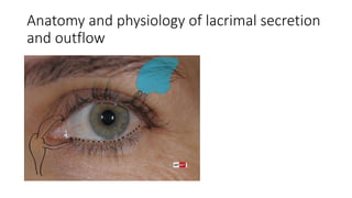
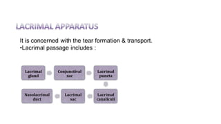
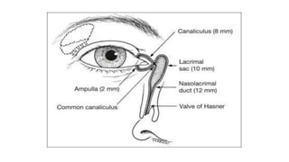
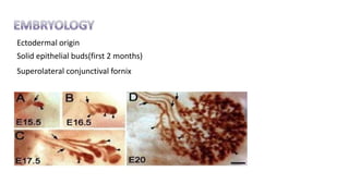
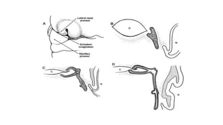
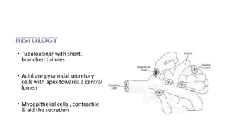
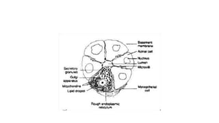
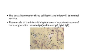
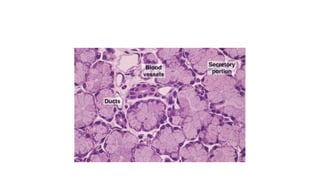
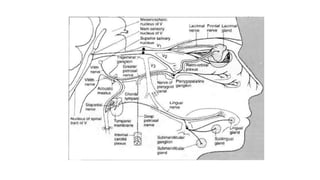
ANATOMY,DEVELOPMENT AND PHYDIOLOGY OF
THE LACRIMAL SECRETORY AND DRAINAGE
SYSTEMS
PRESENTER: DR. MUGABI BARNABAS MUKAABYA
MODULATOR: DR. NAGAWA ELIZABETH
DEPARTMENT OF OPHTHALMOLOGY
MAKCHS
The following components of the lacrimal apparatus are discussed :
 Embryology
 Osteology
 Secretory system
 Excretory system
 Physiology
Anatomy and physiology of lacrimal secretion
and outflow
Lacrimal
gland
Conjunctival
sac
Lacrimal
puncta
Lacrimal
canaliculi
Lacrimal
sac
Nasolacrimal
duct
Ectodermal origin
Solid epithelial buds(first 2 months)
Superolateral conjunctival fornix
• Lacrimal sac and nasolacrimal duct : ectoderm of the naso-optic
furrow or nasolacrimal furrow
• The ectoderm of the furrow buries and forms a solid cord .
• Canalization : begins at 4 months and may continue after birth.
• The lacrimal sac fossa is a depression in the inferomedial orbital rim,
• Maxillary and lacrimal bones.
• Bordered by the anterior lacrimal crest (maxillary bone) & posterior
lacrimal crest (lacrimal bone).
• The fossa is approximately 16-mm high, 4- to 9mm wide, and 2-mm
deep.
• The medial orbital wall : Frontal process of maxilla, lacrimal , ethmoid
, lesser wing of sphenoid bone.
• The frontoethmoidal suture is important in lacrimal surgery
• It marks the roof of the ethmoid sinus. Bony dissection superior to
this suture may expose the dura of the cranial cavity.
• The nasolacrimal canal originates at base of lacrimal fossa.
• Formed by the maxillary bone laterally and the lacrimal and inferior
turbinate bones medially.
• The width of superior opening is 4–6 mm.
• The duct courses posteriorly and laterally in the bone for 12 mm to
drain into the inferior meatus of the nasal cavity.
• It includes lacrimal gland, accessory glands
• Lacrimal gland is above & anterolateral to globe.
• Secretes tears into superior fornix.
• Tears moisten & lubricates the : cornea , conjunctiva.
• It contributes 43D of 50D of refractive power of eye .
• It consists of
• Large Orbital Part
• Smaller Palpebral Part
• Lateral expansion of levator separates the parts
Paired almond-shapedglands.
It is present in a fossa on the anterolateral area of orbit
It has 2 surfaces, 2 borders, 2 extremities
• Superior surface
• Frontal bone
• Inferior surface
• Levator palpebrae superioris & lateral rectus
• Anterior border
• Septum orbitale
• Posterior border
• Contact with orbital fat , level with posterior pole.
• Lateral extremity
• Rest on lateral rectus
• Medial extremity
• On levator
• 1/3rd size of orbital part
• Superior fornix , seen on lid eversion.
• It is situated upon the course of ducts
• Related to levator superiorly, inferiorly to superior fornix
• Posteriorly it continues with orbital part.
• Are small, compound, branched, tubular glands
• Located in the middle of lid (Wolfring glands) or superior & inferior
fornices (Krause glands).
• Ectopic portions of lacrimal gland tissue.
• It is with connective tissue coat and excretory duct.
• The excretory duct splits & form intralobular ducts, connected to
secretory glandular epithelia.
• Secretory epithelia have elongated tubules.
• True acini are absent.
• Tubuloacinar with short,
branched tubules
• Acini are pyramidal secretory
cells with apex towards a central
lumen
• Myoepithelial cells., contractile
& aid the secretion
• In acinus, secretory cells are joined by junctional complexes
• Apical microvilli extend into the lumen
• Nucleus and rough endoplasmic reticulum are basal in the cells.
• Abundant secretory granules, at apex .
• The ducts have two or three cell layers and microvilli at luminal
surface.
• Plasma cells of the interstitial space are an important source of
immunoglobulins secrete IgA(and fewer lgG, lgM, IgE)
• Artery supply : Lacrimal artery , branch of ophthalmic artery.
• Venous drainages : Ophthalmic Vein.
• Lymphatic drainage : Joins that of conjunctiva & drain into the
preauricular lymph nodes.
• Sensory nerve supply : lacrimal nerve , branch of ophthalmic division
of Vth nerve
• Sympathetic nerve supply : carotid plexus
• Secretomotor fibers : superior salivary nucleus
• A small, round or oval orifice on the elevation, the papilla lacrimalis.
• At medial end of lid margin at the junction of its ciliated and non-ciliated
parts.
• Upper punctum medial to lower, from the medial canthus being 6 and 6.5
mm.
• The upper punctum opens inferoposteriorly, the lower superoposteriorly.
• First vertical and then horizontal
• Vertical part is 2 mm & turns medially at right-angle to become horizontal 8 mm
• At angle -dilatation or ampulla.
• The canaliculi pierce the fascia (i.e. the periorbita covering the lacrimal sac)
separately,
• Uniting to enter lacrimal sac.
• Stratified squamous epithelium supported by elastic tissue.
• Lacrimal fossa, formed by lacrimal bone and frontal process of
maxilla.
• The sac, closed above and open below, is continuous with the
nasolacrimal duct.
• The sac is enclosed by a periorbita, splits &form the lacrimal fascia .
• Relations
• Medial : periorbita and bone, arc of ethmoid sinuses.
• Lateral : skin, orbicularis oculi, and lacrimal fascia.
• Anterior: medial palpebral ligament and angular vein.
• Posterior : lacrimal fascia and muscle
• The nasolacrimal duct, continuation of lacrimal sac to the inferior
meatus.
• 15 mm.
• It lies in a canal formed by the maxilla, lacrimal bone and lacrimal
process of inferior concha.
• It descends posterolaterally, a surface indication a line from medial
canthus to first upper molar.
• The valves
• They are folds of mucous
• membrane with no
• valvular function.
• The most constant is the
• 'valve' of Hasner at the lower
• end.
• It prevents sudden blast of air
• (when blowing the nose) from
• entenng the lacrimal sac.
• Structure
• Double-layered Epithelium
• The superficial layer composed of columnar cells, the deeper cells
being flatter.
• The membranous wall of the sac is of fibroelastic tissue, the elastic
element being continued around the canaliculi.
• Around the nasolacrimal duct is plexus of vessels, forming erectile
tissue like that on the inferior concha.
• Engorgement of these vessel obstruct the duct.
• The course of the lacrimal sac and duct can be demonstrated by
dacryocystography
• Vessels
• Artery supply : palpebral branches of the ophthalmic, angular and
infraorbital arteries and nasal branch of the sphenopalatine.
• Venous drainages : Angular and infraorbital vessels above, below into
the nasal veins
• Lymphatic drainage: submandibular and deep cervical nodes.
• Nerves
• Infratrochlear and anterior superior alveolar nerves.
• The tear film overlays corneal and conjunctival epithelia.
• Tears produced by the ocular surface epithelia and adnexa.
• Thickness of up to 40 µm,
• Volume of tears covering the ocular surface range from 2.74 ±2.0µL to
7 µL
• For mucous and aqueous layers, secretion is regulated by neural reflexes.
• For the lipid layer, the blink itself regulates release of pre-secreted
meibomian gland .
• Tear secretion is balanced by drainage and evaporation.
• Drainage is regulated by neural reflexes ,causing vasodilation and
vasoconstriction of blood sinus.
• Evaporation depends blink rate and temperature, humidity, and wind
speed.
• To maintain the refractive power of the cornea;
• To defend against eye infection;
• To allow gas to move between the air and the avascular cornea;
• To support corneal dehydration (assisted by the tear film
hyperosmolality).
 Glycocalyx Mucous
layer
 Aqueous layer.
 Lipid layers
• Structure
• The glycocalyx is a network of polysaccharides that project from cellular surfaces.
• Mucins are classified into secreted and membrane-spanning mucin.
• Secreted mucins are either gel-forming or small soluble
• Function
• The membrane-spanning mucins function to hydrate the ocular surface and serve
as a barrier to pathogens.
• Membrane-spanning mucins appear to be altered in dry eye
• Structure
• The mucous layer backbone is the gel-forming mucin , synthesized
and secreted by conjunctival goblet cells.
• Function
• To resistance of the eye to infection by providing protection against
microorganisms. Mucins serve as wetting agents that keep the
apical epithelia hydrated.
• Lacrimal gland produce aqueous layer.
• Other ocular surface epithelia also contribute to the aqueous layer, eg.
conjunctiva, accessory lacrimal glands
• 7µm thick.
• Without the lubrication , the shearing forces produced on blinking will
cause accumulative ocular surface damage.
• Composed of water, with many solutes, including dissolved mucins,
electrolyte sand proteins.
• The osmotic pressure : concentrations of sodium, potassium and chloride
ions.
• The tear film’s osmotic pressure is important in the control of cornea–tear
film water flux.
• Bicarbonate and carbonate : pH buffering, maintaining the pH at 7.3–7.6
when the eyes open & 6.8 eyes closed.
• Aqueous layer function
• Aqueous deficiency dry eye.
• Protection from bacterial infection
• Reflex secretion washes away noxious substances.
• Protects against changes in pH.
• Meibomian glands, modified sebaceous glands, that line the upper and
lower eyelids.
• Meibomian gland lipids are stored in vesicles.
• The secretory product contains a complex mixture of lipids and proteins
and is termed meibum.
• Meibum is released on to the ocular surface in small amounts with each
blink. 0.1m in thickness
• Function
• Hydrophobic barrier to prevent tear overflow.
• The meibomforms a water-tight seal of the apposed lid margins during
sleep.
• Reduce tear evaporation .
• Lipids enhance the stability of the tear film and provide a smooth optical.
• Conjunctival fornices, preocular tear film, and marginal tear strips.
• Marginal tear strips are wedge shaped tear menisci, borders of upper and
lower lids.
• Apposed lacrimal puncta dip into marginal strip of tears
• Anterior limit of the marginal strip is the mucocutaneousjunction of the lid,
• Tears are lost from the conjunctiva sac by absorption, evaporation,
and nasolacrimal system.
• This is related to the size of the palpebral aperture, the blink rate,
ambient temperature and humidity.
• Tears flow
• the upper and lower marginal strips →upper and lower canaliculi
(capillarity+suction)
• Eyes close
• Pretarsalorbicularis oculi compresses the ampullae+ shortens and
compresses canaliculi+punctamedially.
• Lacrimal part of the orbicularis oculi, contracts → compresses the
sac,(positive pressure) tears → nasolacrimal duct → nose.
• Eyes open
• Muscles relax →canaliculi and sac expand(negative
pressure)+capillarity= tears into sac.
REFERENCES
• Wolff'sAnatomy of the Eye and Orbit.
• Adler'sPhysiology of the Eye .
• The Lacrimal System Diagnosis, Management, and Surgery by Adam J. Cohen, Michael
Mercandetti & Brian G. Brazzo.
• The dry eye , a practical approach by SudiPatel & Kenny J Blades.
• Jack J Kanski’sclinical ophthalmology
• Clinical Anatomy of the Eye by Richard S. Snell & Michael A. Lemp.
lacrimal secretion.pptx

lacrimal secretion.pptx

  • 1.
    ANATOMY,DEVELOPMENT AND PHYDIOLOGYOF THE LACRIMAL SECRETORY AND DRAINAGE SYSTEMS PRESENTER: DR. MUGABI BARNABAS MUKAABYA MODULATOR: DR. NAGAWA ELIZABETH DEPARTMENT OF OPHTHALMOLOGY MAKCHS
  • 2.
    The following componentsof the lacrimal apparatus are discussed :  Embryology  Osteology  Secretory system  Excretory system  Physiology
  • 3.
    Anatomy and physiologyof lacrimal secretion and outflow
  • 4.
  • 6.
    Ectodermal origin Solid epithelialbuds(first 2 months) Superolateral conjunctival fornix
  • 7.
    • Lacrimal sacand nasolacrimal duct : ectoderm of the naso-optic furrow or nasolacrimal furrow • The ectoderm of the furrow buries and forms a solid cord . • Canalization : begins at 4 months and may continue after birth.
  • 9.
    • The lacrimalsac fossa is a depression in the inferomedial orbital rim, • Maxillary and lacrimal bones. • Bordered by the anterior lacrimal crest (maxillary bone) & posterior lacrimal crest (lacrimal bone). • The fossa is approximately 16-mm high, 4- to 9mm wide, and 2-mm deep.
  • 11.
    • The medialorbital wall : Frontal process of maxilla, lacrimal , ethmoid , lesser wing of sphenoid bone. • The frontoethmoidal suture is important in lacrimal surgery • It marks the roof of the ethmoid sinus. Bony dissection superior to this suture may expose the dura of the cranial cavity.
  • 12.
    • The nasolacrimalcanal originates at base of lacrimal fossa. • Formed by the maxillary bone laterally and the lacrimal and inferior turbinate bones medially. • The width of superior opening is 4–6 mm. • The duct courses posteriorly and laterally in the bone for 12 mm to drain into the inferior meatus of the nasal cavity.
  • 13.
    • It includeslacrimal gland, accessory glands • Lacrimal gland is above & anterolateral to globe. • Secretes tears into superior fornix. • Tears moisten & lubricates the : cornea , conjunctiva. • It contributes 43D of 50D of refractive power of eye .
  • 14.
    • It consistsof • Large Orbital Part • Smaller Palpebral Part • Lateral expansion of levator separates the parts
  • 15.
    Paired almond-shapedglands. It ispresent in a fossa on the anterolateral area of orbit It has 2 surfaces, 2 borders, 2 extremities • Superior surface • Frontal bone • Inferior surface • Levator palpebrae superioris & lateral rectus
  • 16.
    • Anterior border •Septum orbitale • Posterior border • Contact with orbital fat , level with posterior pole. • Lateral extremity • Rest on lateral rectus • Medial extremity • On levator
  • 17.
    • 1/3rd sizeof orbital part • Superior fornix , seen on lid eversion. • It is situated upon the course of ducts • Related to levator superiorly, inferiorly to superior fornix • Posteriorly it continues with orbital part.
  • 18.
    • Are small,compound, branched, tubular glands • Located in the middle of lid (Wolfring glands) or superior & inferior fornices (Krause glands). • Ectopic portions of lacrimal gland tissue.
  • 19.
    • It iswith connective tissue coat and excretory duct. • The excretory duct splits & form intralobular ducts, connected to secretory glandular epithelia. • Secretory epithelia have elongated tubules. • True acini are absent.
  • 22.
    • Tubuloacinar withshort, branched tubules • Acini are pyramidal secretory cells with apex towards a central lumen • Myoepithelial cells., contractile & aid the secretion
  • 23.
    • In acinus,secretory cells are joined by junctional complexes • Apical microvilli extend into the lumen • Nucleus and rough endoplasmic reticulum are basal in the cells. • Abundant secretory granules, at apex .
  • 25.
    • The ductshave two or three cell layers and microvilli at luminal surface. • Plasma cells of the interstitial space are an important source of immunoglobulins secrete IgA(and fewer lgG, lgM, IgE)
  • 27.
    • Artery supply: Lacrimal artery , branch of ophthalmic artery. • Venous drainages : Ophthalmic Vein. • Lymphatic drainage : Joins that of conjunctiva & drain into the preauricular lymph nodes.
  • 28.
    • Sensory nervesupply : lacrimal nerve , branch of ophthalmic division of Vth nerve • Sympathetic nerve supply : carotid plexus • Secretomotor fibers : superior salivary nucleus
  • 31.
    • A small,round or oval orifice on the elevation, the papilla lacrimalis. • At medial end of lid margin at the junction of its ciliated and non-ciliated parts. • Upper punctum medial to lower, from the medial canthus being 6 and 6.5 mm. • The upper punctum opens inferoposteriorly, the lower superoposteriorly.
  • 33.
    • First verticaland then horizontal • Vertical part is 2 mm & turns medially at right-angle to become horizontal 8 mm • At angle -dilatation or ampulla. • The canaliculi pierce the fascia (i.e. the periorbita covering the lacrimal sac) separately, • Uniting to enter lacrimal sac. • Stratified squamous epithelium supported by elastic tissue.
  • 34.
    • Lacrimal fossa,formed by lacrimal bone and frontal process of maxilla. • The sac, closed above and open below, is continuous with the nasolacrimal duct. • The sac is enclosed by a periorbita, splits &form the lacrimal fascia .
  • 35.
    • Relations • Medial: periorbita and bone, arc of ethmoid sinuses. • Lateral : skin, orbicularis oculi, and lacrimal fascia. • Anterior: medial palpebral ligament and angular vein. • Posterior : lacrimal fascia and muscle
  • 36.
    • The nasolacrimalduct, continuation of lacrimal sac to the inferior meatus. • 15 mm. • It lies in a canal formed by the maxilla, lacrimal bone and lacrimal process of inferior concha. • It descends posterolaterally, a surface indication a line from medial canthus to first upper molar.
  • 37.
    • The valves •They are folds of mucous • membrane with no • valvular function. • The most constant is the • 'valve' of Hasner at the lower • end. • It prevents sudden blast of air • (when blowing the nose) from • entenng the lacrimal sac.
  • 38.
    • Structure • Double-layeredEpithelium • The superficial layer composed of columnar cells, the deeper cells being flatter. • The membranous wall of the sac is of fibroelastic tissue, the elastic element being continued around the canaliculi.
  • 39.
    • Around thenasolacrimal duct is plexus of vessels, forming erectile tissue like that on the inferior concha. • Engorgement of these vessel obstruct the duct. • The course of the lacrimal sac and duct can be demonstrated by dacryocystography
  • 40.
    • Vessels • Arterysupply : palpebral branches of the ophthalmic, angular and infraorbital arteries and nasal branch of the sphenopalatine. • Venous drainages : Angular and infraorbital vessels above, below into the nasal veins • Lymphatic drainage: submandibular and deep cervical nodes. • Nerves • Infratrochlear and anterior superior alveolar nerves.
  • 41.
    • The tearfilm overlays corneal and conjunctival epithelia. • Tears produced by the ocular surface epithelia and adnexa. • Thickness of up to 40 µm, • Volume of tears covering the ocular surface range from 2.74 ±2.0µL to 7 µL
  • 42.
    • For mucousand aqueous layers, secretion is regulated by neural reflexes. • For the lipid layer, the blink itself regulates release of pre-secreted meibomian gland . • Tear secretion is balanced by drainage and evaporation. • Drainage is regulated by neural reflexes ,causing vasodilation and vasoconstriction of blood sinus. • Evaporation depends blink rate and temperature, humidity, and wind speed.
  • 43.
    • To maintainthe refractive power of the cornea; • To defend against eye infection; • To allow gas to move between the air and the avascular cornea; • To support corneal dehydration (assisted by the tear film hyperosmolality).
  • 44.
     Glycocalyx Mucous layer Aqueous layer.  Lipid layers
  • 46.
    • Structure • Theglycocalyx is a network of polysaccharides that project from cellular surfaces. • Mucins are classified into secreted and membrane-spanning mucin. • Secreted mucins are either gel-forming or small soluble • Function • The membrane-spanning mucins function to hydrate the ocular surface and serve as a barrier to pathogens. • Membrane-spanning mucins appear to be altered in dry eye
  • 47.
    • Structure • Themucous layer backbone is the gel-forming mucin , synthesized and secreted by conjunctival goblet cells. • Function • To resistance of the eye to infection by providing protection against microorganisms. Mucins serve as wetting agents that keep the apical epithelia hydrated.
  • 48.
    • Lacrimal glandproduce aqueous layer. • Other ocular surface epithelia also contribute to the aqueous layer, eg. conjunctiva, accessory lacrimal glands • 7µm thick. • Without the lubrication , the shearing forces produced on blinking will cause accumulative ocular surface damage.
  • 49.
    • Composed ofwater, with many solutes, including dissolved mucins, electrolyte sand proteins. • The osmotic pressure : concentrations of sodium, potassium and chloride ions. • The tear film’s osmotic pressure is important in the control of cornea–tear film water flux. • Bicarbonate and carbonate : pH buffering, maintaining the pH at 7.3–7.6 when the eyes open & 6.8 eyes closed.
  • 50.
    • Aqueous layerfunction • Aqueous deficiency dry eye. • Protection from bacterial infection • Reflex secretion washes away noxious substances. • Protects against changes in pH.
  • 51.
    • Meibomian glands,modified sebaceous glands, that line the upper and lower eyelids. • Meibomian gland lipids are stored in vesicles. • The secretory product contains a complex mixture of lipids and proteins and is termed meibum. • Meibum is released on to the ocular surface in small amounts with each blink. 0.1m in thickness
  • 52.
    • Function • Hydrophobicbarrier to prevent tear overflow. • The meibomforms a water-tight seal of the apposed lid margins during sleep. • Reduce tear evaporation . • Lipids enhance the stability of the tear film and provide a smooth optical.
  • 54.
    • Conjunctival fornices,preocular tear film, and marginal tear strips. • Marginal tear strips are wedge shaped tear menisci, borders of upper and lower lids. • Apposed lacrimal puncta dip into marginal strip of tears • Anterior limit of the marginal strip is the mucocutaneousjunction of the lid,
  • 56.
    • Tears arelost from the conjunctiva sac by absorption, evaporation, and nasolacrimal system. • This is related to the size of the palpebral aperture, the blink rate, ambient temperature and humidity.
  • 57.
    • Tears flow •the upper and lower marginal strips →upper and lower canaliculi (capillarity+suction) • Eyes close • Pretarsalorbicularis oculi compresses the ampullae+ shortens and compresses canaliculi+punctamedially. • Lacrimal part of the orbicularis oculi, contracts → compresses the sac,(positive pressure) tears → nasolacrimal duct → nose. • Eyes open • Muscles relax →canaliculi and sac expand(negative pressure)+capillarity= tears into sac.
  • 59.
    REFERENCES • Wolff'sAnatomy ofthe Eye and Orbit. • Adler'sPhysiology of the Eye . • The Lacrimal System Diagnosis, Management, and Surgery by Adam J. Cohen, Michael Mercandetti & Brian G. Brazzo. • The dry eye , a practical approach by SudiPatel & Kenny J Blades. • Jack J Kanski’sclinical ophthalmology • Clinical Anatomy of the Eye by Richard S. Snell & Michael A. Lemp.