Downloaded 627 times


![Blood Flow Meter
• Blood flow is nothing but the volume of blood per time
[ml/min].
Typical values for blood flow [cm/s]:
1. Aorta 100 – 250
2. Abdominal 100
3. Vena Cava 5 – 40
69 DEEPAK.P](https://image.slidesharecdn.com/unit4biomedical-160822181636/85/Unit-4-biomedical-69-320.jpg)


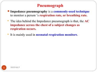
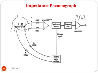
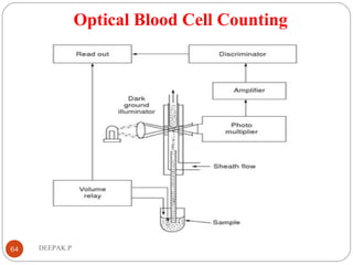
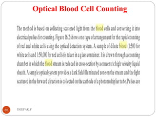
The document discusses various instruments used for respiratory and blood measurements. It describes pneumographs which detect respiration through chest movements. Spirometers are used to measure lung volumes and capacities. Impedance pneumography monitors respiration rate using changes in chest impedance during breathing. Other topics covered include blood cell counting methods like Coulter and optical techniques, electromagnetic and ultrasonic blood flow meters, and measuring blood pH using glass electrodes in blood gas analyzers.


