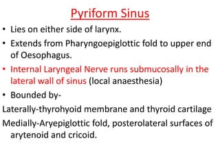
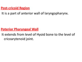
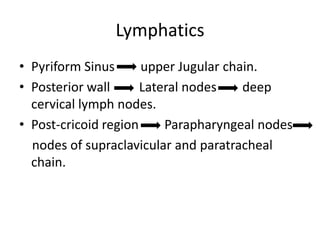
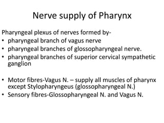
Downloaded 2,923 times


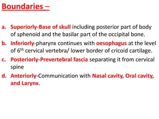
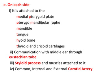
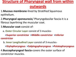
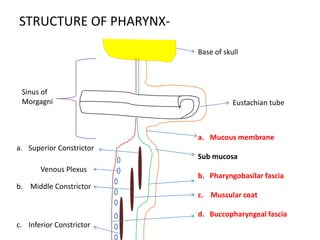
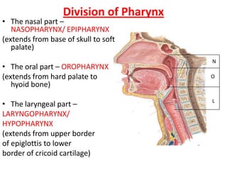


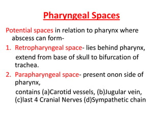


The document provides details on the anatomy of the pharynx, including its boundaries, structure, and divisions. It can be summarized as follows: 1. The pharynx is divided into three parts - nasopharynx, oropharynx, and laryngopharynx. 2. It has mucosa, pharyngeal aponeurosis, muscular coat, and buccopharyngeal fascia in its wall structure. 3. Important structures include the tonsils, lymphatic drainage sites, and nerves that supply the pharynx. 4. Spaces near the pharynx where abscesses can form are the retropharyngeal and parapharyngeal


