Downloaded 21 times

![Constrictor muscles
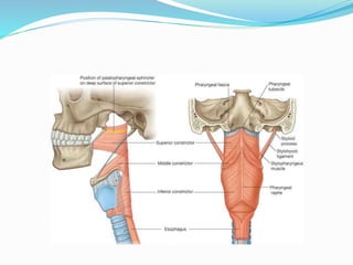
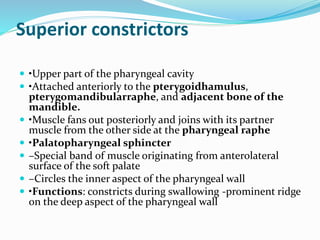
•The 3 constrictor muscle on each side -major contributors
to the structure of the pharyngeal wall
–Superior, Middle and inferior
•Posteriorly, the muscles from each side are joined together
by the pharyngeal raphe
•Anteriorly, these muscles attach to bones and ligaments
related to the lateral margins of the nasal and oral cavities
and the larynx.
•Walls of three flower pots stacked one on the other
•Constrict or narrow the pharyngeal cavity.
•Innervated by the pharyngeal branch of the vagus nerve
[X]](https://image.slidesharecdn.com/pharynxnew-200709052937/85/Pharynx-Anatomy-and-physiology-16-320.jpg)
![Nerves
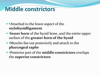
•Motor and most sensory
innervation-branches of the
vagus[X] &
glossopharyngeal[IX] nerves
•This 2 nerved forms a plexus in
the outer fascia of the pharyngeal
wall, consisting of
–the pharyngeal branch of the
vagusnerve [X];
–branches from the external
laryngeal nervefrom the
superior laryngeal branchof the
vagusnerve [X];
–pharyngeal branches of the
glossopharyngealnerve [IX]](https://image.slidesharecdn.com/pharynxnew-200709052937/85/Pharynx-Anatomy-and-physiology-25-320.jpg)
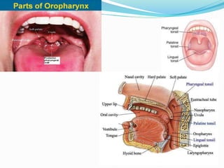


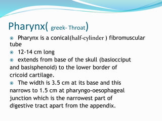
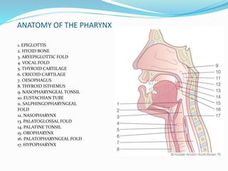
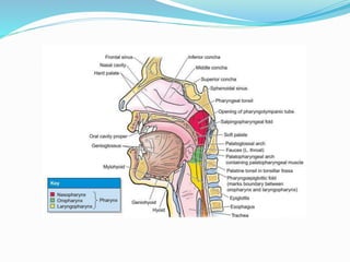
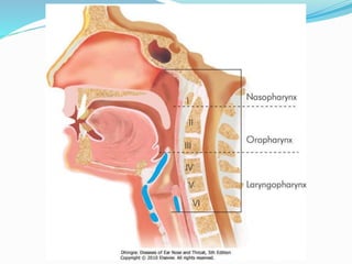
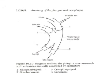
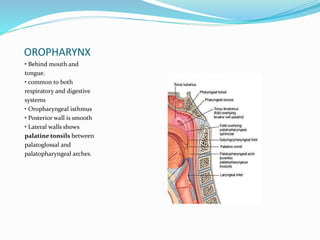
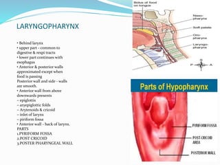
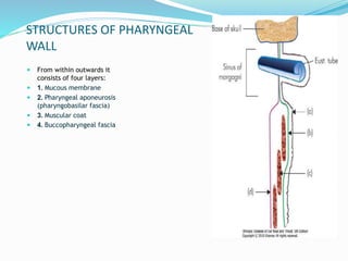
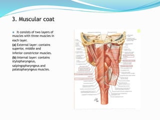
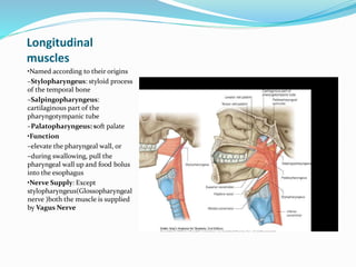
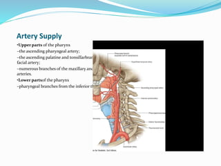
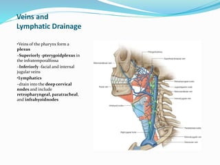
The document summarizes the anatomy and functions of the pharynx. It is a fibromuscular tube approximately 12-14 cm long located behind the nasal cavity, mouth, and larynx. It functions in respiration, swallowing, and sound resonance. The pharynx has three parts - nasopharynx, oropharynx, and laryngopharynx. Its walls consist of mucosa, pharyngeal aponeurosis, a muscular coat with three constrictor muscles, and an outer buccopharyngeal fascia. The pharynx is supplied by branches of the vagus and glossopharyngeal nerves and drains into deep cervical lymph nodes.


