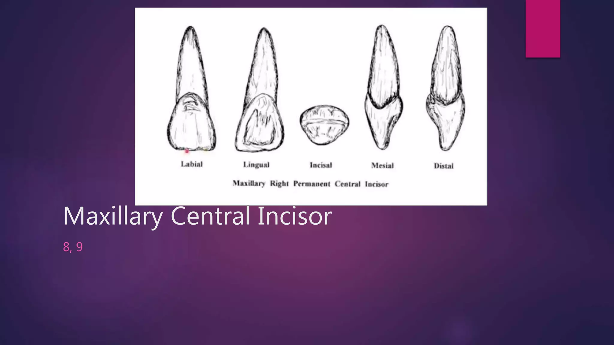
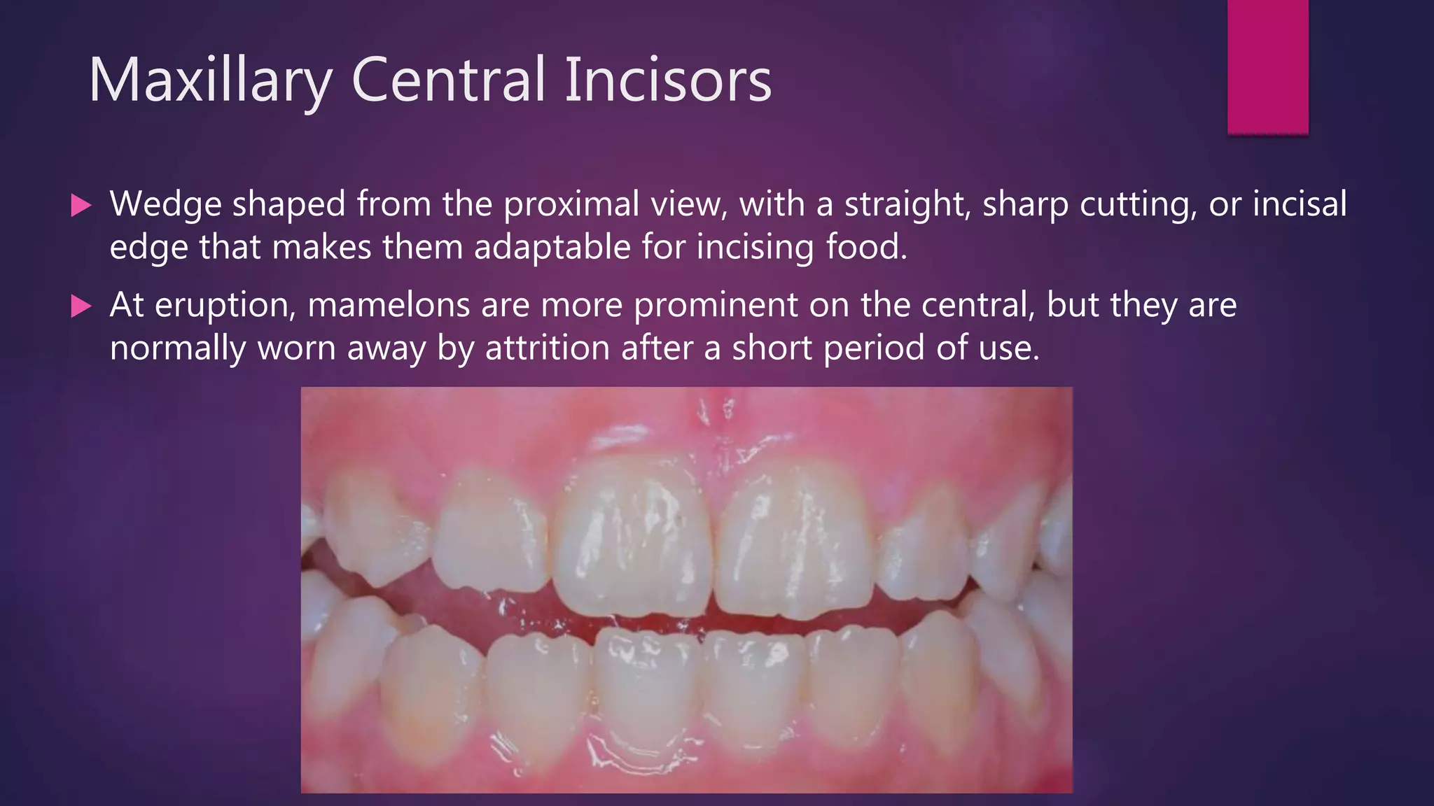
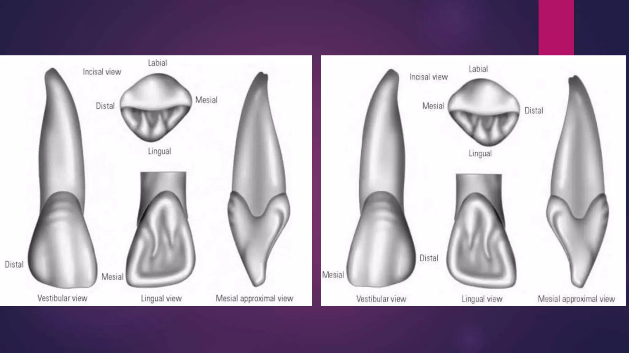
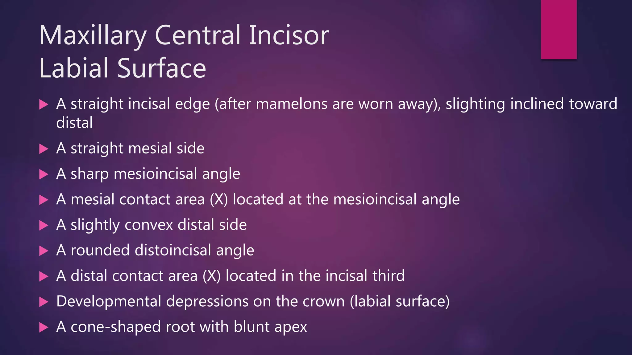
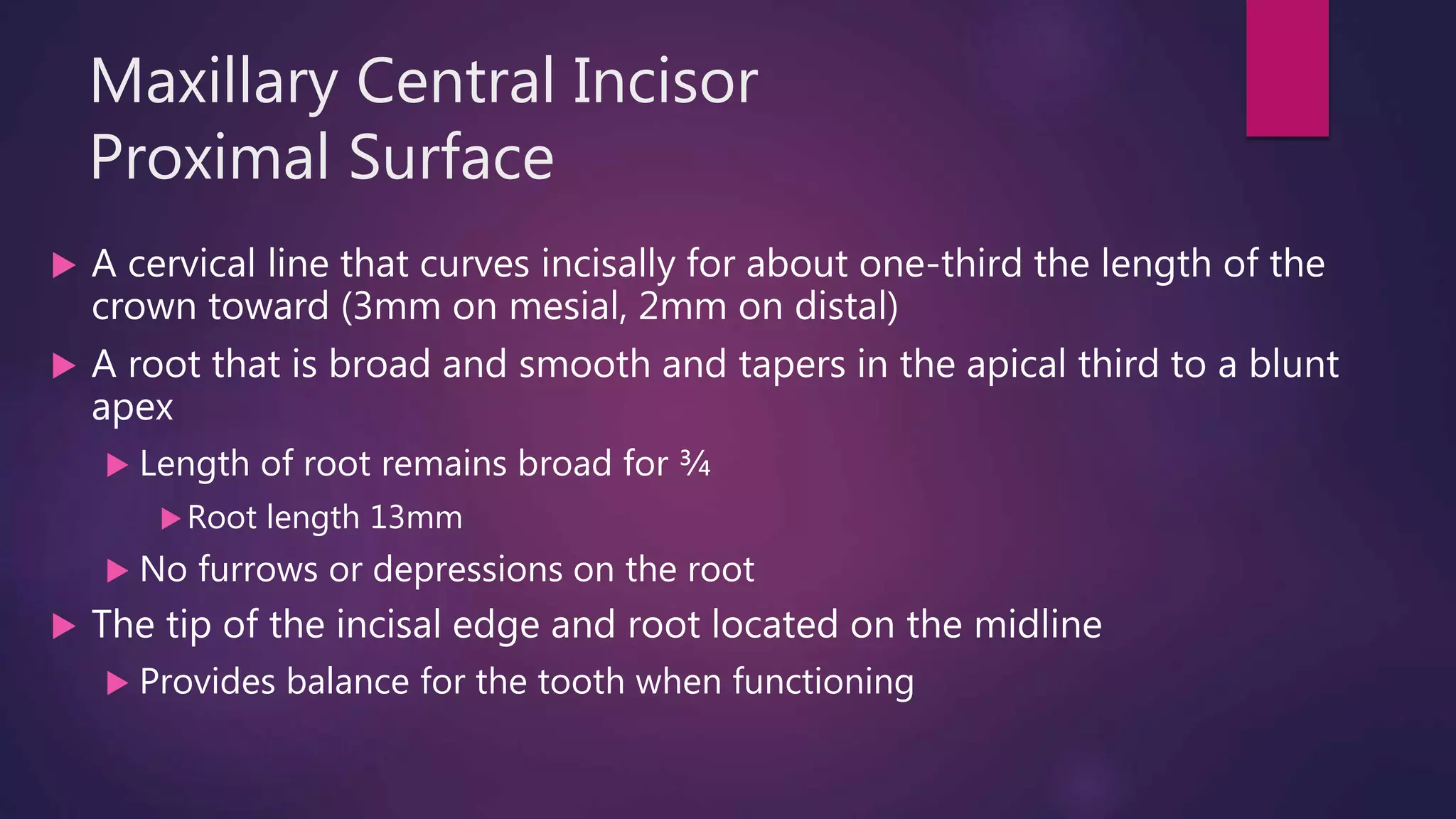
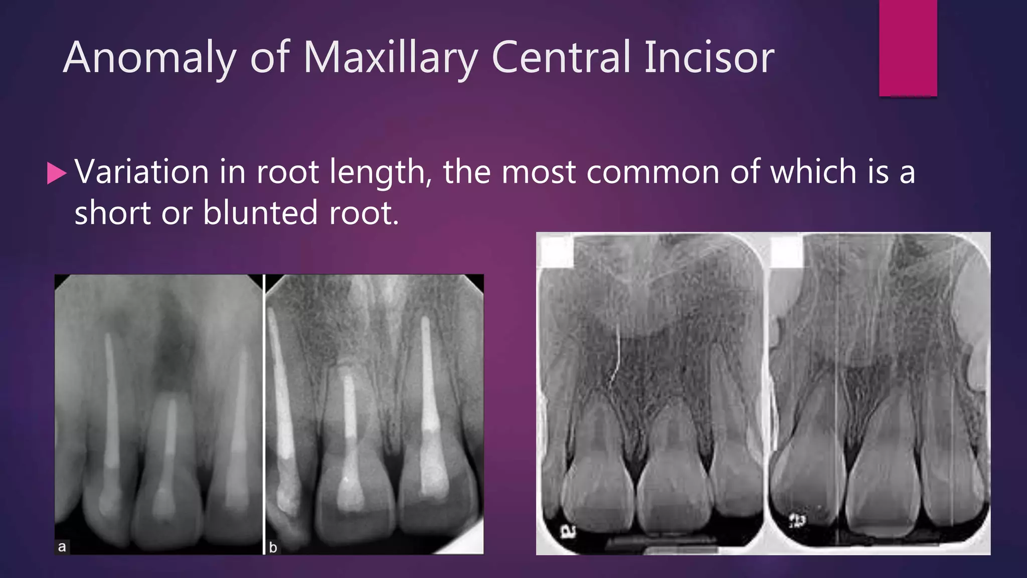
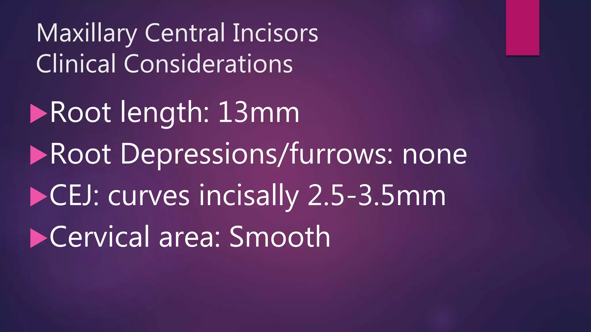
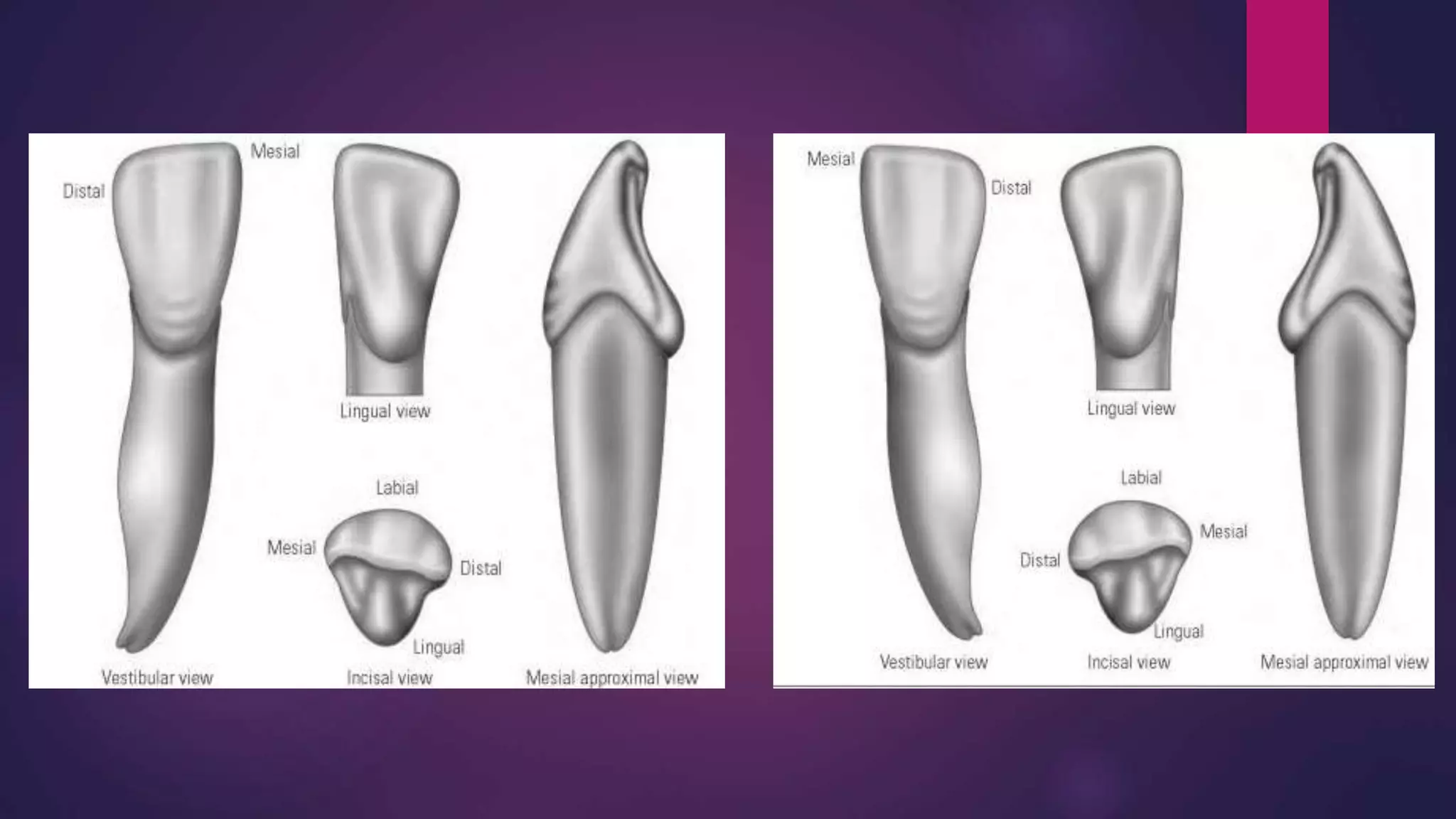
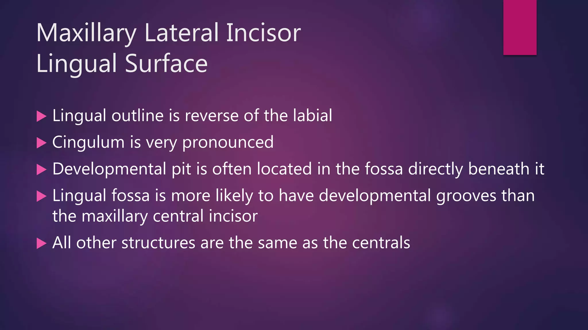
Permanent Anterior Teeth
MAXILLARY/MANDIBULAR CENTRAL & LATERAL INCISOR AND CANINES
Key Terms
 The text and PowerPoint describe “ideal” morphology of each tooth.
 Actual teeth with show variation from the norm, this is called an Anomaly.
 Antagonist: A structure that opposes or counteracts another
structure; in the coral cavity, the teeth that meet those in the other
jaw.
 Concave: curving inward; a depression (a bowl)
 Convex: curving outward; a bump.
 Furrow: long, shallow depression present
Key Terms
 Mamelons: A rounded prominence on the incisal edge of newly erupted incisors
that are remnants of lobe formation.
 Developmental Depressions: A concavity in a surface that formed while the tooth
was developing.
 Lingual Fossa: Shallow depression in the lingual surface of the tooth.
 Marginal Ridges: a linear elevation located around the perimeter of surface.
 Cingulum: an eminence or raised area on the lingual surface of the anterior teeth
resulting from the lingual lobe formation.
 Cervical Line: where the anatomic crown and root join together
 Pit: a pinpoint depression
Maxillary Central Incisor
8, 9
Maxillary Central Incisors
 Wedge shaped from the proximal view, with a straight, sharp cutting, or incisal
edge that makes them adaptable for incising food.
 At eruption, mamelons are more prominent on the central, but they are
normally worn away by attrition after a short period of use.
Maxillary Central Incisor
#8 & #9
Eruption date: 7-8 years old
First Evidence of calcification: 3-4 months old
Crown Completion: 4-5 years old
Root Completion: 10 years old
Maxillary Central Incisor
Function: Biting, incising
Length of crown: 10.5mm
Length of root: 13mm
Antagonist: mandibular central and lateral incisors
Maxillary central incisor is larger than maxillary lateral
incisor
Maxillary Central Incisor
Identifying Features
Widest anterior tooth, mesiodistally
Straight mesial side with sharp mesioincisal angle
and convex distal side
Straight incisal edge
Wedge shape of tooth from proximal view
Maxillary Central Incisor
Labial Surface
 A straight incisal edge (after mamelons are worn away), slighting inclined toward
distal
 A straight mesial side
 A sharp mesioincisal angle
 A mesial contact area (X) located at the mesioincisal angle
 A slightly convex distal side
 A rounded distoincisal angle
 A distal contact area (X) located in the incisal third
 Developmental depressions on the crown (labial surface)
 A cone-shaped root with blunt apex
Maxillary Central Incisor
Lingual Surface
 An outline that is reverse of the labial
 A well-defined concave lingual fossa
 Cingulum merges with lingual fossa
 Mesial and distal marginal ridges that outline the lingual fossa
 A convex cingulum that comprises the cervical third of the crown
 Pronounced cingulum dominates cervical third of crown.
 Shallow grooves found in the fossa
Maxillary Central Incisor
Proximal Surface
 A cervical line that curves incisally for about one-third the length of the
crown toward (3mm on mesial, 2mm on distal)
 A root that is broad and smooth and tapers in the apical third to a blunt
apex
 Length of root remains broad for ¾
Root length 13mm
 No furrows or depressions on the root
 The tip of the incisal edge and root located on the midline
 Provides balance for the tooth when functioning
Anomaly of Maxillary Central Incisor
 Variation in root length, the most common of which is a
short or blunted root.
Maxillary Central Incisors
Clinical Considerations
Root length: 13mm
Root Depressions/furrows: none
CEJ: curves incisally 2.5-3.5mm
Cervical area: Smooth
Maxillary Lateral Incisor
7, 10
Maxillary Lateral Incisor
#7 and #10
Eruption date: 8-9 years old
First Evidence of Calcification: 1 year old
Crown completion: 4-5 years old
Root Completion: 11 years old
Smaller than maxillary central incisor
More convex than maxillary central incisor
Maxillary Lateral Incisor
Function: Biting, incising
Length of crown: 9mm
Length of root: 13mm
Antagonists: Mandibular lateral incisor and
canine
Maxillary Lateral Incisor
Identifying Features
Resembles maxillary central incisor, but is
smaller with more convex mesial and distal
sides and incisal angles
Developmental variations are frequent
Maxillary Lateral Incisor
Labial Surface
 Incisal edge inclines toward distal
 Both mesial and distal incisal angles are rounded, and both mesial
and distal sides of the tooth are convex
 Distal outline is more convex and the angle is more rounded than the
mesial
 Developmental depressions may be present, but usually smooth
 Contact areas are located more cervically than on the central incisor
 Cone-shaped root tapers gradually toward the apex
Maxillary Lateral Incisor
Lingual Surface
 Lingual outline is reverse of the labial
 Cingulum is very pronounced
 Developmental pit is often located in the fossa directly beneath it
 Lingual fossa is more likely to have developmental grooves than
the maxillary central incisor
 All other structures are the same as the centrals
Maxillary Lateral Incisor
Proximal Surface
From this view, the same as central (except for furrow)
Broad root with furrows on both mesial and distal
surfaces
Cervical line curves one-third of the tooth toward the
incisal
Maxillary Lateral Incisors
Clinical Considerations
Root length: 13mm
Root Depressions/furrows: mesial and distal
vertical depressions are usually present
CEJ: curves incisally 2-3mm
Cervical area: may have shallow concavity
Anomaly of Maxillary Lateral Incisor
 Most commonly forms as anomaly (other than 3rd molars)
 Most frequent variation is a ‘peg-shaped’ lateral
Canines
Canine
 Two canines in each arch, one per quadrant
 Maxillary and mandibular canines are similar, with variations noted in
the descriptions to follow.
 Both maxillary and mandibular canines have a sharp, pointed cusp
 Also known as cuspids
 Long, strong and stable teeth
 Note their size and length as compared to the adjacent teeth
 Usually have one root, mandibular could have two.. Discussed later
Canine
 Located at the corner of the arch, so they are referred to as “cornerstone” teeth
 Important to aesthetics because they provide shape to the face
 If dentures are needed and the size or position of the canine is varied, the entire facial
appearance can change.
 Canines are self-cleansing, because of their convexity and location at the corners
of the mouth
 Food easily rolls off of them
 Usually last teeth to be lost be dental decay
 Shape of this tooth, particularly the sharp cusp, make it adaptable for holding and
tearing food
Maxillary Canine
6, 11
Maxillary Canine
#6 and #11
Eruption Date: 11-12 years old
First Evidence of Calcification: 4-5 months old
Crown Completion: 6-7 years old
Root Completion: 13-15 years old
Maxillary Canine
Function: tearing, holding
Length of Crown: 10mm
Length of Root: 17mm
Antagonist: Mandibular Canine and first premolar
Maxillary Canine
Identifying Characteristics
Longest maxillary tooth
Stable, sturdy tooth
Cornerstone of the arch
Sharp, pointed cusp
Bulky cingulum and defined lingual ridges
Maxillary Canine
Labial Surface
 Mesial cusp slope that is shorter than the distal cusp slope
 A mesial contact area that is at the junction of the incisal and middle third
 A distal contact area that is in the middle third of the tooth
 A slightly convex mesial side
 Slightly concave from cervix to contact area on distal side
 A well-developed middle lobe, called the labial ridge
 Depressions that are either side of the labial ridge
 A cusp tip that is centered over the root
 A cone-shape, smooth root
Maxillary Canine
Lingual Surface
An outline that is the reverse of the labial view
A crown and root that converge so that both mesial
and distal surfaces can be seen
A fossa that is divided, by a lingual ridge, into
mesiolingual and distolingual fossa
A pronounced cingulum and marginal ridges
Maxillary Canine
Proximal Surface
A broad crown and root
A cervical line that curves toward the incisal
A deep depression in the root surface
Maxillary Canine
Clinical Considerations
Root length: 17mm
Root Depressions/furrows: deep depressions on both
mesial and distal surfaces
CEJ: curves incisally 1.5-2.5 mm on mesial and distal
surfaces
Cervical area: concave on both mesial and distal
surfaces
Mandibular Central Incisor
24, 25
Mandibular Central Incisor
#24 and #25
Eruption Date: 6-7 years old
First Evidence of Calcification: 3-4 months old
Crown Completion: 4-5 years old
Root Completion: 9 years old
Mandibular Central Incisor
Function: incising, biting
Length of Crown: 9mm
Length of Root: 12.5mm
Antagonist: Maxillary central incisor
Mandibular Central Incisor
Identifying Characteristics
 Smallest tooth in the oral cavity
 Bilaterally symmetric (same on both sides) from labial or
lingual view
 Smooth tooth; no developmental grooves or
depressions in crown
 Only one antagonist
 Hardest tooth to identify as right or left outside of the
mouth
Mandibular Central Incisor
Labial Surface
 Incisal edge is straight, joining the mesial and distal sides at sharp
incisal angles
 Contact areas
 Both mesial and distal sides are straight, tapering evenly to the cervix
where the cervical line forms an arc
 The tooth is bilaterally symmetric
 Root is straight, tapering gradually to the apex (only way to identify
as right or left)
Mandibular Central Incisor
Lingual Surface
 Outline from lingual is reverse of the labial view
 Lingual surface is very smooth, no pits or grooves
 Fossa and cingulum merge with a gentle curve
 Marginal ridges are not evident, unlike maxillary centrals
 Cingulum covers the cervical third of the crown
The smooth, shallow fossa occupies the remaining two-thirds
Mandibular Central Incisor
Proximal Surface
 Width from labial to lingual, is broad to compensate for the
narrow mesial distal measurements
Width is necessary to provide stability in the bone for such a small
tooth
 Root remains broad for two-thirds its length, tapering only at
the apical third
 Both mesial and distal root surfaces have a deep depression
extending most of the root length
Mandibular Central Incisor
Incisal Surface
This aspect helps assists in differentiating
from mandibular lateral incisor
The central has a straight incisal edge that is
bilaterally symmetric
Mandibular Central Incisors
Clinical Considerations
Root length: 12.5mm
Root Depressions/furrows: deep vertical depressions
on both mesial and distal surfaces
CEJ: curves incisally 2-3mm
Cervical area: may have shallow concavity on both
mesial and distal surfaces
Mandibular Lateral Incisor
23, 26
Mandibular Lateral Incisor
#23 and #26
Eruption Date: 7-8 years old
First Evidence of Calcification: 3-4 months old
Crown Completion: 4-5 years old
Root Completion: 10 years old
Mandibular Lateral Incisor
Function: biting, incising
Length of Crown: 9.5mm
Length of Root: 14mm
Antagonist: Maxillary central and lateral incisors
Mandibular Lateral Incisor
Identifying Characteristics
Slightly larger than the central incisor
Slightly more convex than the central incisor
Incisal ridge curves to the distal
Crown distally displaced
Mandibular Lateral Incisor
Labial Surface
 The lateral looks like the central in overall appearance, but is slightly larger and
not bilaterally symmetric
 Incisal edge of the lateral declines toward the distal and forms a rounded incisal
angle with the distal side
 The distal side is slightly convex
 The mesial incisal angle is sharp
 The mesial side can be straight or slightly convex as it tapers toward the cervical
line
 Both the mesial and distal contact areas are at the incisal angle
 The cervical line forms a narrow arch
 The root is straight, tapering at the apical third
Mandibular Lateral Incisor
Lingual/Proximal Surface
Except for size and length, the
mandibular lateral incisor is similar to the
central incisor and has the same
characteristics
The cingulum is distally displaced as it
starts to curve toward the canine
Mandibular Lateral Incisor
Incisal Surface
 The incisal edge of the lateral incisor curves toward the distal,
following contour of the mandibular arch
 The incisal edge of the central is straight
 The curvature on the lateral incisor creates a distally displaced
cingulum as compared to the centrally situated cingulum of the
central
 This structure assists in distinguishing the two teeth from each other
Mandibular Lateral Incisors
Clinical Considerations
Root length: 14mm
Root Depressions/furrows: both mesial and distal
with deeper distal depressions
CEJ: curves incisally 2-3mm
Cervical area: may have shallow concavity
Mandibular Canine
22, 27
Mandiubular Canine
#22 and #27
Eruption Date: 9-10 years old
First Evidence of Calcification: 4-5 months old
Crown Completion: 6-7 years old
Root Completion: 12-14 years old
Mandibular Canine
Function: tearing, holding
Length of Crown: 11mm
Length of Root: 16mm
Antagonist: Maxillary lateral incisor and canine
Mandibular Canine
Identifying Features
Longest mandibular tooth
Well-developed middle lobe called the labial
ridge
Slightly narrower and smoother than maxillary
canine
Mandibular Canine
Labial Surface
 Narrower than the maxillary by 0.5mm
 A mesial cusp slope that is shorter than the distal
 Contact areas that are more incisal than on maxillary canine
 Straight mesial side
 Slightly convex distal side
 A tapered root, mesial inclination
 Labial ridge is not as prominent
 Depressions on either side of ridge
Mandibular Canine
Lingual Surface
The outline is reversed of the labial view, but
narrower because the lingual converges
Structures are the same as maxillary canine, but less
pronounced
Cingulum and marginal ridges are smoother than the
maxillary canine
Two fossae divided by lingual ridge
Fossa are shallower than maxillary canine
Mandibular Canine
Proximal Surface
Tooth is broad from the proximal view
Greater width provides stability
Deep depressions on both mesial and
distal root surfaces
Mandibular Canine
Clinical Considerations
Root length: 16mm
Root Depressions/furrows: deep depressions on both
mesial and distal surfaces
CEJ: curves incisally 1-2.5mm on mesial and distal
surfaces
Cervical area: no concavity on either mesial or distal
surface
Anomaly of Mandibular Canine
 Two roots; two root canals

Permanent anterior teeth

  • 1.
    Permanent Anterior Teeth MAXILLARY/MANDIBULARCENTRAL & LATERAL INCISOR AND CANINES
  • 2.
    Key Terms  Thetext and PowerPoint describe “ideal” morphology of each tooth.  Actual teeth with show variation from the norm, this is called an Anomaly.  Antagonist: A structure that opposes or counteracts another structure; in the coral cavity, the teeth that meet those in the other jaw.  Concave: curving inward; a depression (a bowl)  Convex: curving outward; a bump.  Furrow: long, shallow depression present
  • 3.
    Key Terms  Mamelons:A rounded prominence on the incisal edge of newly erupted incisors that are remnants of lobe formation.  Developmental Depressions: A concavity in a surface that formed while the tooth was developing.  Lingual Fossa: Shallow depression in the lingual surface of the tooth.  Marginal Ridges: a linear elevation located around the perimeter of surface.  Cingulum: an eminence or raised area on the lingual surface of the anterior teeth resulting from the lingual lobe formation.  Cervical Line: where the anatomic crown and root join together  Pit: a pinpoint depression
  • 4.
  • 5.
    Maxillary Central Incisors Wedge shaped from the proximal view, with a straight, sharp cutting, or incisal edge that makes them adaptable for incising food.  At eruption, mamelons are more prominent on the central, but they are normally worn away by attrition after a short period of use.
  • 6.
    Maxillary Central Incisor #8& #9 Eruption date: 7-8 years old First Evidence of calcification: 3-4 months old Crown Completion: 4-5 years old Root Completion: 10 years old
  • 7.
    Maxillary Central Incisor Function:Biting, incising Length of crown: 10.5mm Length of root: 13mm Antagonist: mandibular central and lateral incisors Maxillary central incisor is larger than maxillary lateral incisor
  • 8.
    Maxillary Central Incisor IdentifyingFeatures Widest anterior tooth, mesiodistally Straight mesial side with sharp mesioincisal angle and convex distal side Straight incisal edge Wedge shape of tooth from proximal view
  • 10.
    Maxillary Central Incisor LabialSurface  A straight incisal edge (after mamelons are worn away), slighting inclined toward distal  A straight mesial side  A sharp mesioincisal angle  A mesial contact area (X) located at the mesioincisal angle  A slightly convex distal side  A rounded distoincisal angle  A distal contact area (X) located in the incisal third  Developmental depressions on the crown (labial surface)  A cone-shaped root with blunt apex
  • 11.
    Maxillary Central Incisor LingualSurface  An outline that is reverse of the labial  A well-defined concave lingual fossa  Cingulum merges with lingual fossa  Mesial and distal marginal ridges that outline the lingual fossa  A convex cingulum that comprises the cervical third of the crown  Pronounced cingulum dominates cervical third of crown.  Shallow grooves found in the fossa
  • 12.
    Maxillary Central Incisor ProximalSurface  A cervical line that curves incisally for about one-third the length of the crown toward (3mm on mesial, 2mm on distal)  A root that is broad and smooth and tapers in the apical third to a blunt apex  Length of root remains broad for ¾ Root length 13mm  No furrows or depressions on the root  The tip of the incisal edge and root located on the midline  Provides balance for the tooth when functioning
  • 14.
    Anomaly of MaxillaryCentral Incisor  Variation in root length, the most common of which is a short or blunted root.
  • 15.
    Maxillary Central Incisors ClinicalConsiderations Root length: 13mm Root Depressions/furrows: none CEJ: curves incisally 2.5-3.5mm Cervical area: Smooth
  • 16.
  • 17.
    Maxillary Lateral Incisor #7and #10 Eruption date: 8-9 years old First Evidence of Calcification: 1 year old Crown completion: 4-5 years old Root Completion: 11 years old Smaller than maxillary central incisor More convex than maxillary central incisor
  • 18.
    Maxillary Lateral Incisor Function:Biting, incising Length of crown: 9mm Length of root: 13mm Antagonists: Mandibular lateral incisor and canine
  • 19.
    Maxillary Lateral Incisor IdentifyingFeatures Resembles maxillary central incisor, but is smaller with more convex mesial and distal sides and incisal angles Developmental variations are frequent
  • 21.
    Maxillary Lateral Incisor LabialSurface  Incisal edge inclines toward distal  Both mesial and distal incisal angles are rounded, and both mesial and distal sides of the tooth are convex  Distal outline is more convex and the angle is more rounded than the mesial  Developmental depressions may be present, but usually smooth  Contact areas are located more cervically than on the central incisor  Cone-shaped root tapers gradually toward the apex
  • 22.
    Maxillary Lateral Incisor LingualSurface  Lingual outline is reverse of the labial  Cingulum is very pronounced  Developmental pit is often located in the fossa directly beneath it  Lingual fossa is more likely to have developmental grooves than the maxillary central incisor  All other structures are the same as the centrals
  • 23.
    Maxillary Lateral Incisor ProximalSurface From this view, the same as central (except for furrow) Broad root with furrows on both mesial and distal surfaces Cervical line curves one-third of the tooth toward the incisal
  • 25.
    Maxillary Lateral Incisors ClinicalConsiderations Root length: 13mm Root Depressions/furrows: mesial and distal vertical depressions are usually present CEJ: curves incisally 2-3mm Cervical area: may have shallow concavity
  • 26.
    Anomaly of MaxillaryLateral Incisor  Most commonly forms as anomaly (other than 3rd molars)  Most frequent variation is a ‘peg-shaped’ lateral
  • 27.
  • 28.
    Canine  Two caninesin each arch, one per quadrant  Maxillary and mandibular canines are similar, with variations noted in the descriptions to follow.  Both maxillary and mandibular canines have a sharp, pointed cusp  Also known as cuspids  Long, strong and stable teeth  Note their size and length as compared to the adjacent teeth  Usually have one root, mandibular could have two.. Discussed later
  • 29.
    Canine  Located atthe corner of the arch, so they are referred to as “cornerstone” teeth  Important to aesthetics because they provide shape to the face  If dentures are needed and the size or position of the canine is varied, the entire facial appearance can change.  Canines are self-cleansing, because of their convexity and location at the corners of the mouth  Food easily rolls off of them  Usually last teeth to be lost be dental decay  Shape of this tooth, particularly the sharp cusp, make it adaptable for holding and tearing food
  • 30.
  • 31.
    Maxillary Canine #6 and#11 Eruption Date: 11-12 years old First Evidence of Calcification: 4-5 months old Crown Completion: 6-7 years old Root Completion: 13-15 years old
  • 32.
    Maxillary Canine Function: tearing,holding Length of Crown: 10mm Length of Root: 17mm Antagonist: Mandibular Canine and first premolar
  • 33.
    Maxillary Canine Identifying Characteristics Longestmaxillary tooth Stable, sturdy tooth Cornerstone of the arch Sharp, pointed cusp Bulky cingulum and defined lingual ridges
  • 35.
    Maxillary Canine Labial Surface Mesial cusp slope that is shorter than the distal cusp slope  A mesial contact area that is at the junction of the incisal and middle third  A distal contact area that is in the middle third of the tooth  A slightly convex mesial side  Slightly concave from cervix to contact area on distal side  A well-developed middle lobe, called the labial ridge  Depressions that are either side of the labial ridge  A cusp tip that is centered over the root  A cone-shape, smooth root
  • 36.
    Maxillary Canine Lingual Surface Anoutline that is the reverse of the labial view A crown and root that converge so that both mesial and distal surfaces can be seen A fossa that is divided, by a lingual ridge, into mesiolingual and distolingual fossa A pronounced cingulum and marginal ridges
  • 37.
    Maxillary Canine Proximal Surface Abroad crown and root A cervical line that curves toward the incisal A deep depression in the root surface
  • 39.
    Maxillary Canine Clinical Considerations Rootlength: 17mm Root Depressions/furrows: deep depressions on both mesial and distal surfaces CEJ: curves incisally 1.5-2.5 mm on mesial and distal surfaces Cervical area: concave on both mesial and distal surfaces
  • 40.
  • 41.
    Mandibular Central Incisor #24and #25 Eruption Date: 6-7 years old First Evidence of Calcification: 3-4 months old Crown Completion: 4-5 years old Root Completion: 9 years old
  • 42.
    Mandibular Central Incisor Function:incising, biting Length of Crown: 9mm Length of Root: 12.5mm Antagonist: Maxillary central incisor
  • 43.
    Mandibular Central Incisor IdentifyingCharacteristics  Smallest tooth in the oral cavity  Bilaterally symmetric (same on both sides) from labial or lingual view  Smooth tooth; no developmental grooves or depressions in crown  Only one antagonist  Hardest tooth to identify as right or left outside of the mouth
  • 45.
    Mandibular Central Incisor LabialSurface  Incisal edge is straight, joining the mesial and distal sides at sharp incisal angles  Contact areas  Both mesial and distal sides are straight, tapering evenly to the cervix where the cervical line forms an arc  The tooth is bilaterally symmetric  Root is straight, tapering gradually to the apex (only way to identify as right or left)
  • 46.
    Mandibular Central Incisor LingualSurface  Outline from lingual is reverse of the labial view  Lingual surface is very smooth, no pits or grooves  Fossa and cingulum merge with a gentle curve  Marginal ridges are not evident, unlike maxillary centrals  Cingulum covers the cervical third of the crown The smooth, shallow fossa occupies the remaining two-thirds
  • 47.
    Mandibular Central Incisor ProximalSurface  Width from labial to lingual, is broad to compensate for the narrow mesial distal measurements Width is necessary to provide stability in the bone for such a small tooth  Root remains broad for two-thirds its length, tapering only at the apical third  Both mesial and distal root surfaces have a deep depression extending most of the root length
  • 48.
    Mandibular Central Incisor IncisalSurface This aspect helps assists in differentiating from mandibular lateral incisor The central has a straight incisal edge that is bilaterally symmetric
  • 50.
    Mandibular Central Incisors ClinicalConsiderations Root length: 12.5mm Root Depressions/furrows: deep vertical depressions on both mesial and distal surfaces CEJ: curves incisally 2-3mm Cervical area: may have shallow concavity on both mesial and distal surfaces
  • 51.
  • 52.
    Mandibular Lateral Incisor #23and #26 Eruption Date: 7-8 years old First Evidence of Calcification: 3-4 months old Crown Completion: 4-5 years old Root Completion: 10 years old
  • 53.
    Mandibular Lateral Incisor Function:biting, incising Length of Crown: 9.5mm Length of Root: 14mm Antagonist: Maxillary central and lateral incisors
  • 54.
    Mandibular Lateral Incisor IdentifyingCharacteristics Slightly larger than the central incisor Slightly more convex than the central incisor Incisal ridge curves to the distal Crown distally displaced
  • 56.
    Mandibular Lateral Incisor LabialSurface  The lateral looks like the central in overall appearance, but is slightly larger and not bilaterally symmetric  Incisal edge of the lateral declines toward the distal and forms a rounded incisal angle with the distal side  The distal side is slightly convex  The mesial incisal angle is sharp  The mesial side can be straight or slightly convex as it tapers toward the cervical line  Both the mesial and distal contact areas are at the incisal angle  The cervical line forms a narrow arch  The root is straight, tapering at the apical third
  • 57.
    Mandibular Lateral Incisor Lingual/ProximalSurface Except for size and length, the mandibular lateral incisor is similar to the central incisor and has the same characteristics The cingulum is distally displaced as it starts to curve toward the canine
  • 58.
    Mandibular Lateral Incisor IncisalSurface  The incisal edge of the lateral incisor curves toward the distal, following contour of the mandibular arch  The incisal edge of the central is straight  The curvature on the lateral incisor creates a distally displaced cingulum as compared to the centrally situated cingulum of the central  This structure assists in distinguishing the two teeth from each other
  • 60.
    Mandibular Lateral Incisors ClinicalConsiderations Root length: 14mm Root Depressions/furrows: both mesial and distal with deeper distal depressions CEJ: curves incisally 2-3mm Cervical area: may have shallow concavity
  • 62.
  • 63.
    Mandiubular Canine #22 and#27 Eruption Date: 9-10 years old First Evidence of Calcification: 4-5 months old Crown Completion: 6-7 years old Root Completion: 12-14 years old
  • 64.
    Mandibular Canine Function: tearing,holding Length of Crown: 11mm Length of Root: 16mm Antagonist: Maxillary lateral incisor and canine
  • 65.
    Mandibular Canine Identifying Features Longestmandibular tooth Well-developed middle lobe called the labial ridge Slightly narrower and smoother than maxillary canine
  • 67.
    Mandibular Canine Labial Surface Narrower than the maxillary by 0.5mm  A mesial cusp slope that is shorter than the distal  Contact areas that are more incisal than on maxillary canine  Straight mesial side  Slightly convex distal side  A tapered root, mesial inclination  Labial ridge is not as prominent  Depressions on either side of ridge
  • 68.
    Mandibular Canine Lingual Surface Theoutline is reversed of the labial view, but narrower because the lingual converges Structures are the same as maxillary canine, but less pronounced Cingulum and marginal ridges are smoother than the maxillary canine Two fossae divided by lingual ridge Fossa are shallower than maxillary canine
  • 69.
    Mandibular Canine Proximal Surface Toothis broad from the proximal view Greater width provides stability Deep depressions on both mesial and distal root surfaces
  • 71.
    Mandibular Canine Clinical Considerations Rootlength: 16mm Root Depressions/furrows: deep depressions on both mesial and distal surfaces CEJ: curves incisally 1-2.5mm on mesial and distal surfaces Cervical area: no concavity on either mesial or distal surface
  • 72.
    Anomaly of MandibularCanine  Two roots; two root canals