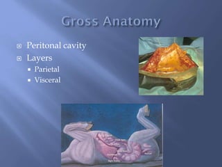
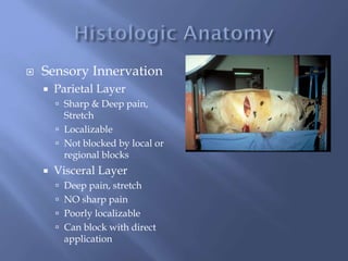
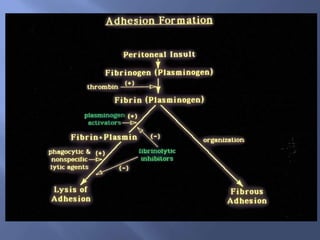
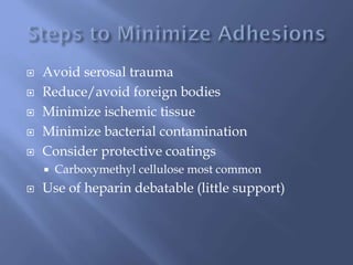
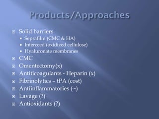
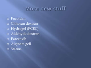
Micky Trent DVM DACVS
 What are the histologic layers of normal
peritoneum?
 Which vary by species?
 Layers
 Mesothelial Cells
 Basement membrane
 Submesothelial
connective tissue
 Loose c.t.
 Species variation
 What are the layers of peritoneum?
 What is the peritoneal space?
 Peritonal cavity
 Layers
 Parietal
 Visceral
 How is peritoneum innervated?
 Why should we as surgeons care?
 Sensory Innervation
 Parietal Layer
 Sharp & Deep pain,
Stretch
 Localizable
 Not blocked by local or
regional blocks
 Visceral Layer
 Deep pain, stretch
 NO sharp pain
 Poorly localizable
 Can block with direct
application
 What abdominal structures are considered
retroperitoneal?
 Why should we as surgeons care?
 List 4 critical functions that depend on the
peritoneum.
 Cell nutrition
 Peritoneal fluid production
 Maintain gliding surfaces
 Waste control
 Cell Nutrition
 Diffusion
 Produce Peritoneal
Fluid
 Transudate from
blood
 Inflammation
changes volume and
character
 Maintain Gliding
Surface
 Surface villi trap
fluid
 Peritoneal fluid
 Plasminogen
activators
 Mesothelial cells
 Fibrin break down
 Depressed with
peritoneal trauma
 How is waste removed from the peritoneal
cavity?
 Low MW compounds?
 Cells?
 Foreign bodies?
 Physical Removal
 Transmesothelial
 Small molecules
 H20
 Stomata/Lymphatics
 Peritoneal flow to
diaphragm 10 route
 Diaphram lymphatics
 Thoracic duct to vena
cava
 Larger molecules
 Particles/cells
 Fluid
 Omental alternative
 Functional Removal
 Localization
 Omental Trapping
 Fibrin Trapping
 Phagocytosis
 PMNs
 Macrophages
 Bacterial kill
 Functional Removal
 Localization
 Omental Trapping
 Fibrin Trapping
 Phagocytosis
 PMNs
 Macrophages
 Bacterial kill
 How does the peritoneum respond initially
(24hrs) to trauma?
 Vascular phase?
 Inflammatory cascade?
 Coagulation cascade?
 Link between inflammation and coagulation?
 Brief vasoconstriction
 Histamine, PGE2 release
 Local vasodilation & cell influx
 Procoagulant factors
 Platelets adhere to wound bed
 Alpha corpuscles degranulate
 Release PDGF, TGF-β, dense corpuscles
 Release epinephrine, serotonin
 Contribute to PG & leukotriene release
 Local release of cytokines
 Cell migration to wound bed
 Platelet aggregation
 Coagulation cascade activation
 Initial clot formation
 Fibrin deposition
 Temporary matrix for signaling molecules &
inflammatory cells
 Temporary bridge between tissues
 Close overlap with Inflammatory response
 Two pathways converging to one
 Intrinsic pathway
 BM damage & collagen exposure
 Factor XII (Hagemen factor)
 Activation of factor II (prothrombin)
 Activation of factor IIa (thrombin precursor)
 Thrombin production
 Thrombin
 Cleaves soluble circulating fibrinogen into insoluble
fibrin clots
 Factor XII stimulates clot formation and
activation of the fibrinolytic system
 Fibrinolysis determined by:
 tPA (mesothelial cells, leukocytes, tissue)
 PAI1&2
 How does a peritoneal defect heal?
 What is the timing?
 Where do new cells come from?
 Are there differences in cells shed in a dialysate?
 How is ECM formed?
 12-24 hrs – PMNs
 24 hrs + macrophages
 Cytokine release
 Day 3
 Mesothelial and fibroblast cells appear
 Area of lesion regresses
 Day 7-10
 Mesothelial layer reestablished
 ECM follows cell migration
 Species variation?
 tPA vs PAI baseline
 Human, horses, dogs – tPA high
 Cows – PAI high
 Post-injury
 Most: tPA down, PAI up
 Cow: PAI up
 Describe the steps in adhesion formation.
 What key enzymes are involved?
 How are these enzymes affected by trauma?
 What is the incidence of post-op adhesions?
 In people?
 In horses?
 Which structures are most commonly involved
in identified cases in horses?
 Why?
 Humans
 67%-93%
 Horses
 9%-32%
 Caveats?
 Canine/Feline
 Low
 Equine
 SI procedures
 Regardless of procedure
 SI
 Ventral incision
 Why??
 Are there any beneficial effects of peritoneal
adhesions?
Beneficial Effects
 Seal Breaks in
continuity
 Prevent visceral
leakage
 Neovascularization
 Stabilize mobile
viscera
 Bovine, canine,
equine
Beneficial Effects
 Seal Breaks in
continuity
 Prevent visceral
leakage
 Neovascularization
 Stabilize mobile
viscera
 Bovine, canine,
equine
 What are common stimuli for adhesions?
 Suturing
Peritoneum!!
 Ischemia
 Serosal trauma
 Drying
 Abrasion
 Others
 Foreign bodies
 Bacteria
 List preventative methods (and mechanisms)
for reduction of adhesions
 Avoid serosal trauma
 Reduce/avoid foreign bodies
 Minimize ischemic tissue
 Minimize bacterial contamination
 Consider protective coatings
 Carboxymethyl cellulose most common
 Use of heparin debatable (little support)
 Solid barriers
 Seprafilm (CMC & HA)
 Interceed (oxidized cellulose)
 Hyaluronate membranes
 CMC
 Omentectomy(x)
 Antiticoagulants - Heparin (x)
 Fibrinolytics – tPA (cost)
 Antiinflammatories (~)
 Lavage (?)
 Antioxidants (?)
 Fucoidan
 Chitosan dextran
 Hydrogel (PCEC)
 Aldehyde dextran
 Parecoxib
 Alginate gell
 Statins

Peritoneal Healing, Cow/Horse

  • 1.
  • 2.
     What arethe histologic layers of normal peritoneum?  Which vary by species?
  • 3.
     Layers  MesothelialCells  Basement membrane  Submesothelial connective tissue  Loose c.t.  Species variation
  • 4.
     What arethe layers of peritoneum?  What is the peritoneal space?
  • 5.
     Peritonal cavity Layers  Parietal  Visceral
  • 6.
     How isperitoneum innervated?  Why should we as surgeons care?
  • 7.
     Sensory Innervation Parietal Layer  Sharp & Deep pain, Stretch  Localizable  Not blocked by local or regional blocks  Visceral Layer  Deep pain, stretch  NO sharp pain  Poorly localizable  Can block with direct application
  • 8.
     What abdominalstructures are considered retroperitoneal?  Why should we as surgeons care?
  • 9.
     List 4critical functions that depend on the peritoneum.
  • 10.
     Cell nutrition Peritoneal fluid production  Maintain gliding surfaces  Waste control
  • 11.
     Cell Nutrition Diffusion  Produce Peritoneal Fluid  Transudate from blood  Inflammation changes volume and character
  • 12.
     Maintain Gliding Surface Surface villi trap fluid  Peritoneal fluid  Plasminogen activators  Mesothelial cells  Fibrin break down  Depressed with peritoneal trauma
  • 13.
     How iswaste removed from the peritoneal cavity?  Low MW compounds?  Cells?  Foreign bodies?
  • 14.
     Physical Removal Transmesothelial  Small molecules  H20  Stomata/Lymphatics  Peritoneal flow to diaphragm 10 route  Diaphram lymphatics  Thoracic duct to vena cava  Larger molecules  Particles/cells  Fluid  Omental alternative
  • 15.
     Functional Removal Localization  Omental Trapping  Fibrin Trapping  Phagocytosis  PMNs  Macrophages  Bacterial kill
  • 16.
     Functional Removal Localization  Omental Trapping  Fibrin Trapping  Phagocytosis  PMNs  Macrophages  Bacterial kill
  • 17.
     How doesthe peritoneum respond initially (24hrs) to trauma?  Vascular phase?  Inflammatory cascade?  Coagulation cascade?  Link between inflammation and coagulation?
  • 18.
     Brief vasoconstriction Histamine, PGE2 release  Local vasodilation & cell influx  Procoagulant factors  Platelets adhere to wound bed  Alpha corpuscles degranulate  Release PDGF, TGF-β, dense corpuscles  Release epinephrine, serotonin  Contribute to PG & leukotriene release
  • 19.
     Local releaseof cytokines  Cell migration to wound bed  Platelet aggregation  Coagulation cascade activation  Initial clot formation  Fibrin deposition  Temporary matrix for signaling molecules & inflammatory cells  Temporary bridge between tissues
  • 20.
     Close overlapwith Inflammatory response  Two pathways converging to one  Intrinsic pathway  BM damage & collagen exposure  Factor XII (Hagemen factor)  Activation of factor II (prothrombin)  Activation of factor IIa (thrombin precursor)  Thrombin production
  • 21.
     Thrombin  Cleavessoluble circulating fibrinogen into insoluble fibrin clots  Factor XII stimulates clot formation and activation of the fibrinolytic system  Fibrinolysis determined by:  tPA (mesothelial cells, leukocytes, tissue)  PAI1&2
  • 22.
     How doesa peritoneal defect heal?  What is the timing?  Where do new cells come from?  Are there differences in cells shed in a dialysate?  How is ECM formed?
  • 23.
     12-24 hrs– PMNs  24 hrs + macrophages  Cytokine release  Day 3  Mesothelial and fibroblast cells appear  Area of lesion regresses  Day 7-10  Mesothelial layer reestablished  ECM follows cell migration
  • 24.
  • 25.
     tPA vsPAI baseline  Human, horses, dogs – tPA high  Cows – PAI high  Post-injury  Most: tPA down, PAI up  Cow: PAI up
  • 26.
     Describe thesteps in adhesion formation.  What key enzymes are involved?  How are these enzymes affected by trauma?
  • 28.
     What isthe incidence of post-op adhesions?  In people?  In horses?  Which structures are most commonly involved in identified cases in horses?  Why?
  • 29.
     Humans  67%-93% Horses  9%-32%  Caveats?  Canine/Feline  Low
  • 30.
     Equine  SIprocedures  Regardless of procedure  SI  Ventral incision  Why??
  • 31.
     Are thereany beneficial effects of peritoneal adhesions?
  • 32.
    Beneficial Effects  SealBreaks in continuity  Prevent visceral leakage  Neovascularization  Stabilize mobile viscera  Bovine, canine, equine
  • 33.
    Beneficial Effects  SealBreaks in continuity  Prevent visceral leakage  Neovascularization  Stabilize mobile viscera  Bovine, canine, equine
  • 34.
     What arecommon stimuli for adhesions?
  • 35.
     Suturing Peritoneum!!  Ischemia Serosal trauma  Drying  Abrasion  Others  Foreign bodies  Bacteria
  • 36.
     List preventativemethods (and mechanisms) for reduction of adhesions
  • 37.
     Avoid serosaltrauma  Reduce/avoid foreign bodies  Minimize ischemic tissue  Minimize bacterial contamination  Consider protective coatings  Carboxymethyl cellulose most common  Use of heparin debatable (little support)
  • 38.
     Solid barriers Seprafilm (CMC & HA)  Interceed (oxidized cellulose)  Hyaluronate membranes  CMC  Omentectomy(x)  Antiticoagulants - Heparin (x)  Fibrinolytics – tPA (cost)  Antiinflammatories (~)  Lavage (?)  Antioxidants (?)
  • 39.
     Fucoidan  Chitosandextran  Hydrogel (PCEC)  Aldehyde dextran  Parecoxib  Alginate gell  Statins