Download as PPSX, PPTX


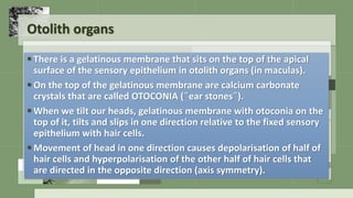

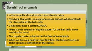

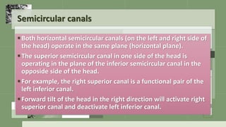


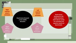


The document summarizes key aspects of peripheral vestibular function and mechanisms. It describes how the vestibular labyrinth senses head movements and positions via hair cells in the semicircular canals and otolith organs. Signals are relayed to the brainstem and cerebellum to control eye movements and posture. The specific roles of the semicircular canals and otolith organs in sensing rotational and linear accelerations are detailed, as well as the sensory transduction processes in the hair cells and how they encode movement.


![Kejis Presentation (1)[1].pptx ..........](https://cdn.slidesharecdn.com/ss_thumbnails/kejispresentation11-250317085159-8af3be0d-thumbnail.jpg?width=640&height=640&fit=bounds)


