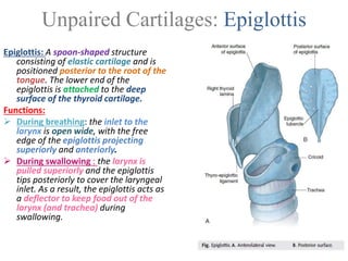
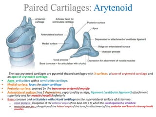
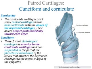
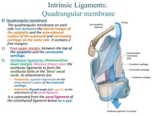
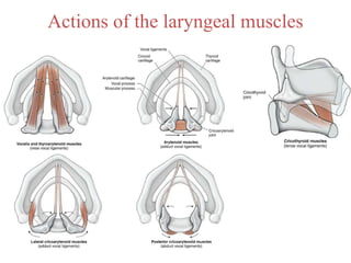
Larynx
Card- 5
Item- 10
Introduction
Description: The larynx is a hollow musculo-ligamentous structure with a cartilaginous
framework that caps the lower respiratory tract.
Location: anterior to the esophagus and at the level between C3 & C6 in its normal position.
Cartilages that make up larynx:
• Unpaired (3): Cricoid (hyaline cartilage), thyroid (hyaline cartilage), and epiglottis (elastic
cartilage)
• Paired (3): Arytenoid, corniculate, and cuneiform ( all from elastic cartilage)
Unpaired Cartilages: Thyroid Cartilage
The thyroid cartilage is the largest of the laryngeal cartilages. Its features are:
 Laryngeal prominence (Adam’s apple): (palpable at C4 level) most superior point of the site
of fusion between the two broad flat laminae. It is 90° in men and 120° in women
 Superior thyroid notch: (palpable at C4 level) point of separation of the 2 laminae as they
diverge laterally.
 Superior horn: Attaches to the tip of the greater horn of the hyoid bone by a lateral
thyrohyoid ligament .
 Inferior horn: Articulates with the cricoid cartilage, forming the cricothyroid joint.
 Oblique line: a site of attachment for the extrinsic muscles of the larynx
Unpaired Cartilages: Cricoid Cartilage
Cricoid cartilage: Shaped like a signet ring (the only complete ring of cartilage in the airways). The lower
border marks the inferior limits of the larynx and pharynx. Provides attachments for laryngeal
muscles, cartilages, and ligaments involved in opening and closing of the airway to produce sound.
The cricoid cartilage has 2 articular facets on each side for articulation with other laryngeal cartilages:
1) One facet is on the sloping superolateral surface of the lamina and articulates with the base of an
arytenoid cartilage.
2) The other facet is on the lateral surface of the lamina near its base and is for articulation with the
medial surface of the inferior horn of the thyroid cartilage.
Unpaired Cartilages: Epiglottis
Epiglottis: A spoon-shaped structure
consisting of elastic cartilage and is
positioned posterior to the root of the
tongue. The lower end of the
epiglottis is attached to the deep
surface of the thyroid cartilage.
Functions:
 During breathing: the inlet to the
larynx is open wide, with the free
edge of the epiglottis projecting
superiorly and anteriorly.
 During swallowing : the larynx is
pulled superiorly and the epiglottis
tips posteriorly to cover the laryngeal
inlet. As a result, the epiglottis acts as
a deflector to keep food out of the
larynx (and trachea) during
swallowing.
Paired Cartilages: Arytenoid
The two arytenoid cartilages are pyramid-shaped cartilages with 3 surfaces, a base of arytenoid cartilage and
an apex of arytenoid cartilage.
 Apex: articulates with a corniculate cartilage.
 Medial surface: faces the other cartilage
 Posterior surface: covered by the transverse arytenoid muscle
 Anterolateral surface: has 2 depressions, separated by a ridge, ligament (vestibular ligament) attachment
superiorly and for muscle (vocalis) inferiorly
 Base: concave and articulates with cricoid cartilage on the superolateral surface of its lamina
– vocal process : elongation of the anterior angle of the base into a to which the vocal ligament is attached.
– muscular process : elongation of the lateral angle of the base for attachment of the posterior and lateral crico-arytenoid
muscles.
Paired Cartilages:
Cuneiform and corniculate
Corniculate
• The corniculate cartilages are 2
small conical cartilages whose
bases articulate with the apices of
the arytenoid cartilages. Their
apices project posteromedially
toward each other.
Cuneiform
• These 2 small club-shaped
cartilages lie anterior to the
corniculate cartilages and are
suspended in the part of the
fibroelastic membrane of the
larynx that attaches the arytenoid
cartilages to the lateral margin of
the epiglottis.
Extrinsic Ligaments
The extrinsic ligaments act to attach the
components of the larynx to external
structures:
Extrinsic:
1. Thyrohyoid membrane – Spans
between the superior aspect of the
thyroid cartilage and the hyoid bone. It is
pierced laterally by the superior
laryngeal vessels and internal laryngeal
nerve (branch of the superior laryngeal
nerve).
– Median thyrohyoid ligament –
Anteromedial thickening of the
membrane.
– Lateral thyrohyoid ligaments –
Posterolateral thickenings of the
membrane.
2. Hyo-epiglottic ligament – Connects the
hyoid bone to the anterior aspect of the
epiglottis.
3. Cricotracheal ligament – Connects the
cricoid cartilage to the trachea.
Intrinsic Ligaments: Cricothyroid
1) Cricothyrioid ligament:
It consists of 3 parts:
– Conus elasticus: attached to the arch of
cricoid cartilage and extends superiorly
to end in a free upper margin within the
space enclosed by the thyroid cartilage.
On each side, this upper free margin
attaches:
• anteriorly: thyroid cartilage
• posteriorly: vocal processes of the
arytenoid cartilages.
– Vocal ligaments – thickening of the free
upper margin of conus elasticus.
Mucosa drapes over the vocal ligaments
to form the vocal folds or the ‘true’
vocal cords.
– Median cricothyroid ligament-
thickening of the conus elasticus in the
anterior mid line
Intrinsic Ligaments:
Quadrangular membrane
2) Quadrangular membrane
The quadrangular membrane on each
side runs between the lateral margin of
the epiglottis and the anterolateral
surface of the arytenoid and corniculate
cartilage on the same side. It contains 2
free margins:
1) Free upper margin: between the top of
the epiglottis and the corniculate
cartilage.
2) Vestibular ligaments: thickened free
lower margin. Mucosa drapes over the
vestibular ligaments to form the
vestibular folds or the ‘false’ vocal
cords. Its attachments are:
– Posteriorly: superior depression on the
anterolateral surface of the arytenoid
cartilage
– Anteriorly: thyroid angle just superior to the
attachment of the vocal ligament
It is separated from the vocal ligament of
the cricothyroid ligament below by a gap.
Laryngeal joints
Crico-arytenoid joints
Articulating surfaces: The crico-arytenoid joints
between are synovial joints:
• Superolateral articular facets on the of the cricoid
cartilage
• Bases of the arytenoid cartilages
Movements: enable the arytenoid cartilages to
– slide away or
– toward each other and to
– rotate so that the vocal processes pivot either
toward or away from the midline.
These movements abduct and adduct the vocal
ligaments
Cricothyroid joints
Articulating surfaces: This synovial joint is between:
• Inferior horns of the thyroid cartilage
• Lateral facet of the cricoid cartilage
Each is surrounded by a capsule and is reinforced by
associated ligaments.
Movements :The cricothyroid joints enable the thyroid
cartilage to:
– move forward
– tilt downward on the cricoid cartilage
Laryngeal Folds: Vocal Folds
There are 2 important soft tissue folds located within the larynx – the
vestibular folds and vocal folds. They play a crucial role in protection
of the airway, breathing, and phonation.
Vocal Folds
The vocal folds (true vocal cords) are the more important of the two
sets. Under the control of the muscles of phonation, they are
abducted, adducted, relaxed and tensed to control the pitch of the
sound created.
Histologically, they are structured as follows (superficial to deep):
 Non-keratinised stratified squamous epithelium – Stratified layer
provides extensive protection against foreign bodies which may
accidentally enter the larynx.
 Reinke’s space – This watery, amorphous layer is rich in
glycosaminoglycans. Due to its fluidity, the epithelium is able to
vibrate freely above it to create sound.
 Vocal ligament – Lies at the free upper edge of the cricothryoid
ligament.
 Vocalis muscle – Exceptionally fine muscle fibres that lie lateral to the
vocal ligaments.
The vocal folds are relatively avascular, and appear white in colour. The
space between the vocal folds is known as the rima glottidis.
Laryngeal Folds: Vestibular Folds
Vestibular Folds
• The vestibular folds (false vocal cords) lie superiorly to the
true vocal cords. They consist of the vestibular ligament
(free lower edge of the quadrangular membrane) covered by
a mucous membrane, and are pink in colour. They are fixed
folds, which act to provide protection to the larynx.
• It is continuous with the posterior surface of the epiglottis.
And is thus, lined with a pseudostratified ciliated columnar
epithelium with goblet cells.
• Rima vestibuli: (When viewed from above) the triangular
opening between the 2 adjacent vestibular folds at the
entrance to the middle chamber of the laryngeal cavity. It is
separated from the ‘true’ vocal cord below by a gap.
Laryngeal inlet
The superior aperture of the cavity (laryngeal
inlet) opens into the anterior aspect of the
pharynx just below and posterior to the tongue
 Anterior border is formed by mucosa covering
the superior margin of the epiglottis.
 Lateral borders are formed by mucosal folds
(aryepiglottic folds), which of the quadrangular
membranes and adjacent soft tissues, and
cuneiform and corniculate cartilages on the more
posterolateral margin of the laryngeal inlet.
 Posterior border in the midline is formed by a
mucosal fold that forms a depression
(interarytenoid notch) between the 2 corniculate
tubercles.
It is lined with non - keratinised stratified
squamous epithelium provides extensive
protection against foreign bodies and abrasion of
the flowing air.
Laryngeal Cavity
Anatomically, the internal cavity of the larynx can
be divided into 3 sections:
 Supraglottis / Vestibule– From the inferior surface
of the epiglottis to the vestibular folds.
 Glottis – Contains vocal cords and 1cm below them.
The opening between the vocal cords is known as
rima glottidis, the size of which is altered by the
muscles of phonation.
 Infraglottis – From inferior border of the glottis to
the inferior border of the cricoid cartilage.
The interior surface of the larynx is lined
by pseudostratified ciliated columnar epithelium.
An important exception to this is the true vocal
cords, which are lined by a stratified squamous
epithelium.
Laryngeal ventricles: On each side, the mucosa of the
middle cavity bulges laterally through the gap
between the vestibular and vocal ligaments to
produce an expanded trough-shaped space .
Laryngeal saccule: An elongate tubular extension of
each ventricle. Within its walls of are numerous
mucous glands. Mucus secreted into the saccules
lubricates the vocal folds.
Intrinsic muscles of larynx
Actions of the laryngeal muscles
Function of the larynx:
Breathing
Quiet respiration:
– The laryngeal inlet, vestibule, rima
vestibuli, and rima glottidis are open.
– The arytenoid cartilages are
abducted and
– the rima glottidis is triangular
shaped.
Forced inspiration:
– Arytenoid cartilages are rotated
laterally, mainly by the action of the
posterior crico-arytenoid muscles.
– As a result, the vocal folds are
abducted
– Rima glottidis widens into a
rhomboid shape, which effectively
increases the diameter of the
laryngeal airway.
Function of the larynx: Phonation
General Mechanism: Phonation, or the production of sound, involves
the intermittent release of expired air coordinated with opening
and closing the rima glottis. The length of the true vocal folds and
the size of the rima glottis are altered by the action of the intrinsic
laryngeal muscles
Pitch: As the length and tension of the vocal folds change, the pitch of
the sound is altered. Generally, the more tense the vocal folds, the
faster they vibrate and thus the higher the pitch. The rima glottis
is:
 wide when low-pitched sounds are produced
 narrows to a slit when high-pitched sounds are produced.
Deepening of voice in men: As a young boy's larynx enlarges during
puberty, the vocal folds become both longer and thicker, causing
them to vibrate more slowly and thus the voice becomes deeper.
Loudness: Loudness of the voice depends proportionally on the force
with which air rushes across the vocal folds.
Pharynx, oral and nasal cavities and the paranasal sinuses acts as
a resonating chamber to amplify and enhance the quality of the
sound.
Articulation: Articulation, or the production of intelligible sounds,
involves the actions the pharyngeal muscles, the tongue the
muscles of facial expression, mandibular movements, soft palate,
hard palate and the teeth.
Function of the larynx:
Effort closure & Swallowing
Effort closure
Effort closure of the larynx occurs when air is retained in the
thoracic cavity to stabilize the trunk, for example during
heavy lifting, or as part of the mechanism for increasing
intra-abdominal pressure.
During effort closure, the
– rima glottidis, rima vestibuli and lower parts of the vestibule is
completely closed.
– The result is to completely and forcefully shut the airway.
Swallowing
 Rima glottidis, rima vestibuli, and vestibule are closed and
the laryngeal inlet is narrowed.
 In addition, the larynx moves up and forward.
 This action causes the epiglottis to swing downward toward
the arytenoid cartilages
 Epiglottis effectively narrow or close the laryngeal inlet.
 The up and forward movement of the larynx also opens the
esophagus.
All these actions together prevent solids and liquids from
entry into the airway and facilitate their movement through
the piriform fossae into the esophagus.
Larynx: Vasculature
Arterial supply:
 Superior laryngeal artery: a branch of the
superior thyroid artery (derived from the
external carotid). It supplies the interior of the
larynx (tissues above the vocal folds and
laryngeal muscles).
 Inferior laryngeal artery: a branch of the
inferior thyroid artery (derived from the
thyrocervical trunk). It supplies the region
below the vocal folds.
Venous drainage:
• Superior laryngeal veins: drain into
superior thyroid veins, which in turn drain
into the internal jugular veins.
• Inferior laryngeal veins :drain into inferior
thyroid veins, which drain into the left
brachiocephalic vein.
Larynx: Innervation
The larynx receives both motor and
sensory innervation via branches of
the vagus nerve:
 Recurrent laryngeal nerve –
provides sensory innervation to the
infraglottis, and motor innervation to
all the internal muscles of larynx
(except the cricothyroid).
 Superior laryngeal nerve – Arises from
the inferior vagal ganglion and divides
into 2 branches
– External laryngeal nerve. innervate the
cricothyroid muscle.
– Internal laryngeal nerve. Provides
general sensory innervation to the
mucosa above the vocal folds.
Cough reflex: The cough reflex mediates
coughing in response to irritation of the
laryngeal mucosa above the vocal folds.
The internal laryngeal nerve provides
the sensory limb of the cough reflex
above the vocal folds.
Embryological Origins
 The connective tissue elements of the 4th and 6th
arches combine and fuse to form all the laryngeal
cartilages, except for the epiglottis.
 Muscle cells of the 4th and 6th arches form the
muscles of the larynx and pharynx
 The 4th arch is associated with the superior
laryngeal branch of the vagus, and the 6th arch is
associated with the recurrent laryngeal nerve. The
muscles of the 6th arch are the intrinsic muscles of
the larynx except cricothyroid which is from the 4th
arch.
Looping of the Recurrent Laryngeal nerves
Clinical anatomy: Cricothyrotomy
A cricothyrotomy is an incision made through the skin between the cricoid and thyroid
cartilages, Then through the cricothyroid membrane a small tube is inserted to establish a
patent airway when the airway is blocked above the vocal folds (i.e., angioedema, serious
facial trauma, or a foreign body). With the exception of the occasional pyramidal lobe of the
thyroid gland or some small vessels, few structures are located in the tissue superficial to
the cricothyroid ligament. A cricothyrotomy is a last resort in a life-threatening situation
when an endotracheal tube is not feasible due to trauma or foreign body obstruction
Clinical anatomy: Tracheostomy
Definition: A tracheostomy is a surgical procedure in which a hole is
made in the trachea and a tube is inserted to enable ventilation.
Reasons: A tracheostomy is typically performed when there is
obstruction to the larynx as a result of inhalation of a foreign body,
severe edema secondary to anaphylactic reaction, or severe head
and neck trauma.
Procedure: The typical situation in which a tracheostomy is performed is
in the calm atmosphere of an operating theater.
– A small transverse incision at the level of the 2nd and 3rd tracheal rings
is placed in the lower third of the neck anteriorly.
– The strap muscles are deviated laterally and the trachea can be easily
visualized.
– Occasionally it is necessary to divide the isthmus of the thyroid gland.
– A small tracheostomy tube is then inserted.
Short term tracheostomies: After the tracheostomy has been in situ for
the required length of time, it is simply removed. The hole through
which it was inserted almost inevitably closes without any
intervention.
Long-term tracheostomies: Patients with long-term tracheostomies are
unable to vocalize because no air is passing through the vocal
cords.
Clinical anatomy: Laryngoscopy
Description: Laryngoscopy is a medical procedure that is
used to inspect the larynx. The functions of
laryngoscopy include the evaluation of patients with
difficulty swallowing, assessment of the vocal cords,
and assessment of the larynx for tumors, masses, and
weak voice.
Methods: The larynx is typically visualized using 2 methods.
1. Indirect laryngoscopy involves passage of a small rod-
mounted mirror into the oropharynx permitting
indirect visualization of the larynx.
2. Direct laryngoscopy can be performed using a device
with a curved metal tip that holds the tongue and
epiglottis forward, allowing direct inspection of the
larynx. This procedure can be performed only in the
unconscious patient or in a patient in whom the gag
reflex is not intact.
Other methods of inspection include the passage of
fiberoptic endoscopes through either the oral cavity
or nasal cavity.
Clinical anatomy
Hoarse voice.
 A lesion of the recurrent laryngeal nerve results in paralysis of laryngeal muscles. The voice
is weak (aka hoarse) because the paralyzed vocal fold on the side of the lesion cannot meet
the contralateral vocal fold.
 When bilateral paralysis of the vocal folds occurs, the voice is almost absent.
 Hoarseness is the most common symptom of disorders of the larynx, including inflammation
or carcinoma of the larynx.
Injury to the superior laryngeal nerve:
 causes anesthesia of the laryngeal mucosa superior to the vocal folds.
 As a result, the protective mechanism designed to keep food out of the larynx (the sensory
limb of the cough reflex) is inactive.
Laryngitis.
Description: Inflammation of the vocal folds, or laryngitis, results in hoarseness or inability to
speak above a whisper.
Causes:
 Overuse of the voice,
 very dry air, bacterial infections,
 inhalation of irritating chemicals
Symptoms:
 irritation of the laryngeal tissues causing swelling
 prevents the vocal folds from moving freely.

Anatomy & Functions of the Larynx

  • 1.
  • 2.
    Introduction Description: The larynxis a hollow musculo-ligamentous structure with a cartilaginous framework that caps the lower respiratory tract. Location: anterior to the esophagus and at the level between C3 & C6 in its normal position. Cartilages that make up larynx: • Unpaired (3): Cricoid (hyaline cartilage), thyroid (hyaline cartilage), and epiglottis (elastic cartilage) • Paired (3): Arytenoid, corniculate, and cuneiform ( all from elastic cartilage)
  • 3.
    Unpaired Cartilages: ThyroidCartilage The thyroid cartilage is the largest of the laryngeal cartilages. Its features are:  Laryngeal prominence (Adam’s apple): (palpable at C4 level) most superior point of the site of fusion between the two broad flat laminae. It is 90° in men and 120° in women  Superior thyroid notch: (palpable at C4 level) point of separation of the 2 laminae as they diverge laterally.  Superior horn: Attaches to the tip of the greater horn of the hyoid bone by a lateral thyrohyoid ligament .  Inferior horn: Articulates with the cricoid cartilage, forming the cricothyroid joint.  Oblique line: a site of attachment for the extrinsic muscles of the larynx
  • 4.
    Unpaired Cartilages: CricoidCartilage Cricoid cartilage: Shaped like a signet ring (the only complete ring of cartilage in the airways). The lower border marks the inferior limits of the larynx and pharynx. Provides attachments for laryngeal muscles, cartilages, and ligaments involved in opening and closing of the airway to produce sound. The cricoid cartilage has 2 articular facets on each side for articulation with other laryngeal cartilages: 1) One facet is on the sloping superolateral surface of the lamina and articulates with the base of an arytenoid cartilage. 2) The other facet is on the lateral surface of the lamina near its base and is for articulation with the medial surface of the inferior horn of the thyroid cartilage.
  • 5.
    Unpaired Cartilages: Epiglottis Epiglottis:A spoon-shaped structure consisting of elastic cartilage and is positioned posterior to the root of the tongue. The lower end of the epiglottis is attached to the deep surface of the thyroid cartilage. Functions:  During breathing: the inlet to the larynx is open wide, with the free edge of the epiglottis projecting superiorly and anteriorly.  During swallowing : the larynx is pulled superiorly and the epiglottis tips posteriorly to cover the laryngeal inlet. As a result, the epiglottis acts as a deflector to keep food out of the larynx (and trachea) during swallowing.
  • 6.
    Paired Cartilages: Arytenoid Thetwo arytenoid cartilages are pyramid-shaped cartilages with 3 surfaces, a base of arytenoid cartilage and an apex of arytenoid cartilage.  Apex: articulates with a corniculate cartilage.  Medial surface: faces the other cartilage  Posterior surface: covered by the transverse arytenoid muscle  Anterolateral surface: has 2 depressions, separated by a ridge, ligament (vestibular ligament) attachment superiorly and for muscle (vocalis) inferiorly  Base: concave and articulates with cricoid cartilage on the superolateral surface of its lamina – vocal process : elongation of the anterior angle of the base into a to which the vocal ligament is attached. – muscular process : elongation of the lateral angle of the base for attachment of the posterior and lateral crico-arytenoid muscles.
  • 7.
    Paired Cartilages: Cuneiform andcorniculate Corniculate • The corniculate cartilages are 2 small conical cartilages whose bases articulate with the apices of the arytenoid cartilages. Their apices project posteromedially toward each other. Cuneiform • These 2 small club-shaped cartilages lie anterior to the corniculate cartilages and are suspended in the part of the fibroelastic membrane of the larynx that attaches the arytenoid cartilages to the lateral margin of the epiglottis.
  • 8.
    Extrinsic Ligaments The extrinsicligaments act to attach the components of the larynx to external structures: Extrinsic: 1. Thyrohyoid membrane – Spans between the superior aspect of the thyroid cartilage and the hyoid bone. It is pierced laterally by the superior laryngeal vessels and internal laryngeal nerve (branch of the superior laryngeal nerve). – Median thyrohyoid ligament – Anteromedial thickening of the membrane. – Lateral thyrohyoid ligaments – Posterolateral thickenings of the membrane. 2. Hyo-epiglottic ligament – Connects the hyoid bone to the anterior aspect of the epiglottis. 3. Cricotracheal ligament – Connects the cricoid cartilage to the trachea.
  • 9.
    Intrinsic Ligaments: Cricothyroid 1)Cricothyrioid ligament: It consists of 3 parts: – Conus elasticus: attached to the arch of cricoid cartilage and extends superiorly to end in a free upper margin within the space enclosed by the thyroid cartilage. On each side, this upper free margin attaches: • anteriorly: thyroid cartilage • posteriorly: vocal processes of the arytenoid cartilages. – Vocal ligaments – thickening of the free upper margin of conus elasticus. Mucosa drapes over the vocal ligaments to form the vocal folds or the ‘true’ vocal cords. – Median cricothyroid ligament- thickening of the conus elasticus in the anterior mid line
  • 10.
    Intrinsic Ligaments: Quadrangular membrane 2)Quadrangular membrane The quadrangular membrane on each side runs between the lateral margin of the epiglottis and the anterolateral surface of the arytenoid and corniculate cartilage on the same side. It contains 2 free margins: 1) Free upper margin: between the top of the epiglottis and the corniculate cartilage. 2) Vestibular ligaments: thickened free lower margin. Mucosa drapes over the vestibular ligaments to form the vestibular folds or the ‘false’ vocal cords. Its attachments are: – Posteriorly: superior depression on the anterolateral surface of the arytenoid cartilage – Anteriorly: thyroid angle just superior to the attachment of the vocal ligament It is separated from the vocal ligament of the cricothyroid ligament below by a gap.
  • 11.
    Laryngeal joints Crico-arytenoid joints Articulatingsurfaces: The crico-arytenoid joints between are synovial joints: • Superolateral articular facets on the of the cricoid cartilage • Bases of the arytenoid cartilages Movements: enable the arytenoid cartilages to – slide away or – toward each other and to – rotate so that the vocal processes pivot either toward or away from the midline. These movements abduct and adduct the vocal ligaments Cricothyroid joints Articulating surfaces: This synovial joint is between: • Inferior horns of the thyroid cartilage • Lateral facet of the cricoid cartilage Each is surrounded by a capsule and is reinforced by associated ligaments. Movements :The cricothyroid joints enable the thyroid cartilage to: – move forward – tilt downward on the cricoid cartilage
  • 12.
    Laryngeal Folds: VocalFolds There are 2 important soft tissue folds located within the larynx – the vestibular folds and vocal folds. They play a crucial role in protection of the airway, breathing, and phonation. Vocal Folds The vocal folds (true vocal cords) are the more important of the two sets. Under the control of the muscles of phonation, they are abducted, adducted, relaxed and tensed to control the pitch of the sound created. Histologically, they are structured as follows (superficial to deep):  Non-keratinised stratified squamous epithelium – Stratified layer provides extensive protection against foreign bodies which may accidentally enter the larynx.  Reinke’s space – This watery, amorphous layer is rich in glycosaminoglycans. Due to its fluidity, the epithelium is able to vibrate freely above it to create sound.  Vocal ligament – Lies at the free upper edge of the cricothryoid ligament.  Vocalis muscle – Exceptionally fine muscle fibres that lie lateral to the vocal ligaments. The vocal folds are relatively avascular, and appear white in colour. The space between the vocal folds is known as the rima glottidis.
  • 13.
    Laryngeal Folds: VestibularFolds Vestibular Folds • The vestibular folds (false vocal cords) lie superiorly to the true vocal cords. They consist of the vestibular ligament (free lower edge of the quadrangular membrane) covered by a mucous membrane, and are pink in colour. They are fixed folds, which act to provide protection to the larynx. • It is continuous with the posterior surface of the epiglottis. And is thus, lined with a pseudostratified ciliated columnar epithelium with goblet cells. • Rima vestibuli: (When viewed from above) the triangular opening between the 2 adjacent vestibular folds at the entrance to the middle chamber of the laryngeal cavity. It is separated from the ‘true’ vocal cord below by a gap.
  • 14.
    Laryngeal inlet The superioraperture of the cavity (laryngeal inlet) opens into the anterior aspect of the pharynx just below and posterior to the tongue  Anterior border is formed by mucosa covering the superior margin of the epiglottis.  Lateral borders are formed by mucosal folds (aryepiglottic folds), which of the quadrangular membranes and adjacent soft tissues, and cuneiform and corniculate cartilages on the more posterolateral margin of the laryngeal inlet.  Posterior border in the midline is formed by a mucosal fold that forms a depression (interarytenoid notch) between the 2 corniculate tubercles. It is lined with non - keratinised stratified squamous epithelium provides extensive protection against foreign bodies and abrasion of the flowing air.
  • 15.
    Laryngeal Cavity Anatomically, theinternal cavity of the larynx can be divided into 3 sections:  Supraglottis / Vestibule– From the inferior surface of the epiglottis to the vestibular folds.  Glottis – Contains vocal cords and 1cm below them. The opening between the vocal cords is known as rima glottidis, the size of which is altered by the muscles of phonation.  Infraglottis – From inferior border of the glottis to the inferior border of the cricoid cartilage. The interior surface of the larynx is lined by pseudostratified ciliated columnar epithelium. An important exception to this is the true vocal cords, which are lined by a stratified squamous epithelium. Laryngeal ventricles: On each side, the mucosa of the middle cavity bulges laterally through the gap between the vestibular and vocal ligaments to produce an expanded trough-shaped space . Laryngeal saccule: An elongate tubular extension of each ventricle. Within its walls of are numerous mucous glands. Mucus secreted into the saccules lubricates the vocal folds.
  • 16.
  • 17.
  • 18.
    Actions of thelaryngeal muscles
  • 19.
    Function of thelarynx: Breathing Quiet respiration: – The laryngeal inlet, vestibule, rima vestibuli, and rima glottidis are open. – The arytenoid cartilages are abducted and – the rima glottidis is triangular shaped. Forced inspiration: – Arytenoid cartilages are rotated laterally, mainly by the action of the posterior crico-arytenoid muscles. – As a result, the vocal folds are abducted – Rima glottidis widens into a rhomboid shape, which effectively increases the diameter of the laryngeal airway.
  • 20.
    Function of thelarynx: Phonation General Mechanism: Phonation, or the production of sound, involves the intermittent release of expired air coordinated with opening and closing the rima glottis. The length of the true vocal folds and the size of the rima glottis are altered by the action of the intrinsic laryngeal muscles Pitch: As the length and tension of the vocal folds change, the pitch of the sound is altered. Generally, the more tense the vocal folds, the faster they vibrate and thus the higher the pitch. The rima glottis is:  wide when low-pitched sounds are produced  narrows to a slit when high-pitched sounds are produced. Deepening of voice in men: As a young boy's larynx enlarges during puberty, the vocal folds become both longer and thicker, causing them to vibrate more slowly and thus the voice becomes deeper. Loudness: Loudness of the voice depends proportionally on the force with which air rushes across the vocal folds. Pharynx, oral and nasal cavities and the paranasal sinuses acts as a resonating chamber to amplify and enhance the quality of the sound. Articulation: Articulation, or the production of intelligible sounds, involves the actions the pharyngeal muscles, the tongue the muscles of facial expression, mandibular movements, soft palate, hard palate and the teeth.
  • 21.
    Function of thelarynx: Effort closure & Swallowing Effort closure Effort closure of the larynx occurs when air is retained in the thoracic cavity to stabilize the trunk, for example during heavy lifting, or as part of the mechanism for increasing intra-abdominal pressure. During effort closure, the – rima glottidis, rima vestibuli and lower parts of the vestibule is completely closed. – The result is to completely and forcefully shut the airway. Swallowing  Rima glottidis, rima vestibuli, and vestibule are closed and the laryngeal inlet is narrowed.  In addition, the larynx moves up and forward.  This action causes the epiglottis to swing downward toward the arytenoid cartilages  Epiglottis effectively narrow or close the laryngeal inlet.  The up and forward movement of the larynx also opens the esophagus. All these actions together prevent solids and liquids from entry into the airway and facilitate their movement through the piriform fossae into the esophagus.
  • 22.
    Larynx: Vasculature Arterial supply: Superior laryngeal artery: a branch of the superior thyroid artery (derived from the external carotid). It supplies the interior of the larynx (tissues above the vocal folds and laryngeal muscles).  Inferior laryngeal artery: a branch of the inferior thyroid artery (derived from the thyrocervical trunk). It supplies the region below the vocal folds. Venous drainage: • Superior laryngeal veins: drain into superior thyroid veins, which in turn drain into the internal jugular veins. • Inferior laryngeal veins :drain into inferior thyroid veins, which drain into the left brachiocephalic vein.
  • 23.
    Larynx: Innervation The larynxreceives both motor and sensory innervation via branches of the vagus nerve:  Recurrent laryngeal nerve – provides sensory innervation to the infraglottis, and motor innervation to all the internal muscles of larynx (except the cricothyroid).  Superior laryngeal nerve – Arises from the inferior vagal ganglion and divides into 2 branches – External laryngeal nerve. innervate the cricothyroid muscle. – Internal laryngeal nerve. Provides general sensory innervation to the mucosa above the vocal folds. Cough reflex: The cough reflex mediates coughing in response to irritation of the laryngeal mucosa above the vocal folds. The internal laryngeal nerve provides the sensory limb of the cough reflex above the vocal folds.
  • 24.
    Embryological Origins  Theconnective tissue elements of the 4th and 6th arches combine and fuse to form all the laryngeal cartilages, except for the epiglottis.  Muscle cells of the 4th and 6th arches form the muscles of the larynx and pharynx  The 4th arch is associated with the superior laryngeal branch of the vagus, and the 6th arch is associated with the recurrent laryngeal nerve. The muscles of the 6th arch are the intrinsic muscles of the larynx except cricothyroid which is from the 4th arch. Looping of the Recurrent Laryngeal nerves
  • 25.
    Clinical anatomy: Cricothyrotomy Acricothyrotomy is an incision made through the skin between the cricoid and thyroid cartilages, Then through the cricothyroid membrane a small tube is inserted to establish a patent airway when the airway is blocked above the vocal folds (i.e., angioedema, serious facial trauma, or a foreign body). With the exception of the occasional pyramidal lobe of the thyroid gland or some small vessels, few structures are located in the tissue superficial to the cricothyroid ligament. A cricothyrotomy is a last resort in a life-threatening situation when an endotracheal tube is not feasible due to trauma or foreign body obstruction
  • 26.
    Clinical anatomy: Tracheostomy Definition:A tracheostomy is a surgical procedure in which a hole is made in the trachea and a tube is inserted to enable ventilation. Reasons: A tracheostomy is typically performed when there is obstruction to the larynx as a result of inhalation of a foreign body, severe edema secondary to anaphylactic reaction, or severe head and neck trauma. Procedure: The typical situation in which a tracheostomy is performed is in the calm atmosphere of an operating theater. – A small transverse incision at the level of the 2nd and 3rd tracheal rings is placed in the lower third of the neck anteriorly. – The strap muscles are deviated laterally and the trachea can be easily visualized. – Occasionally it is necessary to divide the isthmus of the thyroid gland. – A small tracheostomy tube is then inserted. Short term tracheostomies: After the tracheostomy has been in situ for the required length of time, it is simply removed. The hole through which it was inserted almost inevitably closes without any intervention. Long-term tracheostomies: Patients with long-term tracheostomies are unable to vocalize because no air is passing through the vocal cords.
  • 27.
    Clinical anatomy: Laryngoscopy Description:Laryngoscopy is a medical procedure that is used to inspect the larynx. The functions of laryngoscopy include the evaluation of patients with difficulty swallowing, assessment of the vocal cords, and assessment of the larynx for tumors, masses, and weak voice. Methods: The larynx is typically visualized using 2 methods. 1. Indirect laryngoscopy involves passage of a small rod- mounted mirror into the oropharynx permitting indirect visualization of the larynx. 2. Direct laryngoscopy can be performed using a device with a curved metal tip that holds the tongue and epiglottis forward, allowing direct inspection of the larynx. This procedure can be performed only in the unconscious patient or in a patient in whom the gag reflex is not intact. Other methods of inspection include the passage of fiberoptic endoscopes through either the oral cavity or nasal cavity.
  • 28.
    Clinical anatomy Hoarse voice. A lesion of the recurrent laryngeal nerve results in paralysis of laryngeal muscles. The voice is weak (aka hoarse) because the paralyzed vocal fold on the side of the lesion cannot meet the contralateral vocal fold.  When bilateral paralysis of the vocal folds occurs, the voice is almost absent.  Hoarseness is the most common symptom of disorders of the larynx, including inflammation or carcinoma of the larynx. Injury to the superior laryngeal nerve:  causes anesthesia of the laryngeal mucosa superior to the vocal folds.  As a result, the protective mechanism designed to keep food out of the larynx (the sensory limb of the cough reflex) is inactive. Laryngitis. Description: Inflammation of the vocal folds, or laryngitis, results in hoarseness or inability to speak above a whisper. Causes:  Overuse of the voice,  very dry air, bacterial infections,  inhalation of irritating chemicals Symptoms:  irritation of the laryngeal tissues causing swelling  prevents the vocal folds from moving freely.