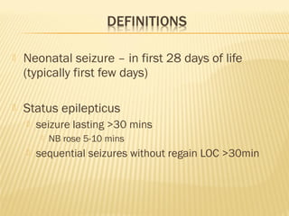
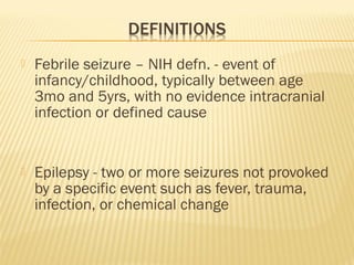
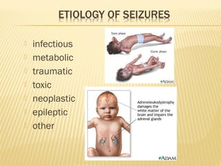
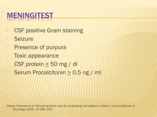
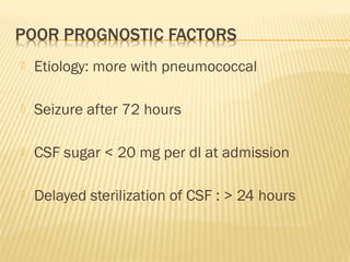
- Neonatal seizures, febrile seizures, CNS infections, and strokes are common neurological conditions seen in pediatrics.
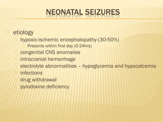
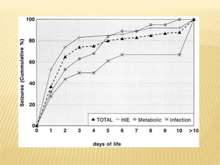
- Common etiologies of neonatal seizures include hypoxic-ischemic encephalopathy, congenital CNS anomalies, intracranial hemorrhage, and electrolyte or metabolic abnormalities.
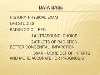
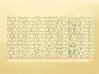
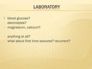
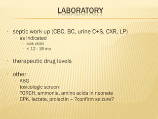
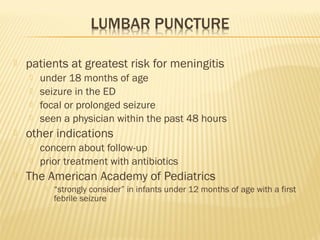
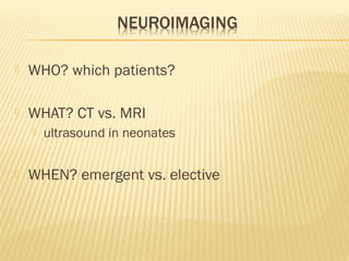
- Evaluation involves history, physical exam, lab studies, and neuroimaging like EEG, ultrasound, CT, or MRI depending on the situation.
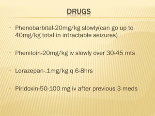
- Treatment involves anticonvulsants tailored to the specific condition, with phenobarbital, phenytoin, and lorazepam as first line options for status epilepticus.
This document discusses various conditions that can mimic epilepsy in children. It notes that epilepsy is sometimes underdiagnosed or overdiagnosed due to unusual symptom presentations or epilepsy mimics. Several common epilepsy mimics are described in detail for different age groups, including breath holding spells in infants, tics and parasomnias in children, and syncope in adolescents. Tables compare features of epilepsy mimics to epileptic seizures during sleep and wakefulness. In conclusion, the document emphasizes taking an age- and state-based approach to differentiating epilepsy from conditions it may imitate.
1. A seizure is caused by abnormal excessive neuronal activity in the brain and can be focal, generalized, or febrile.
2. Evaluation of a first seizure involves obtaining a detailed history, physical exam, EEG, and brain imaging to determine the cause and classify the seizure type.
3. Generalized seizures include absence seizures, myoclonic seizures, and generalized tonic-clonic seizures. Focal seizures can become secondary generalized seizures.
This document provides information on various pediatric neurology emergencies, including recommendations for febrile seizures, status epilepticus, meningitis, encephalitis, and herpes simplex virus encephalitis. For febrile seizures, the AAP guidelines recommend against continuous or intermittent anticonvulsant therapy for children with simple febrile seizures due to risks outweighing benefits. Status epilepticus treatments include benzodiazepines, phenytoin, phenobarbital, and for refractory cases, midazolam or propofol infusion. Meningitis diagnosis and management involves prompt antibiotics, steroids for certain cases, and meningococcemia requires antibiotics within 20 minutes. En
This document discusses approaches to pediatric neurologic emergencies including seizures, headaches, and status epilepticus. It provides definitions, classifications, differential diagnoses, evaluation steps, treatment guidelines, and disposition criteria for various pediatric neurologic presentations. Key points include recommendations for workup of new onset seizures, guidelines for status epilepticus treatment, considerations for neuroimaging and lumbar puncture, and acute treatment options for migraine headaches in children.
Neonatal sepsis is a clinical syndrome characterized by signs and symptoms of infection in the first month of life. It can involve sepsis, meningitis, pneumonia and other infections. The document discusses the definition, classification, risk factors, clinical features, investigations and management of neonatal sepsis. Key points include that sepsis is a leading cause of neonatal mortality, the importance of sepsis screening and blood cultures, and that initial empirical antibiotic therapy typically involves ampicillin and an aminoglycoside.
1. The document discusses different types of seizures including focal onset seizures, generalized onset seizures, and seizures of unknown onset. It provides definitions and examples of motor and non-motor seizures.
2. Etiologies of seizures in the neonatal period and beyond are outlined, including infections, metabolic disorders, brain malformations, drugs/poisons, and epilepsy syndromes.
3. Febrile seizures are defined as seizures associated with fever in children between 6-60 months old. Simple febrile seizures are brief and do not recur within 24 hours, while complex febrile seizures are prolonged or recurrent.
This document discusses pediatric stroke. It begins with definitions, types, epidemiology, etiology, and pathophysiology of pediatric stroke. The main types are ischemic and hemorrhagic stroke. Risk factors in children include structural heart disease, vasculopathies, hematological disorders, and prothrombotic states. Clinical features can include focal neurological deficits like hemiparesis. Diagnosis involves neuroimaging such as MRI and distinguishing stroke from other conditions. Management aims to prevent recurrence and support rehabilitation.
The document discusses various epileptic syndromes categorized by age of onset - neonatal, infancy, childhood, adolescence-adult. For each syndrome, it provides information on defining features, age of onset, seizure types, EEG patterns, treatment and prognosis. The syndromes discussed include benign familial neonatal epilepsy, Ohtahara syndrome, West syndrome, Panayiotopoulos syndrome, Lennox-Gastaut syndrome, juvenile myoclonic epilepsy and others.
This document discusses supraventricular tachycardia (SVT) in pediatric patients. SVT is the most common abnormal heart rhythm seen in children and the most common arrhythmia requiring treatment. It is usually caused by re-entry mechanisms involving an accessory pathway or the atrioventricular node. Diagnosis involves obtaining an electrocardiogram during episodes to identify P wave patterns. Treatment options include vagal maneuvers, medications like adenosine, calcium channel blockers, or beta blockers, and cardioversion. Radiofrequency ablation can provide a cure for refractory or recurrent cases. Proper diagnosis of the underlying SVT mechanism guides selection of the most appropriate treatment approach.
This document discusses bronchopulmonary dysplasia (BPD), a chronic lung disease that occurs in premature infants requiring respiratory support. It covers the definition, risk factors, pathogenesis, clinical features, prevention, and treatment of BPD. The definition has evolved over time from relying solely on oxygen need at 28 days to incorporating factors like oxygen need, pressure support, and gestational age. BPD results from lung injury and disrupted lung development due to prematurity and respiratory support. Management aims to protect the lung from injury through gentle ventilation, optimal oxygen levels, and other strategies.
1) Hypoxic-ischemic encephalopathy (HIE) is brain injury caused by lack of oxygen and blood flow before, during, or after birth. It remains a serious condition that can cause death or long-term disabilities like cerebral palsy or intellectual impairment.
2) The document discusses the definition, risk factors, pathophysiology, clinical features based on the Sarnat staging system, diagnosis using imaging and EEG, and treatment approaches for HIE including supportive care, perfusion management, anti-seizure medications, and therapeutic hypothermia.
3) The goal of treatment is to prevent further brain injury by maintaining appropriate oxygenation, blood pressure, glucose levels, and treating seizures
This document discusses seizures in children, including febrile seizures. It defines seizures and different types, like generalized seizures and focal seizures. It covers the epidemiology, causes, clinical presentation and diagnosis of seizures. Complications, both acute and chronic, are outlined. Investigations and management approaches are also summarized. The document focuses in particular on febrile seizures, their definition, causes, types, evaluation and treatment in children presenting with fever and seizures.
ATAXIA IN CHILDREN -CAUSES, MANAGEMENT, INVESTIGATIONS, TYPES, COMMONEST ATAXIA IN CHILDREN IN DETAIL, HOW WILL YOU FIND OUT THE CAUSE FOR ATAXIA IN CHILDREN FLOWCHART, DEFINITION, TREATMENT
Pediatric traumatic brain injury presentation Robert Parker
This document provides an overview of pediatric traumatic brain injury (TBI) management. It describes the case of a 13-year-old male involved in a motor vehicle collision. Key goals in management include maintaining cerebral perfusion pressure, decreasing intracranial pressure, and preventing secondary injury. Early management focuses on airway protection, oxygenation, imaging, and treating hypotension and elevated ICP. Refractory elevated ICP may be treated with osmotherapy, barbiturates, or decompressive craniectomy. Ongoing intensive care involves careful monitoring and treatment of ICP, CPP, seizures, and other complications. Outcomes depend on age, injury severity, and development of secondary insults like hypot
This presentation focuses on Acute Bacterial Meningitis.
Viral and fungal cause is mentioned but focus is on bacterial meningitis in Pediatrics Patient.
Feel free to correct if there is any error.
Refer to other reference books for clarity.
The document discusses headaches in children and adolescents. It describes the different types of headaches including acute, recurrent, chronic, and secondary headaches. It outlines the evaluation process for headaches including obtaining a thorough history, physical exam, and determining if imaging is needed. Lifestyle factors and various treatment options are also covered such as acute medication, preventative treatment, and lifestyle modifications.
This document provides information on evaluating hypotonia in infants. It defines muscle tone and the differences between hypotonia and weakness. Hypotonia can have central or peripheral causes. The differential diagnosis for a floppy infant is extensive and includes central conditions like genetic syndromes or brain insults and peripheral conditions involving the motor unit, nerve, neuromuscular junction or muscle. A thorough evaluation includes the infant's history, development, family history, examination and potentially genetic or metabolic testing to determine the underlying cause.
1. Febrile seizures are seizures that occur in children between 6 months and 5 years of age associated with a fever over 38°C but without an underlying infection or metabolic imbalance. They are classified as simple or complex based on duration and recurrence.
2. The underlying pathophysiology of febrile seizures is unknown but likely involves a genetic predisposition. Risk factors include a family history of febrile seizures and developmental delays.
3. Management of febrile seizures focuses on treating the underlying cause of fever. For recurrent or prolonged seizures, antiepileptic drugs may be considered.
pediatrics.Seizures and epilepsy.(dr.adnan)student
This document defines and classifies different types of seizures and epilepsy. It discusses various etiologies and syndromes of epilepsy, including idiopathic, symptomatic, partial and generalized seizures. It also covers the diagnosis, treatment and prognosis of conditions like complex partial seizures, absence seizures, tonic-clonic seizures, infantile spasms, febrile seizures, and status epilepticus.
Neonatal sepsis is a clinical syndrome of bacteremia and infection in infants under 4 weeks of age. Common causes are E. coli, Group B Streptococcus, and Listeria. It can be early-onset from transmission during birth or late-onset from hospital-acquired infections. Symptoms are non-specific but include respiratory distress, feeding issues, and temperature instability. Diagnosis involves blood, urine and CSF cultures. Treatment is antibiotics like ampicillin and gentamicin for 10-14 days along with supportive care. Prevention includes good antenatal care, treating maternal infections, early breastfeeding and infection control policies in the NICU.
This document discusses the approach to evaluating and diagnosing a hypotonic infant. It begins by defining hypotonia and noting that determining the cause can be challenging. A detailed history and physical exam are important to localize the cause as central or peripheral. Differential diagnosis involves considering central nervous system, genetic, infectious, metabolic, and muscular causes. Basic lab tests include screening for infection and metabolic/genetic disorders. Imaging, EMG/NCV, muscle biopsy and genetic testing can further evaluate potential peripheral and muscular etiologies. The case presented is of a newborn with hypotonia, absent reflexes, and no family history or dysmorphic features to suggest a cause.
The document discusses hypotonia in infants. It begins by defining tone and describing central and peripheral causes of hypotonia. Central causes account for 60-80% of cases and involve the brain or spinal cord, while peripheral causes involve the motor unit, nerves, neuromuscular junction, or muscles. The document then examines the evaluation and differential diagnosis of hypotonia in infants, highlighting important history, physical exam findings, and potential etiologies to consider. Key tests include the traction response and assessment of tone, strength, and reflexes. Thorough evaluation is needed to identify the underlying cause and guide management.
HIE-Pathophysiology & recent advances in managementViraj Satenahalli
This document discusses hypoxic ischemic encephalopathy (HIE), including its pathophysiology and recent advances in management. It provides details on the brain physiology of newborns, terminology, definition, etiology, pathogenesis through 4 phases, management including hypothermia, and neuroprotective strategies such as maintaining energy stores and growth factors. Hypothermia is highlighted as the most viable neuroprotective strategy, with studies showing it reduces mortality and neurodevelopmental disability in infants with HIE.
1) The document summarizes a seminar on seizure disorders in newborns presented by Dr. Md. Moklesur Rahman and Dr. Sarbari Saha.
2) It discusses the definition, incidence, pathophysiology, etiology, classification, diagnostic approach, management and prognosis of neonatal seizures.
3) Common causes of neonatal seizures presented include hypoxic ischemic encephalopathy, intracranial hemorrhage, infection, hypoglycemia, hypocalcemia and inborn errors of metabolism.
This document discusses neonatal hypocalcemia, including its types, causes, and management. There are two types - early onset within 72 hours requiring calcium supplementation, and late onset after 7 days requiring longer treatment. Hypocalcemia is defined as low serum calcium levels. Causes of early onset include prematurity, maternal conditions, and iatrogenic factors. Management of early onset involves calcium supplementation. Late onset in the first week is usually symptomatic and caused by high phosphate intake. Causes also include hypomagnesemia, vitamin D deficiency, and genetic or metabolic syndromes.
This document provides guidance on evaluating patients presenting with paraplegia. It outlines the key components of the clinical history and neurological examination needed to determine the cause and level of spinal cord injury. The history should ascertain details of onset and any associated symptoms. The exam focuses on assessing sensory and motor function at different dermatomal and myotomal levels to localize the lesion. Together this information can indicate if the injury is acute, subacute, or chronic, and identify potential etiologies like trauma, infection, inflammation, compression, or vascular causes. The goal is to arrive at a diagnosis and localization of injury within the spinal cord or vertebrae.
This document summarizes several pediatric neurologic disorders including hydrocephalus, neural tube defects, cerebral palsy, spinal cord injury, and infections of the central nervous system. It describes the causes, signs and symptoms, diagnostic tests, nursing diagnoses, and treatment approaches for each condition. Nursing priorities for patients include maximizing respiratory function, preventing further injury, promoting mobility, preventing complications, and supporting psychological adjustment.
The document discusses several pediatric neurological emergencies including seizures, meningitis, stroke, traumatic brain injuries, and increased intracranial pressure. It provides details on assessing and initially stabilizing patients, distinguishing different seizure types, approaches to febrile and afebrile seizures, signs of meningitis, evaluating increased intracranial pressure, and causes and features of disorders like hypotonia. Investigations and differential diagnoses for various conditions are also outlined.
This child presents with drooping eyelids and squint that worsens in the evenings.
- Myasthenia gravis.
- Ask about weakness of other muscles, response to rest.
- Tensilon test and acetylcholine receptor antibody levels would help confirm the diagnosis.
Pediatric cardiovascular problems in emergency setting 1 (5 feb- 2011)taem
1) A 7-year-old boy presents with dyspnea and tachypnea for 1 day after an upper respiratory infection, and is found to have tachycardia, dyspnea, and signs of congestive heart failure.
2) The document discusses cardiogenic shock, including the physiology, signs and symptoms, and management with inotropes, vasodilators, and supportive care.
3) Various case presentations are provided demonstrating different emergency cardiovascular problems in pediatrics, such as hypoxic spells, tachyarrhythmias, ventricular tachycardia, and pulmonary hypertensive crisis. Management strategies are outlined for each condition.
This document is a collection of photo credits from various photographers and organizations. There are over 20 different photo credits listed from sources like Pink Sherbet Photography, VinothChandar, digitalbob8, and others. The document ends by encouraging the reader to create their own presentation using photos on SlideShare.
This document discusses several epileptic encephalopathies:
- Early myoclonic encephalopathy presents in neonates and is characterized by fragmentary myoclonus, erratic seizures, and a suppression-burst EEG pattern. Prognosis is poor with over 50% mortality.
- Ohtahara syndrome presents from 10 days to 3 months of age with tonic spasms and a suppression-burst EEG pattern in both waking and sleep states. It can progress to West syndrome and Lennox-Gastaut syndrome.
- Lennox-Gastaut syndrome typically starts between ages 1-7 and is characterized by multiple seizure types, cognitive issues, and EEG showing slow spike-wave dis
Pediatric stroke can be caused by a variety of factors such as cardiac diseases, infections like varicella, sickle cell disease, moyamoy disease, cerebral sinus thrombosis, and genetic conditions like MELAS. The presentation of pediatric stroke depends on the location and size of the lesion in the brain. Diagnosis involves imaging techniques like CT, MRI, MRA and angiography. Early diagnosis and treatment is important to prevent long term neurological deficits in children.
Seizures in children, dr.amit vatkar, pediatric neurologistDr Amit Vatkar
This document provides information about pediatric epilepsy from Dr. Amit Vatkar, a pediatric neurologist. It discusses the types of epilepsy according to Ayurveda, how epilepsy presents differently in children than adults, diagnostic testing and treatment options. Key points include that 70% of epilepsy starts in childhood, initial seizures are often not treated but recurrence risk is reduced with treatment, and around 67% of patients achieve remission over time, with 86% doing so without medication.
Epilepsy recent classification and definitions, dr. amit vatkar, pedaitric ne...Dr Amit Vatkar
This document contains information about Dr. Amit Vatkar's credentials and contact information, as well as summaries of definitions, classifications, and types of seizures and epilepsy. It defines seizures, epilepsy, acute symptomatic seizures, epilepsy syndromes, drug-resistant epilepsy, and describes different types of seizures including myoclonic, tonic, epileptic spasms, clonic, and tonic-clonic seizures. It also discusses recent definitions and classifications of epilepsy from international organizations.
Neuro examination, pediatric neurologist, dr. amit vatkarDr Amit Vatkar
This document contains information about Dr. Amit Vatkar's credentials and specialization in pediatric neurology. It then provides an overview of topics related to clinical neurology examinations, including the cranial nerves, motor and sensory systems, cerebellar function, gait, and signs of meningism. The document outlines examination techniques and disorders for each topic area. It concludes by thanking the reader and providing Dr. Vatkar's contact information.
Children with epilepsy are at risk for cognitive difficulties like lower IQ, learning problems, and academic underachievement due to factors like underlying brain abnormalities, seizures, and medication side effects. While half of children with epilepsy develop normally, certain epilepsy syndromes carry a higher risk of intellectual disability or unfavorable cognitive outcomes. Autism also commonly co-occurs with epilepsy, especially among those with intellectual disabilities, suggesting shared neurological mechanisms. Anti-seizure medications can cause mild to moderate cognitive impairments involving slowed processing, reduced working memory, and decreased mental flexibility, with effects varying between medications.
pediatirc neuroimaging , primer for pediatricians interested in neuroimaging and basic stuff for radiologists.
included examples of normal and abnormal.
when to do what imaging
Assessment And Managment Of Critically Ill Child 1Dang Thanh Tuan
This document discusses the paramedic's role in pediatric emergency care. It describes assessing and managing critically ill children using the Pediatric Assessment Triangle to evaluate appearance, work of breathing, and circulation. Case studies demonstrate applying this technique to identify respiratory distress, failure, shock, or brain dysfunction. The document also outlines general pediatric patient management including airway control, fluids, electrical therapy and transport considerations.
This document discusses the approach to tall stature in children. It defines tall stature and excessive growth. It covers growth charts, predictors of height like genetics and hormones. Common causes of tall stature include familial tallness, endocrine disorders, and genetic syndromes like Marfan's and Klinefelter's. Physical exam focuses on measurements, signs of specific disorders, and Tanner staging. Investigations are aimed at identifying a cause. Reassurance is the key for normal tallness, while treatment may be considered for extreme predicted heights or psychosocial impairment.
This document discusses several topics related to high-risk neonates and their neurodevelopmental outcomes. It begins by defining high-risk neonates as babies exposed to conditions that endanger their survival. Some factors that can contribute to high-risk status include high-risk pregnancies, medical illnesses in the mother, complications during labor, and neonatal medical conditions. The document then discusses several conditions in more detail, including hypothermia, hyperthermia, hypoglycemia, infants of diabetic mothers, and neonatal sepsis. It provides definitions, risk factors, clinical presentations, and management strategies for each of these conditions.
Service providers who receive high nutrition risk referrals, particularly Registered Dietitians, need to be knowledgeable about general and clinical pediatric nutrition as well as counselling skills for working with families and children.
This is the second of five self-directed training modules available in PowerPoint presentations that have been developed and evaluated to respond to this need
- The document provides guidance on performing a respiratory exam, including positioning the patient, inspecting the patient and areas around the bed, examining the hands, face, neck, chest, and auscultating breath sounds.
- Key steps are inspecting for signs of distress, noting peripheral clues, assessing expansion and movements of the chest, percussion findings, breath sounds, and timing of crackles.
- Performing techniques like assessing forced expiratory time can help evaluate for obstructive lung diseases like COPD.
This document discusses seizures in pediatrics, including neonatal seizures, febrile seizures, status epilepticus, and epileptic syndromes. It provides information on etiology, clinical presentation, diagnostic evaluation with EEG, imaging and labs, and treatment with antiepileptic drugs. Causes of seizures in newborns include hypoxic-ischemic encephalopathy, infections, hemorrhage, and metabolic derangements. Febrile seizures typically occur between 3 months and 5 years of age and are usually self-limited. Status epilepticus is defined as continuous seizure activity or recurrent seizures without regaining consciousness lasting over 30 minutes.
Bacterial meningitis is an acute infection of the membranes (meninges) surrounding the brain and spinal cord. It is usually caused by bacteria like Streptococcus pneumoniae, Neisseria meningitidis, and Listeria monocytogenes. Clinical features include fever, headache, and neck stiffness. Diagnosis involves examination of cerebrospinal fluid which shows increased white blood cells and decreased glucose levels. Treatment involves intravenous antibiotics like ceftriaxone or cefotaxime for 10-14 days. Prognosis depends on factors like age, underlying health conditions, and time to treatment initiation. Complications can include neurological deficits, hearing loss, seizures and death in up to 20% of cases of pneumococ
This document provides information on bacterial meningitis in children, including common causes, symptoms, diagnosis, treatment, and prognosis. The main causes of bacterial meningitis in children vary by age, with organisms like S. pneumoniae, N. meningitidis, and Hib being common. Symptoms can include fever, headache, vomiting, and signs of meningeal irritation. Diagnosis involves lumbar puncture and CSF analysis. Treatment involves prompt administration of antibiotics and supportive care. Outcomes depend on causative organism and presence of complications, with potential for hearing loss, neurological deficits, or death.
Bacterial meningitis remains a serious disease in adults, with S. pneumoniae and N. meningitidis causing the majority of cases. The initial approach involves assessing for the classic triad of symptoms, but lumbar puncture may be deferred if neuroimaging is warranted due to concerns for mass lesions or decreased consciousness. Empiric antibiotic therapy should begin as soon as possible, often in combination with vancomycin and dexamethasone, which reduces mortality and morbidity when given early in treatment. Outcomes depend on severity of symptoms and causative organism, with pneumococcal meningitis carrying the highest fatality and long-term complication rates.
A 4 year old boy presented to the emergency room after having a seizure. Possible diagnoses include viral infections like herpes simplex encephalitis, which can cause seizures and disturbances in consciousness. Other potential causes are bacterial or parasitic infections like tuberculosis or toxoplasmosis. It is important to obtain a thorough history, including any recent illnesses, and perform diagnostic tests like a lumbar puncture and CSF analysis to determine the cause and guide treatment.
This document discusses acute bacterial meningitis (BM), including its definition, incidence, risk factors, etiology, clinical presentation, diagnosis, treatment goals and principles, specific treatment recommendations for common causative organisms, prevention through vaccination, and outpatient management considerations. The prognosis of untreated BM is poor, with high treatment failure rates and risk of neurological complications; however, prompt administration of appropriate antibiotic therapy can significantly improve outcomes.
This document discusses non-traumatic coma in children. It begins by providing incidence rates for non-traumatic coma (30 per 100,000 children per year) and traumatic brain injury (670 per 100,000). The rest of the document discusses the definition, causes, evaluation, management, and outcomes of non-traumatic coma in children. Infection is the most common cause. Mortality depends on the underlying etiology and can range from 3-84%. Overall mortality in studies is around 46%.
This document provides an overview of meningitis beyond the neonatal period. It discusses the epidemiology, etiology, pathogenesis, clinical manifestations, diagnosis, treatment, complications and prognosis of meningitis. The most common causative organisms include Streptococcus pneumoniae, Neisseria meningitidis, and Haemophilus influenzae. Clinical features may include fever, headache, vomiting, and signs of meningeal irritation. Diagnosis involves lumbar puncture and culture of CSF. Empiric antibiotic treatment is initiated while awaiting culture results. Complications can be early like seizures or late like hearing loss. Prognosis depends on causative organism, age of presentation, and presence of co-morbidities.
Fever with Fits 22.1.2016 (to print), update.pptxKyawMyoHtet10
This document discusses various causes of fever with fits in children including febrile convulsions, central nervous system infections like meningitis, encephalitis, brain abscess, and cerebral malaria. It provides details on the clinical presentation, investigations, and management of these conditions. Common causes of meningitis in children are discussed along with signs of meningism on examination. Diagnosis of meningitis involves lumbar puncture and analysis of cerebrospinal fluid. Tuberculous meningitis has distinct cerebrospinal fluid findings and requires prolonged antibiotic treatment. Viral encephalitis is usually self-limiting and treatment focuses on controlling symptoms. Brain abscesses require imaging studies for diagnosis and may necessitate
Fever is a common reason for pediatric visits. The hypothalamus regulates body temperature and fever occurs when its set point is elevated. Fever without a source is difficult to diagnose and can be caused by infections, inflammatory disorders, or malignancies. Evaluation involves history, exam, labs including blood cultures, and imaging if indicated. Lower risk children based on criteria like Rochester may be managed as outpatients without antibiotics. Higher risk children receive empiric antibiotics targeting common pathogens until diagnosis is made. Antipyretics and antivirals are also used for symptom relief in some cases.
The document provides an overview of acute bacterial meningitis, including:
- Common causes are Streptococcus pneumoniae, Neisseria meningitidis, and group B Streptococcus.
- Clinical presentation often includes the classic triad of fever, neck stiffness, and altered mental status. Investigations include lumbar puncture and analysis of CSF.
- Differentiating between bacterial and viral meningitis can be done using factors like CSF white blood cell count, glucose and protein levels, and peripheral blood markers.
- Treatment involves early administration of empirical antibiotics like ceftriaxone or penicillin, with duration typically 5 days if the patient responds well. Prognosis depends on factors like
This document provides an overview of bacterial meningitis beyond the neonatal period. It defines meningitis as inflammation of the leptomeninges surrounding the brain and spinal cord. The most common causes of meningitis in this age group are Neisseria meningitidis, Streptococcus pneumoniae, and Haemophilus influenzae type b. Risk factors include young age, recent contact with an infected individual, crowding, and conditions causing impaired immunity. Clinical manifestations may include fever, headache, vomiting, altered mental status, and neck stiffness. Diagnosis involves lumbar puncture and analysis of CSF for pleocytosis, elevated proteins, and low glucose. Management includes supportive care, antibiotics, and corticosteroids
This document discusses the objectives, clinical presentation, diagnosis, and management of central nervous system infections, seizure disorders, and different types of seizures. It covers topics such as viral and bacterial meningitis, brain abscesses, classification of seizures, workup and treatment of epilepsy, and surgical options for refractory seizures. Diagnostic testing includes lumbar puncture, imaging like CT and MRI, and EEG. Management involves antibiotics, anticonvulsants, and sometimes surgery. Complications can include neurological deficits, but prognosis is generally good with appropriate treatment.
- The document summarizes a presentation on bacterial meningitis given by Dr. Marissa B. Lukban. It discusses the epidemiology of bacterial meningitis worldwide and locally in the Philippine General Hospital based on a 5-year survey from 2002-2006. It also reviews clinical practice guidelines for performing lumbar punctures in children presenting with febrile seizures and highlights various diagnostic tests and treatments for bacterial meningitis. Local studies on prevalence and risk factors associated with bacterial meningitis in children with febrile seizures are mentioned. Antibiotic recommendations for different clinical subgroups and causative organisms are provided based on susceptibility patterns. The role of pediatricians in advocating for prevention of meningitis through vaccination initiatives and
This document summarizes a presentation on bacterial meningitis. It discusses the clinical presentation, mortality and risks, diagnostic workup including CSF results, treatment including antibiotics and vaccination, and scripts for bacterial versus viral meningitis. Key points include that untreated bacterial meningitis has nearly 100% mortality, but with treatment mortality is around 25%. Presentation often includes fever, nuchal rigidity, and altered mental status. Diagnostic tests include lumbar puncture and analysis of CSF findings. Treatment involves antibiotics and dexamethasone to improve outcomes.
This document discusses bacterial meningitis in children over 1 month old. It covers the incidence, common causative organisms by age group, clinical features, evaluation, diagnosis and treatment. Some key points:
- The incidence is highest in children under 2 months (80.69 per 100,000) and declines with age.
- Common causative organisms vary by age but include Group B Strep, pneumococcus, meningococcus and gram-negatives.
- Clinical features depend on age but may include fever, irritability, vomiting, headache and nuchal rigidity.
- Evaluation involves blood and CSF tests including cell count, glucose, protein and cultures. Imaging is only needed if signs of
Abir, an 8-year-old boy, was admitted to the hospital with difficulty swallowing, talking, and weakness in his upper and lower limbs. He had a fever 20 days prior. Acute flaccid paralysis (AFP) is characterized by rapid onset weakness that can include respiratory and bulbar weakness. Differential diagnoses for AFP include Guillain-Barré syndrome, transverse myelitis, poliomyelitis, traumatic neuritis, and hypokalemic paralysis. Proper management of AFP requires assessing respiratory function, bulbar weakness, cardiovascular stability, and ruling out electrolyte imbalances or spinal cord compression.
This document provides an overview of neonatal immunology and the epidemiology, clinical presentation, diagnosis, and treatment of common neonatal infections. It discusses how the neonatal immune system is immature and ineffective, putting neonates at high risk for infection. The most common bacterial infections are sepsis, meningitis, and pneumonia, often caused by Group B Streptococcus, E. coli, and other organisms. Signs of early onset sepsis can be nonspecific but include respiratory distress, hypothermia/fever, and poor feeding. Diagnosis involves blood, urine and CSF cultures along with blood tests. Empiric antibiotics such as ampicillin and gentamicin are typically started. Late onset infections from healthcare-associated sources are also
This document discusses neonatal sepsis, including definitions, risk factors, evaluation, treatment and prevention. It provides guidelines for:
- Empiric antibiotic treatment of common organisms like GBS and E. coli based on susceptibility patterns. For GBS, penicillin is recommended. For ampicillin-sensitive E. coli, ampicillin or cefotaxime can be used.
- Duration of treatment based on culture results and clinical response. Treatment is typically 10-14 days for positive cultures and 48 hours for negative cultures if the infant is improving.
- Changing or extending treatment if meningitis is suspected or the infant is not improving on current regimens. Serial monitoring of markers like CRP is advised to
This document discusses recent advances in neonatal septicemia. It begins by defining neonatal septicemia and describing the types, including early onset sepsis within 72 hours of birth and late onset sepsis from 3-90 days of life. It then discusses the epidemiology, risk factors, clinical presentation and diagnostic evaluation. Key diagnostic tests discussed include blood cultures, sepsis screens, lumbar puncture and novel biomarkers. Management of neonatal septicemia includes supportive care and administration of empiric and targeted antibiotic therapy guided by culture results.
June 3, 2024 Anti-Semitism Letter Sent to MIT President Kornbluth and MIT Cor...Levi Shapiro
Letter from the Congress of the United States regarding Anti-Semitism sent June 3rd to MIT President Sally Kornbluth, MIT Corp Chair, Mark Gorenberg
Dear Dr. Kornbluth and Mr. Gorenberg,
The US House of Representatives is deeply concerned by ongoing and pervasive acts of antisemitic
harassment and intimidation at the Massachusetts Institute of Technology (MIT). Failing to act decisively to ensure a safe learning environment for all students would be a grave dereliction of your responsibilities as President of MIT and Chair of the MIT Corporation.
This Congress will not stand idly by and allow an environment hostile to Jewish students to persist. The House believes that your institution is in violation of Title VI of the Civil Rights Act, and the inability or
unwillingness to rectify this violation through action requires accountability.
Postsecondary education is a unique opportunity for students to learn and have their ideas and beliefs challenged. However, universities receiving hundreds of millions of federal funds annually have denied
students that opportunity and have been hijacked to become venues for the promotion of terrorism, antisemitic harassment and intimidation, unlawful encampments, and in some cases, assaults and riots.
The House of Representatives will not countenance the use of federal funds to indoctrinate students into hateful, antisemitic, anti-American supporters of terrorism. Investigations into campus antisemitism by the Committee on Education and the Workforce and the Committee on Ways and Means have been expanded into a Congress-wide probe across all relevant jurisdictions to address this national crisis. The undersigned Committees will conduct oversight into the use of federal funds at MIT and its learning environment under authorities granted to each Committee.
• The Committee on Education and the Workforce has been investigating your institution since December 7, 2023. The Committee has broad jurisdiction over postsecondary education, including its compliance with Title VI of the Civil Rights Act, campus safety concerns over disruptions to the learning environment, and the awarding of federal student aid under the Higher Education Act.
• The Committee on Oversight and Accountability is investigating the sources of funding and other support flowing to groups espousing pro-Hamas propaganda and engaged in antisemitic harassment and intimidation of students. The Committee on Oversight and Accountability is the principal oversight committee of the US House of Representatives and has broad authority to investigate “any matter” at “any time” under House Rule X.
• The Committee on Ways and Means has been investigating several universities since November 15, 2023, when the Committee held a hearing entitled From Ivory Towers to Dark Corners: Investigating the Nexus Between Antisemitism, Tax-Exempt Universities, and Terror Financing. The Committee followed the hearing with letters to those institutions on January 10, 202
How to Add Chatter in the odoo 17 ERP ModuleCeline George
In Odoo, the chatter is like a chat tool that helps you work together on records. You can leave notes and track things, making it easier to talk with your team and partners. Inside chatter, all communication history, activity, and changes will be displayed.
How to Manage Your Lost Opportunities in Odoo 17 CRMCeline George
Odoo 17 CRM allows us to track why we lose sales opportunities with "Lost Reasons." This helps analyze our sales process and identify areas for improvement. Here's how to configure lost reasons in Odoo 17 CRM
How to Build a Module in Odoo 17 Using the Scaffold MethodCeline George
Odoo provides an option for creating a module by using a single line command. By using this command the user can make a whole structure of a module. It is very easy for a beginner to make a module. There is no need to make each file manually. This slide will show how to create a module using the scaffold method.
How to Fix the Import Error in the Odoo 17Celine George
An import error occurs when a program fails to import a module or library, disrupting its execution. In languages like Python, this issue arises when the specified module cannot be found or accessed, hindering the program's functionality. Resolving import errors is crucial for maintaining smooth software operation and uninterrupted development processes.
Main Java[All of the Base Concepts}.docxadhitya5119
This is part 1 of my Java Learning Journey. This Contains Custom methods, classes, constructors, packages, multithreading , try- catch block, finally block and more.
A workshop hosted by the South African Journal of Science aimed at postgraduate students and early career researchers with little or no experience in writing and publishing journal articles.
Macroeconomics- Movie Location
This will be used as part of your Personal Professional Portfolio once graded.
Objective:
Prepare a presentation or a paper using research, basic comparative analysis, data organization and application of economic information. You will make an informed assessment of an economic climate outside of the United States to accomplish an entertainment industry objective.
A review of the growth of the Israel Genealogy Research Association Database Collection for the last 12 months. Our collection is now passed the 3 million mark and still growing. See which archives have contributed the most. See the different types of records we have, and which years have had records added. You can also see what we have for the future.
This slide is special for master students (MIBS & MIFB) in UUM. Also useful for readers who are interested in the topic of contemporary Islamic banking.
3.
brief and subtle
eye blinking
mouth/tongue movements
“bicycling” motion to limbs
typically sz’s can’t be provoked/consoled
autonomic changes
EEG alone less predictable-unless together
with mri.(murray,boylan,r yan-pediatrics-2009 )
7.
Drug withdrawal
CNS trauma – maternal drug toxicity
4 EPILEPTIC SYNDROMES:
1)Benign familial conv.2)fifth day fits
3)Myoclonic encephalopathy
4)epileptic encephalopathy(Ontahara
syn) 5) DE VIVO SYNDROME
8.
HISTORY- PHYSICAL EXAM
LAB STUDIES
RADIOLOGIC – EEG
1)ULTRASOUND- CHOICE
2)CT-LOTS OF RADIATION
BETTER,CONGENITAL, INFARCTION
3)MRI- MORE DEF OF INFARTS
AND MORE ACCURATE FOR PROGNOSIS
9. •
Phenobarbital-20mg/kg slowly(can go up to
40mg/kg total in intractable seizures)
•
Phenitoin-20mg/kg iv slowly over 30-45 mts
•
Lorazepan-.1mg/kg q 6-8hrs
•
Piridoxin-50-100 mg iv after previous 3 meds
11.
Neonatal seizure – in first 28 days of life
(typically first few days)
Status epilepticus
seizure lasting >30 mins
NB rose 5-10 mins
sequential seizures without regain LOC >30min
12.
Febrile seizure – NIH defn. - event of
infancy/childhood, typically between age
3mo and 5yrs, with no evidence intracranial
infection or defined cause
Epilepsy - two or more seizures not provoked
by a specific event such as fever, trauma,
infection, or chemical change
13.
generalized
partial – focal onset
LOC
tonic, clonic, tonic-clonic, myoclonic, atonic, absence
simple partial – no LOC
complex partial – LOC
partial secondarily generalized
unclassified
17.
Epidemiology
age 3mo – 5yrs
peak age 9-20 mo
2-5% children will have before age 5
25-40% will have family history
80 – 97% simple
3 - 20% complex
18.
< 15 mins
no focal features
no greater than 1 episode in 24h
neurologically and developmentally normal
20.
Recurrence
risk recurrence 25-50%
risk recurrence after 2nd – 50%
most recurrences within 6-12 mo
(20% within same febrile illness)
Risk of epilepsy
2-3% (baseline 1%)
increased in
family history of epilepsy
abnormal developmental status
complex febrile seizure
21.
definition
seizure lasting >30 mins
NB Rosen 5-10 mins
sequential seizures without regain LOC >30min
mortality in pediatric status epilepticus 4%
morbidity may be as high as 30%
29. •
septic work-up (CBC, BC, urine C+S, CXR, LP)
–
as indicated
•
•
sick child
< 12 - 18 mo
•
therapeutic drug levels
•
other
–
–
–
–
ABG
toxicologic screen
TORCH, ammonia, amino acids in neonate
CPK, lactate, prolactin – ?confirm seizure?
30.
patients at greatest risk for meningitis
other indications
under 18 months of age
seizure in the ED
focal or prolonged seizure
seen a physician within the past 48 hours
concern about follow-up
prior treatment with antibiotics
The American Academy of Pediatrics
“strongly consider” in infants under 12 months of age with a first
febrile seizure
32.
predictors of abnormal findings of computed tomography of the head
in pediatric patients presenting with seizures
Warden CR - Ann Emerg Med - 01-Apr-1997; 29(4): 518-23
retrospective case series
predicts CT scan results normal if
no underlying high-risk condition
malignancy, NCT, recent CHI, or recent CSF shunt revision
older than 6 months
sustained a seizure of 15 minutes or less
no new-onset focal neurologic deficit
not prospectively validated
33. •
•
•
correct underlying pathology, if any
antipyretics ineffective in febrile seizure
anti-epileptic choice often trial and error
•
•
no anti-epileptic 100% effective
febrile seizure – diazepam, phenobarbital, valproic acid
–
•
•
•
•
•
Currently AAP does not recommend
neonatal - phenobarbital
generalized TC – phenytoin, phenobarbital, carbamazepine, valproic
acid, primidone
absence – ethosuximide, valproic acid
new anti-epileptics – felbamate, gabapentin, lamotrigine,
topiramate, tiagabine, vigabatrine
in consultation with neurologist
35. •
Fever or hypothermia
Poor Feeding
Irritability or lethargy
Seizures
Rash
Tachypnea or apnea
Jaundice
Bulging fontanelle (late)
Vomiting or diarrhea
•
Altered Sleep Pattern
•
•
•
•
•
•
•
•
***INCREASE INTRACRANIAL PRES.3 DE CUSHING
Norris, Cecilia M.R. et al. Aseptic Meningitis in the Newborn and Young Infant. AAFP.
15 May 1999; 59.
36. Affects all age groups
Male = Female
Newborns ( 0 - 4 weeks )
Group b strep ( 50 % )
E. coli ( 25 % )
Other gram - negative rods ( 8 % )
Listeria monocytogenes ( 6 % )
S. pneumoniae ( 5 % )
Stoll BJ, Hansen NI, Sanchez PJ, et al. Early onset neonatal sepsis: the burden of group B
Streptococcal and E. Coli disease continues. Pediatrics 2011; 127: 817.
37.
Infants ( > 1 month - < 3 months )
Group b streptococcus ( 39 % )
Gram-negative bacilli ( 32 % )
S. pneumoniae ( 14 % )
N. meningitidis ( 12 % )
Nigrovic LE, Kuppermann N, Malley R, Bacterial Meningitis Study Group of the Emergency Medicine
Collaborative Research Committee of the American Academy of Pediatrics. Children with bacterial
meningitis presenting to the emergency department during the pneumococcal conjugate vaccine era. Acad
Emerg Med 2008; 15: 522.
38.
39. < 1 month old
Amp + Aminoglycoside
Amp + 3rd Gen Ceph
Amp + aminoglycoside + 3rd Gen Ceph
No Ceftriaxone in above = Kernicterus
1 – 23 months old
Vancomycin + 3rd Gen Ceph
Tunkel, Allan R. Practice guidelines for the management of bacterial meningitis.
Clinical Infectious Disease. 1 November 2004.
40. •
•
GBS + : Pen G or Amp for 14 - 21 days
E. Coli Amp Resistant: 3rd Gen Ceph plus
Aminoglycoside
•
•
•
Must repeat LP with all Gram Neg Bacilli
Min 7 - 14 days combination + total 21 days of 3rd gen
ceph or 14 days after CSF Sterility whichever is longer ( A
III )
L. Monocytogenes: Amp x 14 – 21 days
Tunkel, Allan R. Practice guidelines for the management of bacterial meningitis. Clinical Infectious
Disease. 1 November 2004.
41. Symptoms lasting < 24 hours ( 48 % )
Focal Neurologic Deficit ( 33 % )
Rash ( 26 % )
Petechiae
Palpable purpura
Coma ( 14 % )
Seizure ( 5 % )
Van de Beek D et al. Clinical features and prognostic features in adults with
bacterial meningitis. NEJM. 28 October 2004.
48. Neurological
•
•
•
•
Impaired mental status ( most irritable / lethargic
15 % comatose at admission )
Cerebral edema and increased intracranial pressure
Seizures ( 20 – 30 % )-lorazepan + dilantin
Focal Deficits
•
•
•
•
•
•
Hearing loss ( 11 % )
CN VI - most commonly affected
Cerebrovascular abnormalities
Neuropsychological impairment ( 4 % )
Subdural effusion ( 10 – 33 % )
Hydrocephalus
Kaplan, Sheldon et al. Neurologic complications of bacterial meningitis in children. UpToDate.
24 Jan 2011.
49. 2 - 50 years of age - Empiric
Vancomycin + 3rd gen ceph
For Gram Stain +
N. meningitidis / H. influenzae: 3rd gen ceph
S. pneumoniae: Vancomycin + 3rd gen ceph
Tunkel, Allan R. et al. Practice guidelines for the management of bacterial meningitis. Clin Infect
Dis. 1 November 2004.
50. Inpatient ( often ICU )
Appropriate Antibiotic Therapy
Supportive Care(hemo-dinamic ,respiratory,
renal & electrolytes, myocardial support)
Treat coexisting conditions(seizures ,brain
edema)
Prevent hypothermia and dehydration
Tunkel, Allan R. et al. Practice guidelines for the management of bacterial meningitis.
Clin Infect Dis. 1 November 2004.
51.
CSF positive Gram staining
Seizure
Presence of purpura
Toxic appearance
CSF protein > 50 mg / dl
Serum Procalcitonin > 0.5 ng / ml
Dubos, Francois et al. Clinical decision rules for evaluating meningitis in children. Current Opinion in
Neurology 2009, 22:288–293.
52.
Etiology: more with pneumococcal
Seizure after 72 hours
CSF sugar < 20 mg per dl at admission
Delayed sterilization of CSF : > 24 hours
54.
Estado patologico caracterizado por
1)inconciencia profunda-perdida de via
area , broncoaspiracion.
2)ojos cerrados
3)resistencia a estimulos externos
4)DISFUNCION de ARAS
5)Bien en tronco o hemisferios cer.
6)requiere minimo una hora para
distingirlo de contusion,sincope u otras entidades
de aparicion transitoria.
55.
Management of ABC ,S comes first
Airway clearing comes first
If hx. Of trauma or not CSPINE stabilization
Respiratory effort evaluation + 02 supp or
providing airway
May need assist control respiration plus
volume support
56.
Evaluation of “DERM”
D- depth of coma or response to stimuli
E- pupils equal ,reactive ,dilated ,constricted
R- respiration altered?taquipnea,distress ?
M- paralysis? Motor response? How is the
response ? Decorticate? Decerabrate?
VITAL-SIGNS:SHOCK?ARRYTMIA??FEVER?
CUSHING TRIAD(icp high)
57.
Injuries causing coma?-injuries caused by fall?
What do witness referred?
Causes: not enough 02? Low sugar?
Decreased brain perfusion with decrease 02
and sugar.
Structural causes: trauma plus consequences
Metabolic, toxins, infection, fever?
Editor's Notes
The vital signs provide clues to volume status, presence of shock, and the presence of increased intracranial pressure. The constellation of systemic hypertension, bradycardia, and respiratory depression (Cushing triad) is a late sign of increased intracranial pressure.
Head circumference should be measured at the time of admission in children younger than 18 months of age.
“In prospective surveillance of nearly 400,000 neonates in the United States (2006-2009), 72 percent of those who had sepsis or meningitis within 72 hours of birth had infection caused by GBS and E. coli.”Stoll BJ, Hansen NI, Sánchez PJ, et al. Early onset neonatal sepsis: the burden of group B Streptococcal and E. coli disease continues. Pediatrics 2011; 127:817.
“Nevertheless, even after institution of universal immunization with PCV7, pneumococcus remains the most common etiologic agent of bacterial meningitis in children, accounting for one-third of cases. Importantly, half of the children with pneumococcal meningitis were infected with serotypes not included in the vaccine (so-called “nonvaccine-type” strains). In our study population, many cases of pneumococcal meningitis were due to serotypes that have emerged as important causes of invasive pneumococcal disease in the post–conjugate vaccine era (such as serotypes 19A, 15C, and 33F)”. Nigrovic LE, Kuppermann N, Malley R, Bacterial Meningitis Study Group of the Pediatric Emergency Medicine Collaborative Research Committee of the American Academy of Pediatrics. Children with bacterial meningitis presenting to the emergency department during the pneumococcal conjugate vaccine era. Acad Emerg Med 2008; 15:522.
“In the non-hospitalized neonate who is strongly suspected to have bacterial meningitis, but whose CSF parameters are not immediately available or not entirely consistent with bacterial meningitis (eg, negative Gram stain), ampicillin plus an aminoglycoside plus cefotaxime should be initiated. In one survey of febrile infants younger than 90 days who presented to an emergency center, nearly 80 percent of infants with meningitis had ampicillin-resistant pathogens”. Byington CL, Rittichier KK, Bassett KE, et al. Serious bacterial infections in febrile infants younger than 90 days of age: the importance of ampicillin-resistant pathogens. Pediatrics 2003; 111:964.
“ The authors recommended that the initial regimen contain ampicillin and gentamicin plus a third-generation cephalosporin because of the risk of GBS and L. monocytogenes infection in this age group, as well as for treatment of cefotaxime susceptible organisms. When meningitis resulting from a gram-negative organism is strongly suspected, for example when the CSF Gram stain reveals gram-negative bacilli, the empirical regimen of ampicillin and an aminoglycoside should be expanded to include cefotaxime.”
Clin Microbiol Rev. 2010 July; 23(3): 467–492. doi: Epidemiology, Diagnosis, and Antimicrobial Treatment of Acute Bacterial Meningitis
Matthijs C. Brouwer,1 Allan R. Tunkel,2 and Diederik van de Beek. “Keep HSV in differential if rash, Increase liver transaminases, sepsis like syndrome: controversy over adding acyclovir: can be d/c’d once HSV PCR and cultures are negative or alternative dx is made; enteroviral PCR. Viral isolates usually grow in tissue by 4-8 days. HSV meningitis: neonatal 60mg/kg per day divided in 3 divided doses x 14-21 days.”
“When meningitis resulting from a gram-negative organism is strongly suspected, for example when the CSF Gram stain reveals gram-negative bacilli, the empirical regimen of ampicillin and an aminoglycoside should be expanded to include cefotaxime.”
“Late-onset sepsis without meningitis in neonates who remain hospitalized is treated with vancomycin and an aminoglycoside. When lumbar puncture suggests meningitis, cefotaxime should be added to provide an extended spectrum for gram-negative enterics and for optimal activity in the CSF against pneumococci. Ampicillin should be added to a vancomycin-aminoglycoside regimen if GBS or L. monocytogenes is suspected (eg, on the basis of the Gram stain), because vancomycin concentrations in the CSF are not bactericidal for these organisms”. Albanyan EA, Baker CJ. Is lumbar puncture necessary to exclude meningitis in neonates and young infants: lessons from the group B streptococcus cellulitis- adenitis syndrome. Pediatrics 1998; 102:985.
“Repeat lumbar puncture — Lumbar puncture should be repeated routinely at 24 to 48 hours after initiation of antimicrobial therapy to document CSF sterilization. Gram-positive bacteria usually clear rapidly (within 24 to 48 hours) from the CSF after initiation of appropriate antimicrobial therapy, whereas gram-negative may persist for several days in severe cases.”
“Reevaluation of the CSF 24 to 48 hours after initiation of antimicrobial therapy is important for several reasons:
•Delayed sterilization of the CSF is associated with an increased risk of developing neurologic sequelae.
•The persistent identification of organisms on a Gram-stained smear may be an early indication of inadequacy of antimicrobial therapy (eg, the organism is not susceptible to the concentration of antibiotic that is attained in the CSF. Persistence of viable organisms more than 48 hours after initiation of antimicrobial therapy is an indication for diagnostic neuroimaging because it can indicate a purulent focus (eg, obstructive ventriculitis, multiple small vessel thrombi) that can require additional intervention or increased duration of antimicrobial therapy.
•Sterilization of the CSF is a criterion for discontinuing combination therapy for some pathogens (eg, GBS, Listeria).
In uncomplicated neonatal meningitis, the CSF culture obtained 24 to 48 hours after initiation of therapy is sterile. If there is just a colony or two, it is appropriate to obtain a third CSF culture when it is known that the second is positive (ie, after an additional 24 to 48 hours). However, as indicated above, a positive culture obtained 24 to 48 hours after initiation of therapy with more than one or two colonies raises a concern for ventriculitis. The additional evaluation and management of infants with possible ventriculitis is individualized and should be undertaken in consultation with specialists in pediatric infectious diseases and pediatric neurosurgery. Treatment and outcome of bacterial meningitis in the neonate
Authors
Morven S Edwards, MD
Carol J Baker, MD June 1, 2011. Treatment and outcome of bacterial meningitis in the neonate” Positive CSF culture — The duration of antimicrobial therapy depends upon the causative organism and the clinical course:
•A 14-day course is sufficient for neonates with uncomplicated GBS meningitis and for meningitis caused by other gram-positive organisms, such as L. monocytogenes or Enterococcus.
•A longer course of therapy is required for neonates with GBS meningitis who have a complicated course.
•A 21-day course is the minimum for neonates with meningitis resulting from E. coli or other gram-negative pathogens.
•Prolonged treatment, sometimes for as long as eight weeks, may be required for neonates with ventriculitis, abscesses, or multiple areas of infarction. “
Negative CSF culture — The duration of antibiotic therapy for neonates with negative cultures must be determined on an individual clinical basis.
•For neonates with suspected but unproven bacterial meningitis, we usually suggest discontinuation of antimicrobials after 48 to 72 hours of negative CSF culture.
•For neonates with CSF pleocytosis and bacteremia, but a negative CSF culture, we usually continue meningeal doses of antimicrobial therapy for 10 days for gram-positive bacteremia (ie, GBS) and 14 days for gram-negative bacteremia. “
E. Coli Ampicillin Resistant: 3rd Generation Cephalosporin plus Aminoglycoside
Minimum 7 - 14 days combination plus total 21 days of 3rd
generation cephalosporin or 14 days after CSF Sterility
whichever is longer ( A III )
“Duration of therapy depends on individual patient response, though generalized guidelines according to the responsible pathogen are as follows: Neisseria meningitidis or H. influenzae, seven days; S. pneumoniae, 10 to 14 days; Streptococcus agalactiae, 14 to 21 days; aerobic gram-negative bacilli, 21 days (two weeks beyond the first sterile CSF culture in neonates); Listeria monocytogenes, 21 days or longer. Intravenous therapy is recommended throughout to maintain sufficient CSF concentrations.
In neonates with meningitis caused by gram-negative bacilli, the duration of therapy should be determined in part by repeated lumbar punctures documenting CSF sterilization (A-III). Patients who have not responded clinically after 48 hours of appropriate therapy also should be monitored with repeated CSF analysis (A-III), particularly those with meningitis caused by resistant strains and those who have received adjunctive dexamethasone.”
After Discharge: check audiology screen (also prior to d/c), consider earlier cochlear implant. Hearing test after 4-6 weeks.
Check the following: hearing loss, orthopedic complication, skin complication, psychosocial problems, neurological and developmental problems, renal failure. Do Immune testing if more than 1 episode of meningococcal dz, if serotypes other than ACY W135, plus menigococcal dz plus other bacterial meningitis.
Coagulase-negative staphylococci — Vancomycin is the antimicrobial of choice for proven meningitis caused by coagulase-negative staphylococci. These organisms rarely invade the meninges except as a complication of bacteremia accompanying intraventricular hemorrhage in very LBW infants (birth weight <1500 g) or as a result of surgical manipulations or placement of a ventriculoperitoneal shunt. Such infections invariably are of late onset.
Van de Beek D et al. Clinical features and prognostic features in adults with bacterial meningitis. NEJM. 28 October 2004.
“Sixty-three percent of patients with meningococcal meningitis present with a rash that is usually petechial. Petechial rash may also be caused by Haemophilus influenzae or Streptococcus pneumoniae infection. Pneumococcal meningitis is more likely than meningococcal meningitis to be associated with seizures, focal neurologic findings, and altered consciousness. Clinical or laboratory feature Sensitivity (%) Two of the following features: fever, neck stiffness, altered mental status, and headache 95
Cerebrospinal fluid white blood cell count ≥ 100 per μL (0.10 × 10⁹ per L) 93
Headache 87
Neck stiffness 83
Fever ≥ 100.4°F (38°C)77
Nausea74
Altered mental status (Glasgow Coma Scale score < 14)69
Growth of organism in blood culture 66
Triad of fever, neck stiffness, and altered mental status 44
Focal neurologic signs33
Seizure 5
Papilledema 3”
“5% sensitivity and 95% specificity for kernigs and brudzinski’s signs; 30% sensitivity for Nuchal rigidity-late presentation in children. Meningeal signs — Although meningeal signs are present at the time of admission in the majority of patients, they are not invariably present. In one review of 1064 cases of acute bacterial meningitis in children older than one month, 16 (1.5 percent) had no meningeal signs during their entire period of hospitalization. Nuchal rigidity may not be elicited in comatose patients or those with focal or diffuse neurologic deficits. In addition, nuchal rigidity may occur late in the course, particularly in young children. When meningitis was defined as ≥6 white cells/microL of CSF, the sensitivity was extremely low (5 percent for each sign and 30 percent for nuchal rigidity); the specificity was 95 percent for each sign and 68 percent for nuchal rigidity. Neither Kernig's nor Brudzinski's sign performed much better among the 29 patients with moderate or the four patients with severe meningeal inflammation, defined as ≥100 and ≥1000 white cells/microL, respectively. Nuchal rigidity was present in all four patients with severe meningeal irritation but had a specificity of only 70 percent”: Clinical Features of Bacterial Meningitis UpToDate.
Sadoun, Tania and Amandeep Singh. Adult Bacterial Meningitis in the United States: 2009 Update. Emergency Medicine Practice. September 2009.Volume 11, Number 9.
OBTAIN an extensive travel and exposure history.
--exposure to rodents (LCM), ticks (lyme), TB
--Sexual activity (HSV-2, HIV, Syphillis)
--Contacts with patients with viral exanthems (enterovirus)
--Use of meds
Physical exam: Rash: maculopauluar exanthem; enterovirus, HIV, syphillis, meningococcal, RMSF
--parotitis
---severe vesicular/ulcerative genital lesions HSV-2
---oropharyngeal thrush and cervical lymphadenopathy—HIV
---asymmetric flaccid paralysis: WNV.
Meningitis: uncomfortable, lethargic, ha, cerebral function mostly normal can have seizure
Encephalitis: AMS, as above, motor or sensory deficits, altered behavior, personality changes, speech or movement d/o.
May present with meningoencephalitis a combo of both.
Meds as cuase: A delayed hypersensitivity type reaction or 2 direct meningeal irritation.
CSF with CSF RBC >2000 in nontraumatic tab: HSV Encephalatis: 0-27,000 RBCs also ct with frontal and temporal edema. RESOLVES WITHOUT SPECIFIC THERAPY. PCR for CSF Enterovirus: confirms diagnosis, decreases cost of unnecessary antibiotics, shorten stays in hospital.
LCM: lymphocytic Choriomeningitis Virus. Human Zoonosis—Rodent borne areanovirus. Common in winter: excreted in feces of mice, rats,.
HSV-2: Sexualy Activity: think of HSV- 2, HIV, Syphillis. IF GENITAL LESION present current OR week prior to meningeal symtpoms: Strong suggestion of HSV -2: 85% genital lesion present or preceded by about 1 week. USE ACYCLOVIR 10mg/kg q 8 hour oral agent at D/C x 10-14 days. PCR for HSVDNA in the CSF. IF recurrent HSV2 meningitis: a form of recurrent benign lymphocytic meningitis (RBLSM) : >3 episodes of fever and mengismus lasting 2-5 days caused by HSV2: Recurrent Mollaret’s Meningitis.
“Successful management of intracranial abscess involves the administration of parenteral antibiotics and neurosurgical consultation. The combination of a third-generation cephalosporin (cefotaxime 50 mg/kg IV every 4 hours, with a maximum dose of 2 gm; or ceftriaxone 50 mg/kg IV every 12 hours, with a maximum dose of 2 gm), metronidazole (15 mg/kg IV every 12 hours), and vancomycin (15 mg/kg IV every 6 hours with a maximum dose of 500 mg) can be used in most patients who are presumed to have a contiguous source of infection (eg, an ear, sinus, or dental infection). Although some experimental evidence suggests that corticosteroids (dexamethasone 10 mg IV followed by 4 mg every 6 hours) reduce edema surrounding brain abscesses,36,37 no quality trials in humans have demonstrated clinical benefits from corticosteroid therapy. Emergent neurosurgical drainage should be considered in patients with signs of increased ICP; otherwise, patients may be observed for clinical response to parenteral antibiotics.”
“Prior administration of antimicrobial agents, particularly oral antibiotics, tends to have minimal effects on CSF cytology . However, CSF chemistry results in pretreated patients must be interpreted with caution. In a retrospective review of 231 children with bacterial meningitis in the post-Hib and pneumococcal conjugate vaccine era, 85 children received antibiotics before LP . Receipt of antibiotics for ≥12 hours before LP was associated with increased median CSF glucose concentration (48 versus 29 mg/dL [2.66 versus 1.6 mmol/L]) and decreased median CSF protein concentration (121 versus 178 mg/dL [1.21 versus 1.78 g/L]).
Although the use of antimicrobial therapy before LP affects the CSF culture and perhaps the Gram stain, conventional teaching has been that a pathogen still can be identified in the CSF in the majority of patients up to several hours after the administration of antibiotics .
However, a review of 128 children with bacterial meningitis specifically addressed this question and found that the time interval between antibiotic administration and negative CSF cultures may be shorter than appreciated for children who receive parenteral antibiotics :
Among children with meningococcal meningitis who were treated with a parenteral dose of an extended-spectrum cephalosporin, three of nine LPs were sterile within one hour (occurring as early as 15 minutes), and all were sterile by two hours.
Sterilization of the CSF was slower with pneumococcal meningitis. The first negative culture was obtained four hours after administration of antibiotics, and five of seven were negative by 10 hours.
Rarely, patients with bacterial meningitis may present with normal or near-normal white blood cell counts, glucose levels, and protein levels. This has been observed in young children with neutropenia and other immunocompromised states, and very early in the course of meningococcal meningitis. Blood cultures should be obtained before antibiotic therapy is initiated in patients with suspected meningitis. Positive blood cultures are obtained in approximately two-thirds of patients with bacterial meningitis”:
Van de Beek D, de Gans J, Spanjaard L, Weisfelt M, Reitsma JB, Vermeulen M. Clinical features and prognostic factors in adults with bacterial meningitis. N Engl J Med. 2004;351(18):1849-1859. (Prospective; 696 cases). “CSF culture remains the gold standard for the diagnosis of bacterial meningitis; aerobic culturing techniques are obligatory for community-acquired bacterial meningitis. Anaerobic culture may be important for postneurosurgical meningitis or for the investigation of CSF shunt meningitis. “
Clin Microbiol Rev. 2010 July; 23(3): 467–492. doi: Epidemiology, Diagnosis, and Antimicrobial Treatment of Acute Bacterial MeningitisMatthijs C. Brouwer,1 Allan R. Tunkel,2 and Diederik van de Beek also with these same authors: Blood Culture
Blood cultures are valuable to detect the causative organism and establish susceptibility patterns if CSF cultures are negative or unavailable. Blood culture positivity differs for each causative organism: 50 to 90% of H. influenzae meningitis patients, 75% of pneumococcal meningitis patients, and 40% of children and 60% of adult patients with meningococcal meningitis. The yield of blood cultures was decreased by 20% for pretreated patients in two studies.
Kaplan, Sheldon et al. Neurologic complications of bacterial meningitis in children. UpToDate. 24 Jan 2011.
“In one review of 235 children with bacterial meningitis, approximately 78 percent were irritable or lethargic, 7 percent were somnolent, and 15 percent semicomatose or comatose at the time of admission . Among patients with pneumococcal meningitis, 29 percent were semicomatose or comatose at the time of admission. All of the children with hearing loss had one or more of the following risk factors at presentation:
Symptoms for ≥2 days before admission Absence of petechiae CSF glucose concentration ≤10.8 mg/dL (0.6 mmol/L) S. pneumoniae infection
Ataxia Hearing loss is two to three times more common in children with pneumococcal meningitis than with other forms of bacterial meningitis . Ataxia is commonly associated with hearing loss in children, since both are related to bacterial labyrinthitis.”
Cranial nerve palsy
The abducens (VI) nerve is the cranial nerve most commonly affected in meningitis, probably because its long intracranial segment adjacent to the brainstem is highly vulnerable to elevated intracranial pressure and the inflammatory reaction that can occur with meningitis.
Tunkel, Allan R. et al. Practice guidelines for the management of bacterial meningitis. Clin Infect Dis. 1 November 2004
Empiric regimen — The empiric regimen should include coverage for penicillin-resistant S. pneumoniae and N. meningitidis, the two most common causes of bacterial meningitis in infants and children.
An appropriate empiric regimen (ie, one that covers antibiotic-resistant S. pneumoniae, N. meningitidis, and Hib) includes high doses of a third-generation cephalosporin (eg, cefotaxime, ceftriaxone) and vancomycin :
Cefotaxime (300 mg/kg per day intravenously [IV], maximum dose 12 g/day, in 3 or 4 divided doses), or ceftriaxone (100 mg/kg per day IV, maximum dose 4 g/day, in 1 or 2 divided doses), plus:
Vancomycin (60 mg/kg per day IV, maximum dose 4 g/day, in 4 divided doses)
If ceftriaxone is used, twice daily dosing to avoid the possibility of inadequate treatment in the event of dosing errors, delayed doses, or missed doses . Some experts suggest the addition of rifampin to the empiric regimen if dexamethasone is administered.
Consultation with an expert in pediatric infectious diseases is recommended for children in whom cephalosporins or vancomycin are contraindicated.
Prehospital: GET GCS or AVPU, Finger stick, O2, 2 largebore IV’s; universal precautions; chemoprophylaxis to those who intubate and have contact with mucous membranes as well as confirmed menigococcal meningitis.
Dubos, Francois et al. Clinical decision rules for evaluating meningitis in children. Current Opinion in Neurology 2009, 22:288–293:
“Refined Bacterial Meningitis Score: Introduced clinical variables and deleted less useful variables such as White count (peripheral ANC) and CSF neutrophil counts. So added purpura and added serum procalcitonin. The modified rule recommends antibiotic treatment and hospitalization for
children with meningitis and presenting at least one of the following criteria: seizure, ‘toxic’ appearance (i.e.irritability, lethargy, or low capillary refill), purpura, positive CSF Gram staining, PCT of at least 0.5 ng/ml, or CSF protein at least 50 mg/dl. This modified rule, called the Meningitest, had a sensitivity of 100% in the derivation and internal validation sets (95% CI 78–100 and 65–100, respectively) and a specificity of 62 and 51%, respectively. The external validation of this Meningitest used the same European population (six hospitalbased cohort studies from five European countries) mentioned above. The sensitivity remained 100% (95%CI 96–100) but the specificity was lower [37% (95% CI 28–47)]. This would still make it possible to safely eliminate antibiotic treatments and hospital stays for 37% of the children with acute meningitis. insufficient validation for the Meningitest. Both can be used, though cautiously, bearing in mind the limitations of each rule: some rare false negative patients with the BMS and insufficient validation for the Meningitest.Large prospective multicenter studies are now needed to provide confirmation of their validity before the extensive generalization of their use.
Exclusion Criteria: Neurosurgical history Immunosuppression CSF red blood cell count ≥ 0.01 × 10⁶ per μL Antibiotic use in the previous 48 hours. 100% Sensitivity; 37-51% Specific n=365. Toxic appearance: irritability, lethargy, or low capillary refill.
c The sensitivities of the rules are determined by presence of at least one criterion, and specificities by the absence of any criteria.”