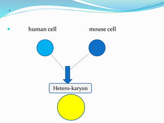
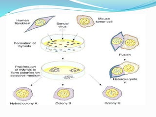
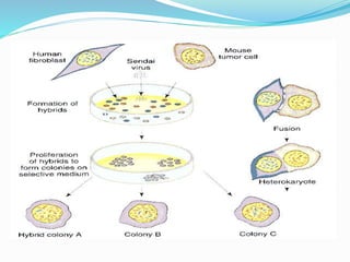
Presented By
ASAD HASHMI
Somatic cell hybridization
 The process of fusion between different somatic
cell (such as human and rodent cells) to produce
hybrid cells in which there is often one fused
nucleus.
 Such a process occurs in cell cultures and the
products are useful in establishing the expression and
location of particular genes.
OR
A technique which allows the manipulation of
cellular genomes by protoplast fusion.
Applications
 Its major contribution to plant breeding is in over
coming common crossing barriers among plant
species.
 The technique of somatic cell hybridization is
extensively used in human genome mapping, but it
can in principle be used in many different animal
systems.
 It is very important technique which develop and most
famous for producing mono-colonal antibodies.
 Now a days researchers develop this technique for
physical mapping of chromosomes.
 We can map very small segment of chromosome with
the help of SCH.
Procedure
 The procedure uses cells growing in culture.
 A virus called the Sendai virus has a useful property that
makes the mapping technique possible.
 Each Sendai virus has several points of attachment, so it
can simultaneously attach to two different cells if they
happen to be close together.
 However, a virus is very small in comparison with a cell, so
the two cells to which the virus is attached are held very
close together indeed.
 In fact, the membranes of the two cells may fuse together
and the two cells become one—a bi-nucleate heterokaryon.
.
 human cell mouse cell
Hetero-karyon
How it can survive
 For this kind of fusion and the survivality of
heterokaryon need to supply the nutrients.
 This experiment must be carried in pettery plate with
supporting material and also some nutrient sources.
 In case of SCH we are deriving a mechanism by which
we are selecting this heterokaryon .
Mechanism
 If suspensions of human and mouse cells are mixed
together in the presence of Sendai virus that has been
inactivated by ultraviolet light, the virus can mediate
fusion of the cells from the different species.
 When the cells have fused, the nuclei subsequently
fuse to form a uni-nucleate cell line composed of both
human and mouse chromosome sets.
.
.
 Because the mouse and human chromosomes
are recognizably different in number and shape,
the two sets in the hybrid cells can be readily
distinguished.
 However, in the course of subsequent cell
divisions, for unknown reasons the human
chromosomes are gradually eliminated from
the hybrid at random.
.
 The loss of human chromosomes can be
arrested in the following way to encourage the
formation of a stable partial hybrid.
 The cells used are mutant for some biochemical
function; so, if the cells are to grow, the missing
function must be supplied by the
other genome.
 This selective technique results in the
maintenance of hybrid cells that have a
complete set of mouse chromosomes and a
small number of human chromosomes, which
vary in number and type from hybrid to hybrid
but which always include the
human chromosome carrying the wild-
type allele defective in the mouse genome.
. Let’s look at the specific genes that make the selective
system work.
 In cells, DNA can be made by two pathways ;
 1. De novo pathway (“from scratch”) or
 2. Salvage pathway that uses molecular skeletons
already available.
.
 The selective technique involves the application
of a chemical, aminopterin, that blocks the de
novo synthetic pathway, confining DNA
synthesis to the salvage pathway.
 Two essential salvage
enzymes, thymidine kinase (TK) and
hypoxanthine-guanine phosphoribosyl
transferase (HGPRT), are relevant to the
system, as shown in the following two
reactions:
.
.
 The mouse cell line to be fused is genetically
unable to make TK because it is homozygous
for the allele tk−,
 whereas the human cell line is genetically
unable to make HGPRT because it is
homozygous at another locus for the
allele hgprt−.
 So the genotypes of the two fusing cell lines are:
.
 Because each is deficient for one enzyme,
neither the mouse nor the human cells are able
to make DNA individually
 In the hybrid cells, however,
the tk+ allele complements the hgprt+ allele, so
the cells can make both enzymes.
 Therefore, DNA is synthesized and the cells can
proliferate.
 Most human chromosomes are eliminated
from the hybrid cell cultures because their loss
has no effect on the cultures’ ability to grow.
HAT as a medium:
 To continue to grow in medium containing
hypoxanthine, aminopterin, and thymidine (HAT
medium), a hybrid culture must retain at least one of
the human chromosomes that carries the tk+ allele.
 The progressive elimination of the human
chromosomes from the fused cell lines can be followed
under the microscope because mouse chromosomes
can easily be distinguished from human
chromosomes.
 Staining process is used for this purpose.
Staining process:
 Usually two stains are used for this purpose
Quinacrine and Giemsa
 Chromosome stains such as quinacrine and Giemsa
reveal a pattern of banding within the chromosomes.
 The size and the position of these bands vary from
chromosome to chromosome, but the banding
patterns are highly specific and invariant for each
chromosome.
 Thus, it is easy to identify the human chromosomes
that are present in any hybrid cell .
.
 Different hybrid cells are grown separately into lines;
eventually a bank of lines is produced that contains, in
total, all the human chromosomes.
 (a) Stained human chromosomes. Under the
microscope, the chromosomes appear as a jumbled
cluster, as shown at the right.
.
 With a complete bank of chromosomes, we can begin
to assign genes or markers to chromosomes.
 If the human chromosome set is homozygous for a
human molecular marker—such as an allele that
controls a cell-surface antigen, drug resistance, a
nutritional requirement, a specific protein, or
a DNA marker—then the presence or absence of this
genetic marker in each line of hybrid cells can be
correlated with the presence or absence of certain
human chromosomes in each line.
.
 Data of this sort are presented in Table in which “+”
means presence and “−” means absence of the genetic
marker.
 We can see that, in the different hybrid cell
lines, genetic markers 1 and 3 are always present or
absent together.
 We can conclude, then, that they are linked.
 Furthermore, the presence or absence of genes 1 and 3
is correlated with the presence or absence of
chromosome 2, so we can assume that these genes are
located on chromosome 2.
.
. By the same reasoning, gene 2 must be on chromosome
1, but the location of gene 4 cannot be assigned. Large
numbers of human genes have now been localized to
specific chromosomes in this way.
.

Somatic cell hybridization

  • 2.
  • 3.
    Somatic cell hybridization The process of fusion between different somatic cell (such as human and rodent cells) to produce hybrid cells in which there is often one fused nucleus.  Such a process occurs in cell cultures and the products are useful in establishing the expression and location of particular genes. OR A technique which allows the manipulation of cellular genomes by protoplast fusion.
  • 4.
    Applications  Its majorcontribution to plant breeding is in over coming common crossing barriers among plant species.  The technique of somatic cell hybridization is extensively used in human genome mapping, but it can in principle be used in many different animal systems.  It is very important technique which develop and most famous for producing mono-colonal antibodies.
  • 5.
     Now adays researchers develop this technique for physical mapping of chromosomes.  We can map very small segment of chromosome with the help of SCH.
  • 6.
    Procedure  The procedureuses cells growing in culture.  A virus called the Sendai virus has a useful property that makes the mapping technique possible.  Each Sendai virus has several points of attachment, so it can simultaneously attach to two different cells if they happen to be close together.  However, a virus is very small in comparison with a cell, so the two cells to which the virus is attached are held very close together indeed.  In fact, the membranes of the two cells may fuse together and the two cells become one—a bi-nucleate heterokaryon.
  • 7.
    .  human cellmouse cell Hetero-karyon
  • 8.
    How it cansurvive  For this kind of fusion and the survivality of heterokaryon need to supply the nutrients.  This experiment must be carried in pettery plate with supporting material and also some nutrient sources.  In case of SCH we are deriving a mechanism by which we are selecting this heterokaryon .
  • 9.
    Mechanism  If suspensionsof human and mouse cells are mixed together in the presence of Sendai virus that has been inactivated by ultraviolet light, the virus can mediate fusion of the cells from the different species.  When the cells have fused, the nuclei subsequently fuse to form a uni-nucleate cell line composed of both human and mouse chromosome sets.
  • 10.
  • 11.
    .  Because themouse and human chromosomes are recognizably different in number and shape, the two sets in the hybrid cells can be readily distinguished.  However, in the course of subsequent cell divisions, for unknown reasons the human chromosomes are gradually eliminated from the hybrid at random.
  • 12.
    .  The lossof human chromosomes can be arrested in the following way to encourage the formation of a stable partial hybrid.  The cells used are mutant for some biochemical function; so, if the cells are to grow, the missing function must be supplied by the other genome.  This selective technique results in the maintenance of hybrid cells that have a complete set of mouse chromosomes and a small number of human chromosomes, which vary in number and type from hybrid to hybrid but which always include the human chromosome carrying the wild- type allele defective in the mouse genome.
  • 13.
    . Let’s lookat the specific genes that make the selective system work.  In cells, DNA can be made by two pathways ;  1. De novo pathway (“from scratch”) or  2. Salvage pathway that uses molecular skeletons already available.
  • 14.
    .  The selectivetechnique involves the application of a chemical, aminopterin, that blocks the de novo synthetic pathway, confining DNA synthesis to the salvage pathway.  Two essential salvage enzymes, thymidine kinase (TK) and hypoxanthine-guanine phosphoribosyl transferase (HGPRT), are relevant to the system, as shown in the following two reactions:
  • 15.
  • 16.
    .  The mousecell line to be fused is genetically unable to make TK because it is homozygous for the allele tk−,  whereas the human cell line is genetically unable to make HGPRT because it is homozygous at another locus for the allele hgprt−.  So the genotypes of the two fusing cell lines are:
  • 17.
    .  Because eachis deficient for one enzyme, neither the mouse nor the human cells are able to make DNA individually  In the hybrid cells, however, the tk+ allele complements the hgprt+ allele, so the cells can make both enzymes.  Therefore, DNA is synthesized and the cells can proliferate.  Most human chromosomes are eliminated from the hybrid cell cultures because their loss has no effect on the cultures’ ability to grow.
  • 18.
    HAT as amedium:  To continue to grow in medium containing hypoxanthine, aminopterin, and thymidine (HAT medium), a hybrid culture must retain at least one of the human chromosomes that carries the tk+ allele.  The progressive elimination of the human chromosomes from the fused cell lines can be followed under the microscope because mouse chromosomes can easily be distinguished from human chromosomes.  Staining process is used for this purpose.
  • 19.
    Staining process:  Usuallytwo stains are used for this purpose Quinacrine and Giemsa  Chromosome stains such as quinacrine and Giemsa reveal a pattern of banding within the chromosomes.  The size and the position of these bands vary from chromosome to chromosome, but the banding patterns are highly specific and invariant for each chromosome.  Thus, it is easy to identify the human chromosomes that are present in any hybrid cell .
  • 20.
    .  Different hybridcells are grown separately into lines; eventually a bank of lines is produced that contains, in total, all the human chromosomes.  (a) Stained human chromosomes. Under the microscope, the chromosomes appear as a jumbled cluster, as shown at the right.
  • 22.
    .  With acomplete bank of chromosomes, we can begin to assign genes or markers to chromosomes.  If the human chromosome set is homozygous for a human molecular marker—such as an allele that controls a cell-surface antigen, drug resistance, a nutritional requirement, a specific protein, or a DNA marker—then the presence or absence of this genetic marker in each line of hybrid cells can be correlated with the presence or absence of certain human chromosomes in each line.
  • 23.
    .  Data ofthis sort are presented in Table in which “+” means presence and “−” means absence of the genetic marker.  We can see that, in the different hybrid cell lines, genetic markers 1 and 3 are always present or absent together.  We can conclude, then, that they are linked.  Furthermore, the presence or absence of genes 1 and 3 is correlated with the presence or absence of chromosome 2, so we can assume that these genes are located on chromosome 2.
  • 24.
  • 25.
    . By thesame reasoning, gene 2 must be on chromosome 1, but the location of gene 4 cannot be assigned. Large numbers of human genes have now been localized to specific chromosomes in this way.
  • 26.