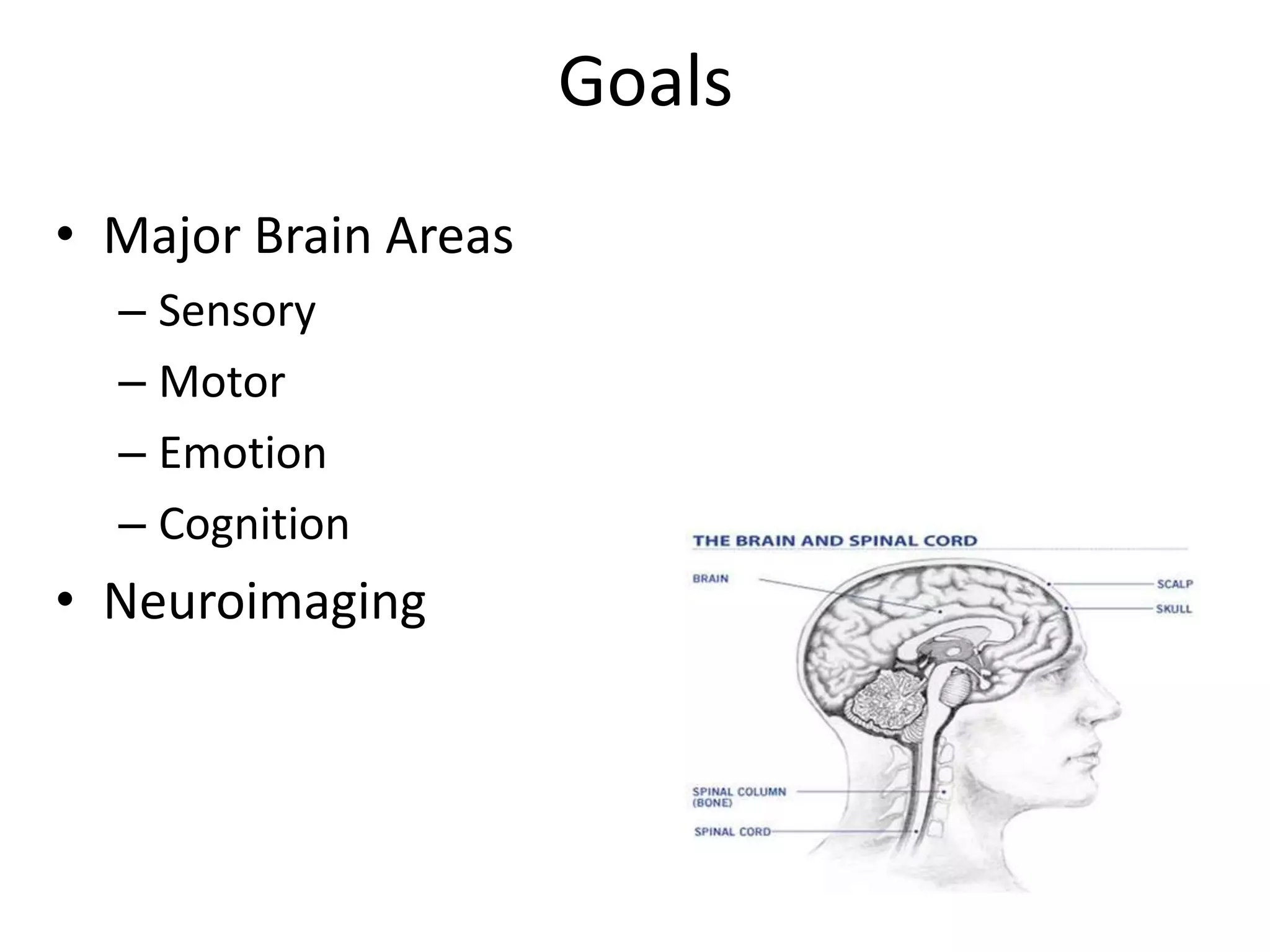
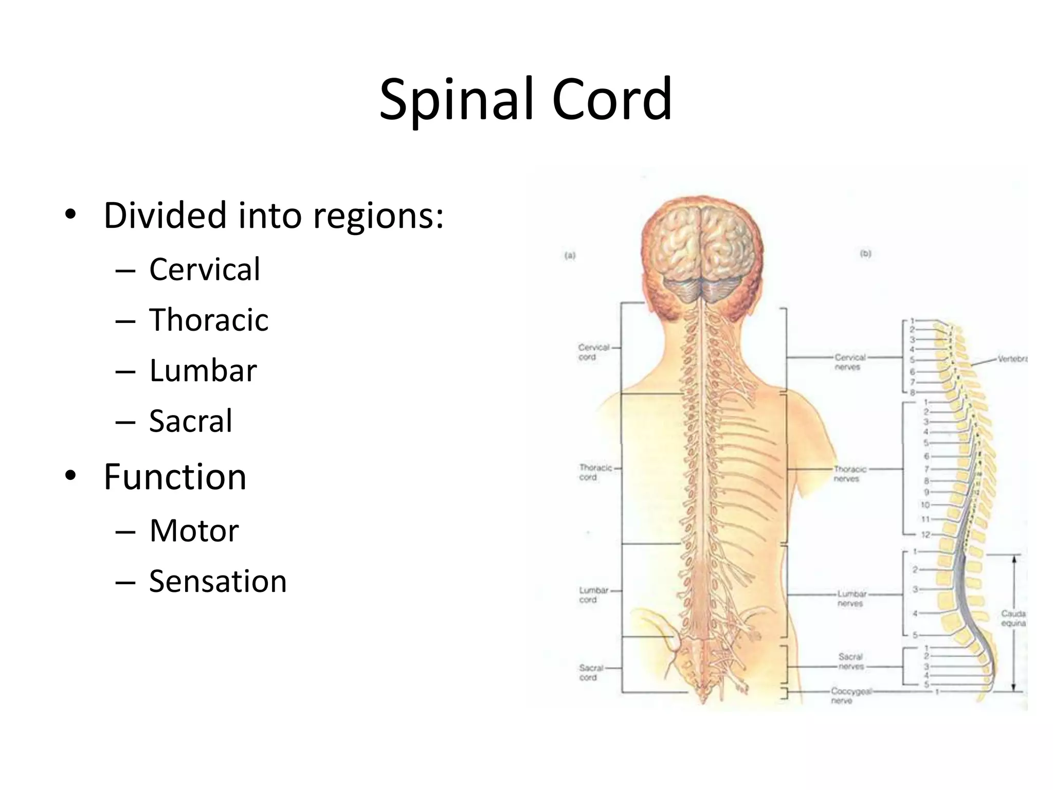
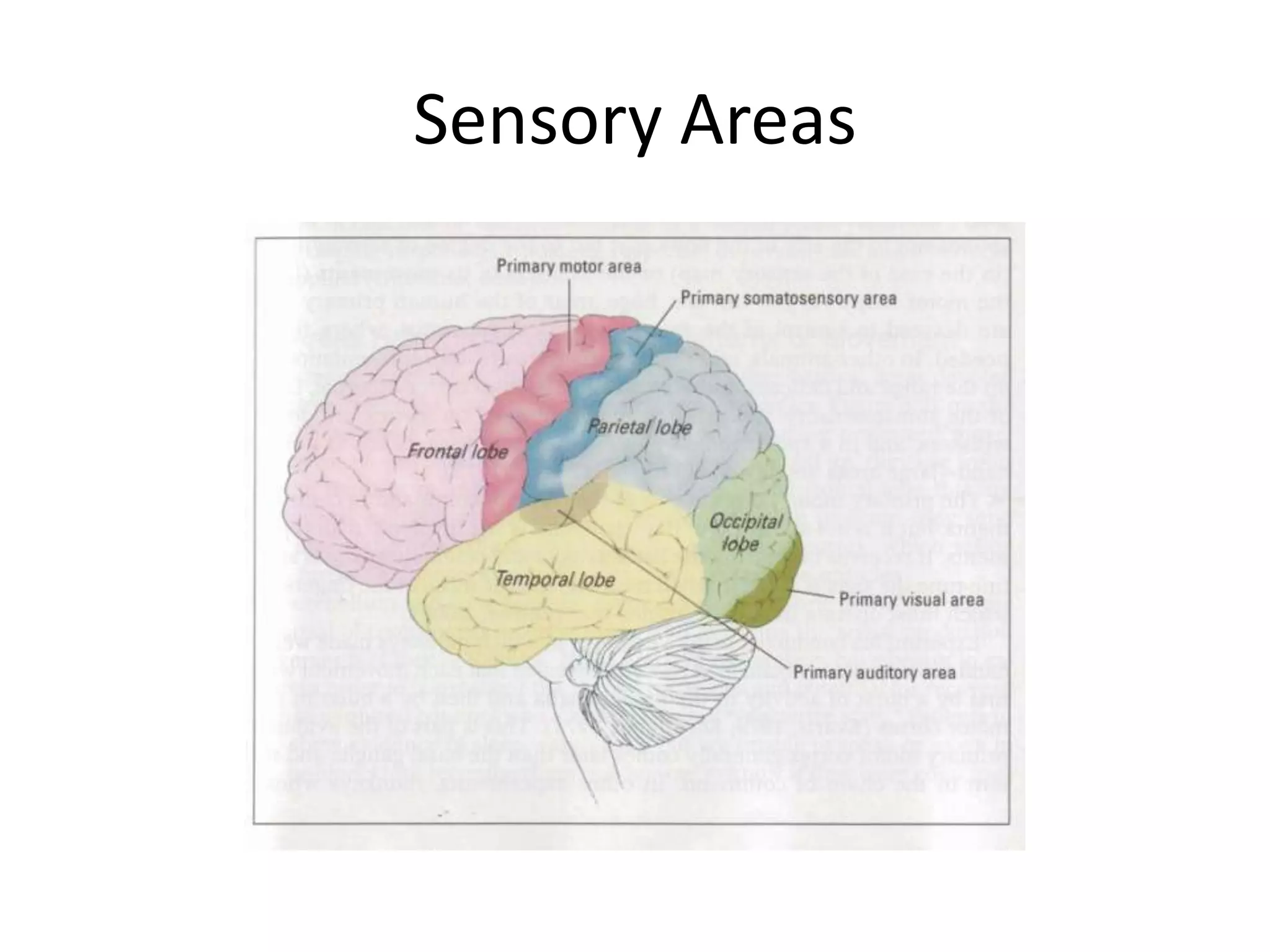
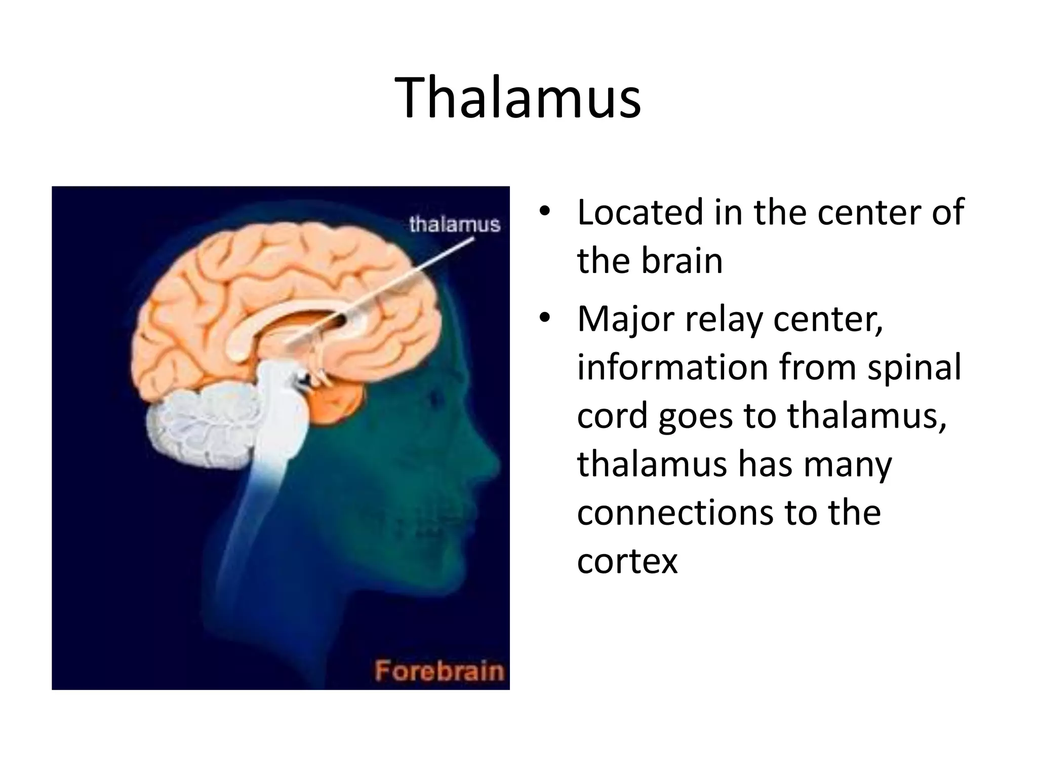
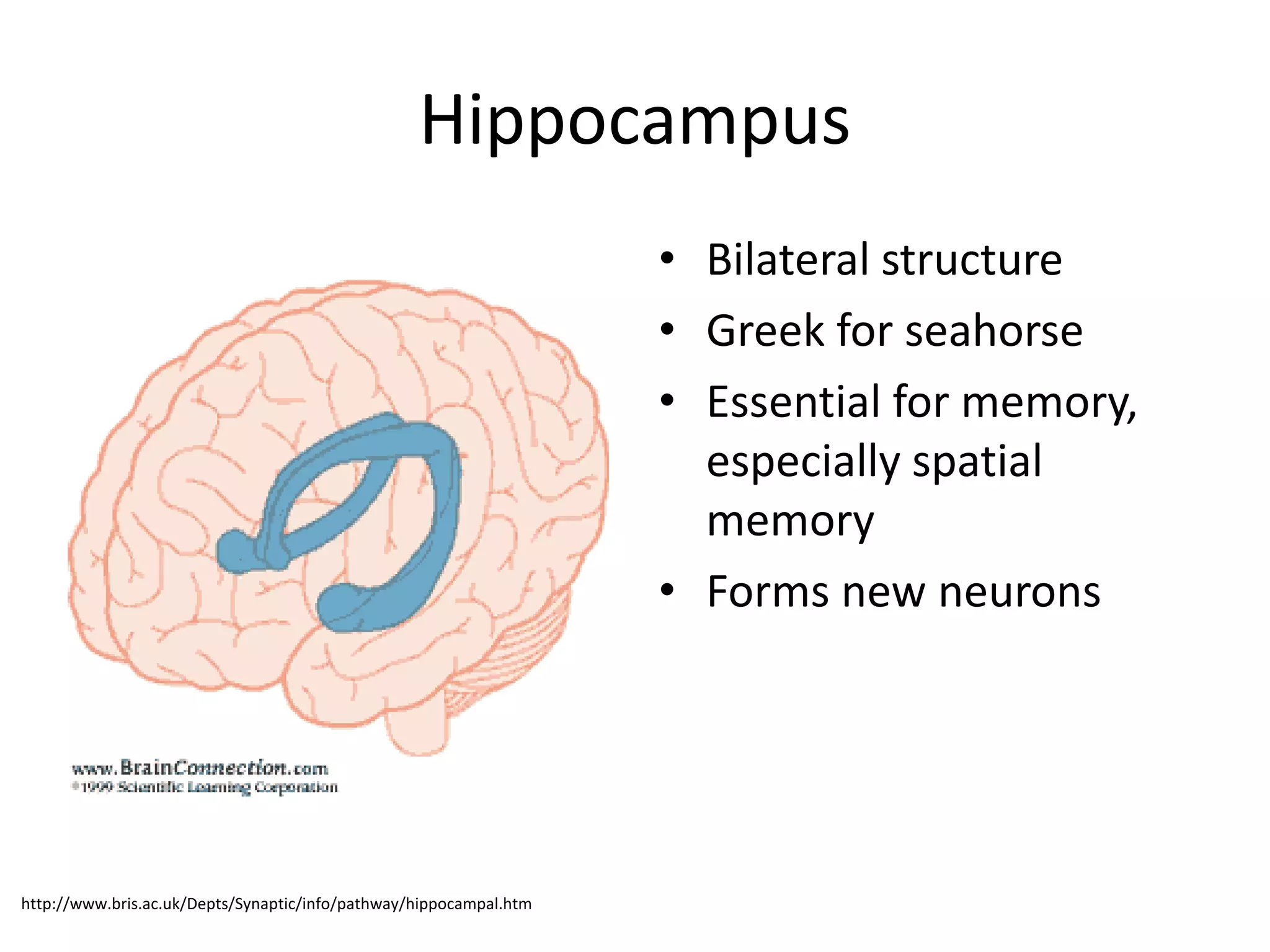
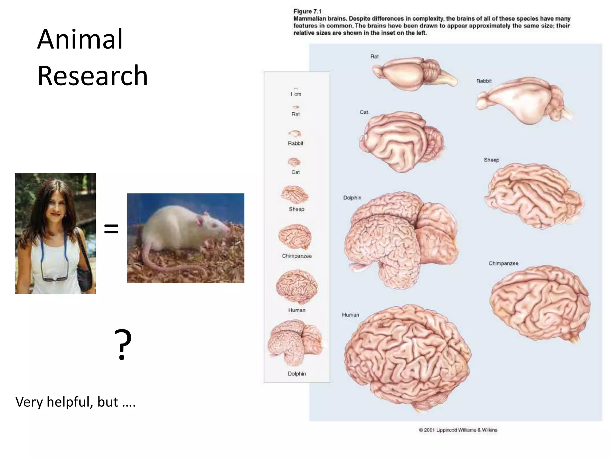
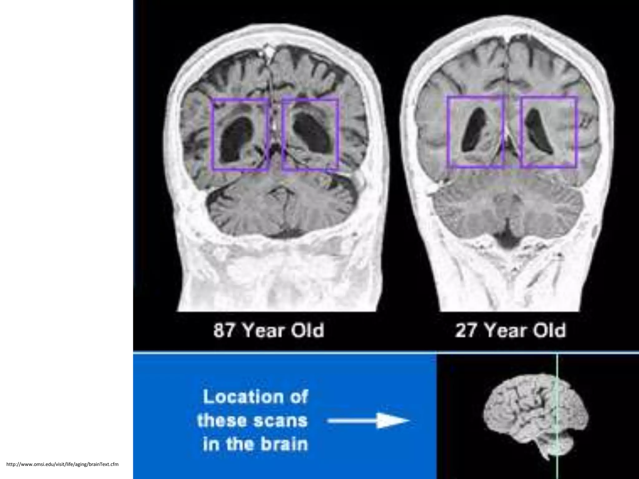
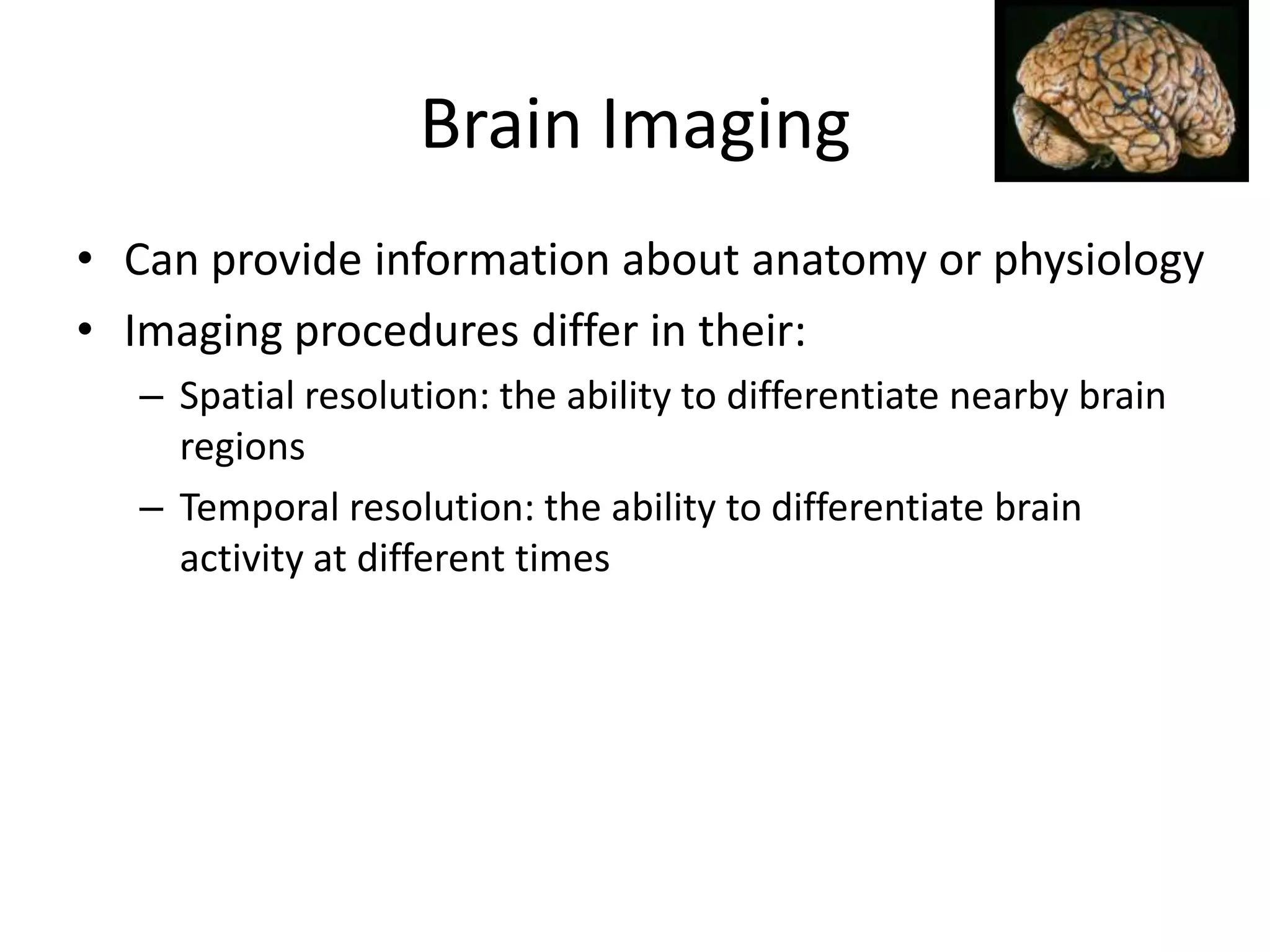
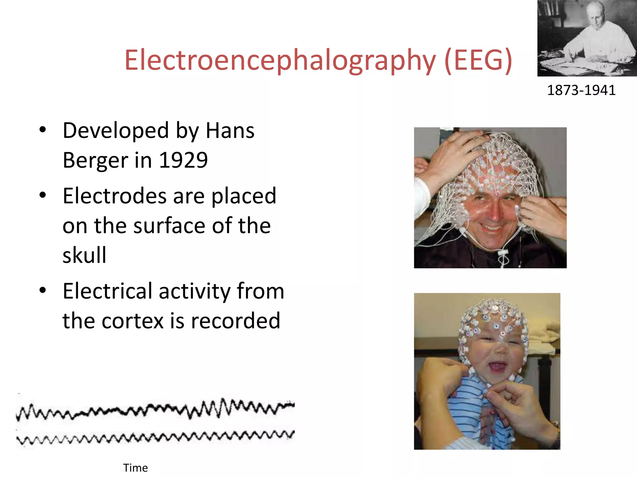
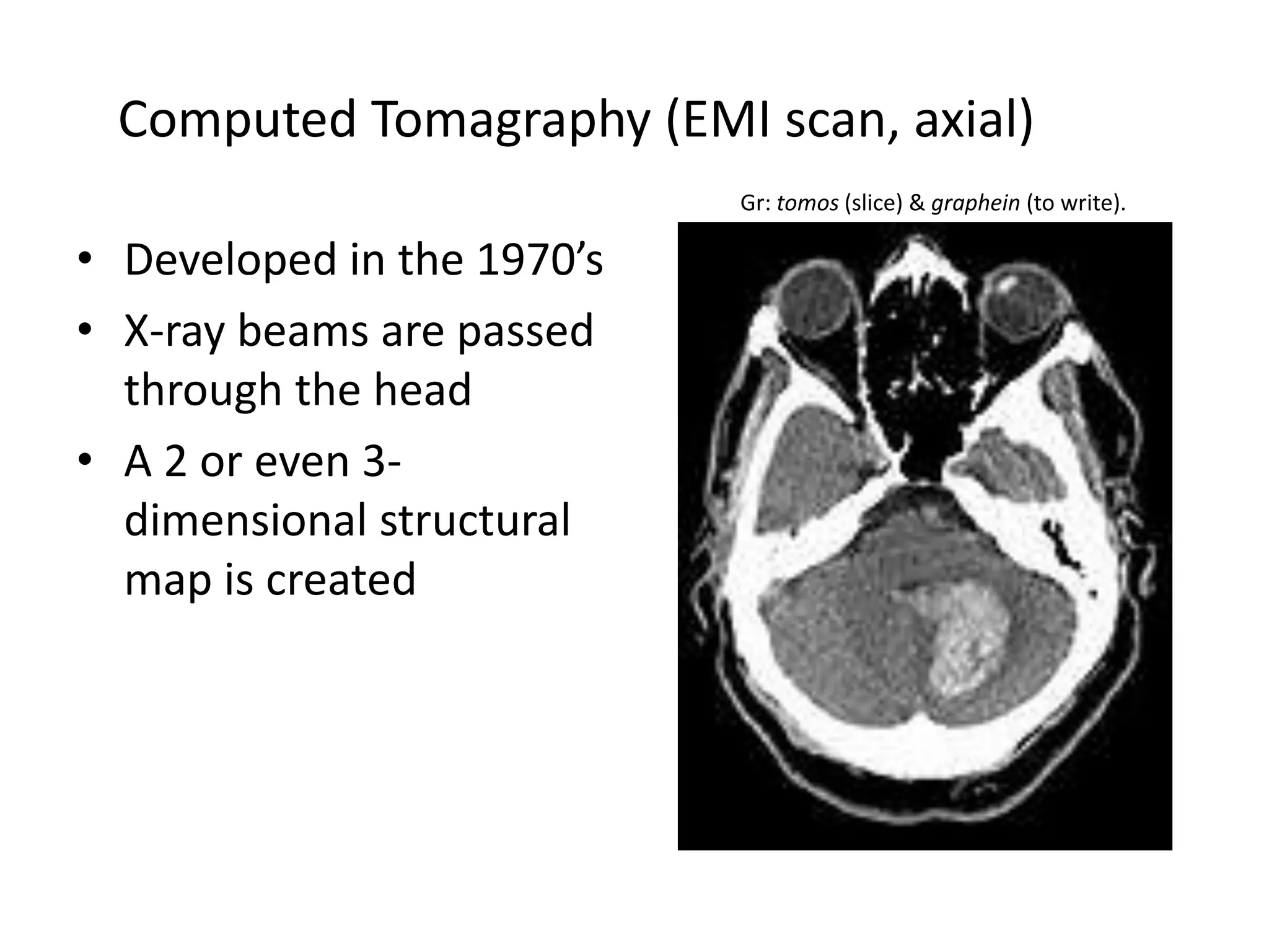
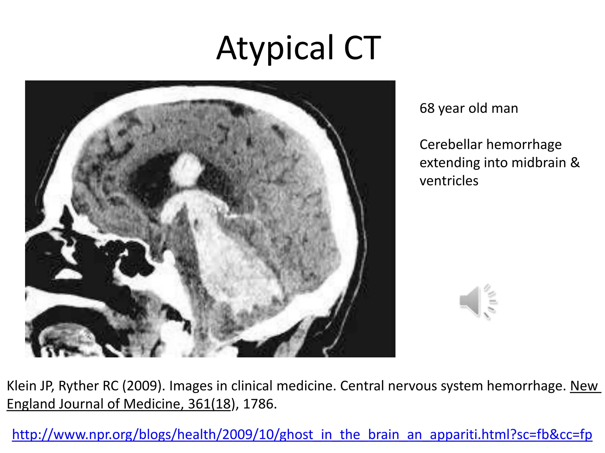
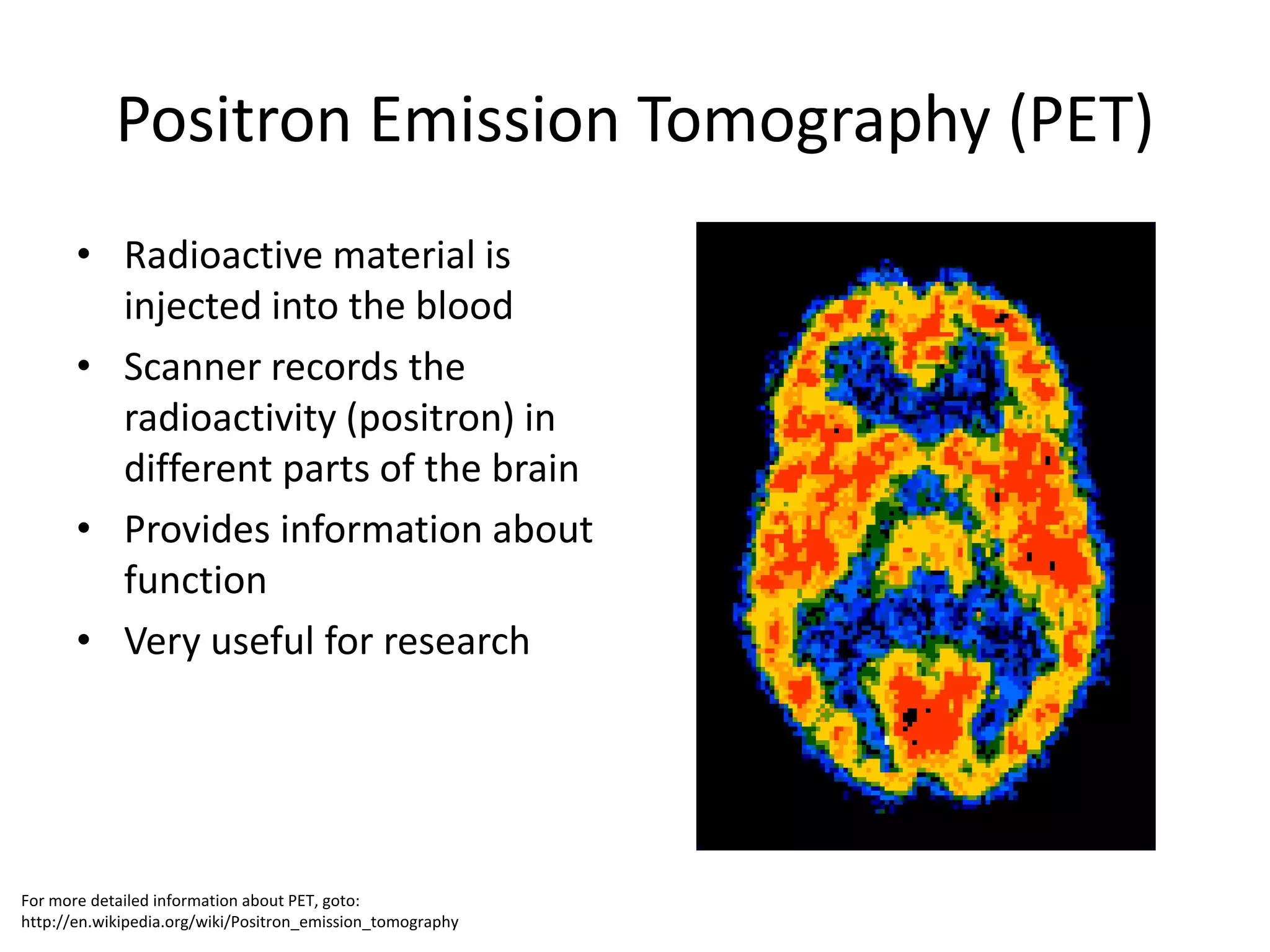
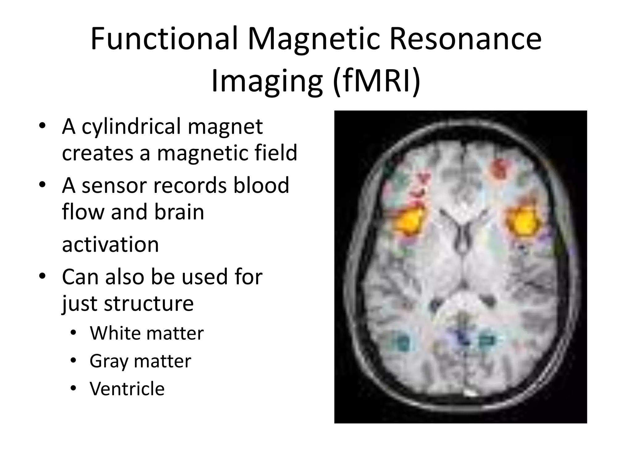
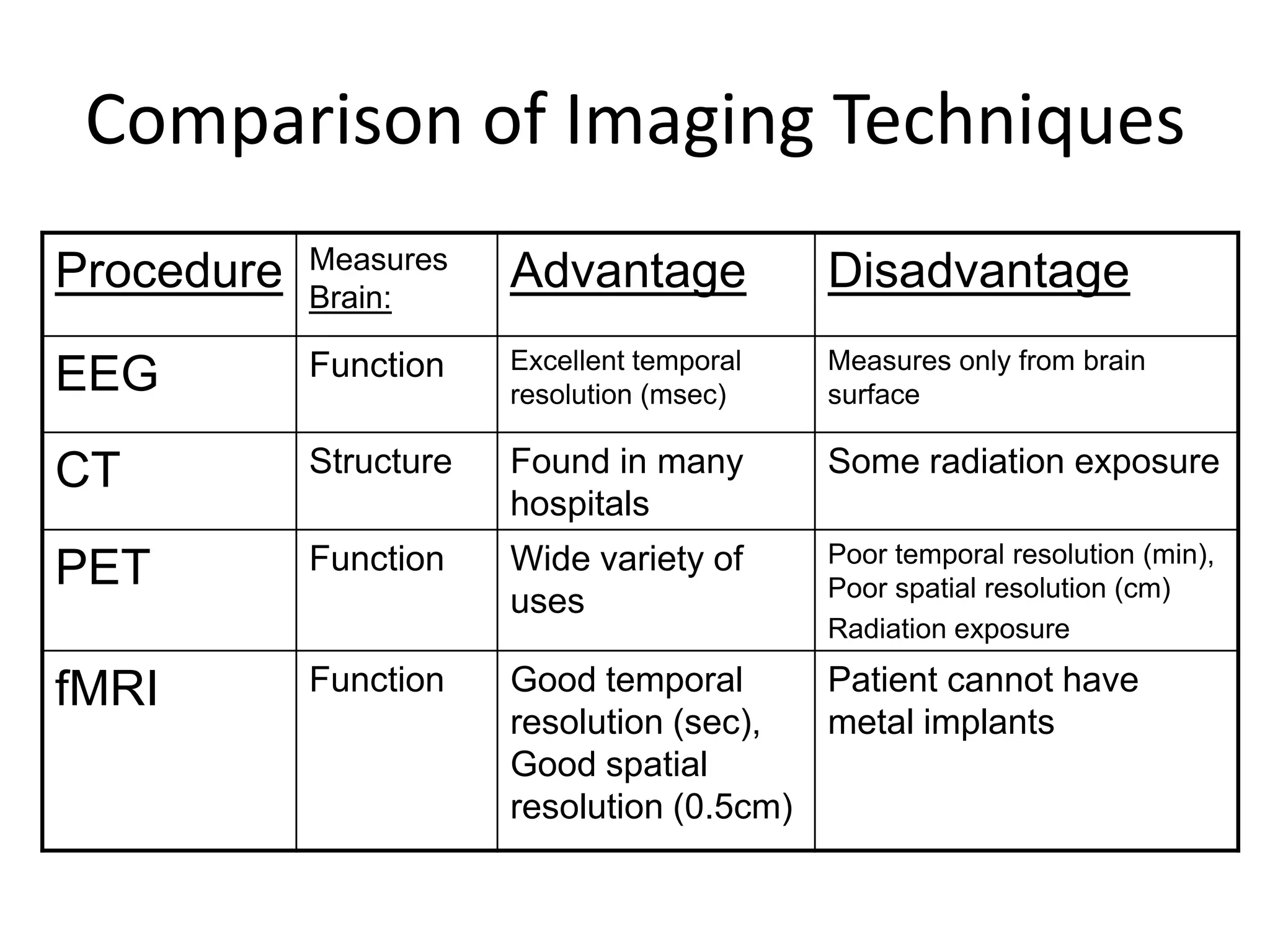
Neuroanatomy & Neurophysiology
       Brian J. Piper, Ph.D., M.S.
Goals
• Major Brain Areas
  – Sensory
  – Motor
  – Emotion
  – Cognition
• Neuroimaging
Spinal Cord
• Divided into regions:
   –   Cervical
   –   Thoracic
   –   Lumbar
   –   Sacral
• Function
   – Motor
   – Sensation
Beneath Skull
• Dura: thick/tough layer
• Arachnoid: contains blood vessels
• Pia: thin layer
______ lobe
Match Color
Description of 3-Dimensional Space
• Coronal:
   – section from ear to ear, like a loaf of bread
• Axial:
   – section that parallels horizon
• Sagittal:
   – section from front to back
   – mid-sagittal shows brain with left and
     right cortex separated
Corpus Callosum
• Fibers that connect left and right cortex
Anatomical Terminology
Cingulate Gyrus
• Tissue surrounding corpus collosum
  – Anterior
  – Posterior
Brainstem

The Medulla is the base
 of the brainstem that
controls heartbeat and
       breathing.

    Example: SIDS
Cerebellum

     • Located below the
       occipital cortex
CC   • Important for motor
       function
BS   • Site of action of alcohol
Cerebellum
       (a mid-sagittal)

      • Located below the
CC
        occipital cortex
      • Important for motor
        function
 BS
      • Site of action of alcohol
Functions of Different Cortical Areas
•   Frontal: cognition, executive function
•   Temporal: hearing, olfaction
•   Occipital: vision
•   Parietal: integration of sensory information
                    Dorsal


    Posterior                         Anterior


                    Ventral
Sensory Areas
Thalamus
    • Located in the center of
      the brain
    • Major relay center,
      information from spinal
      cord goes to thalamus,
      thalamus has many
      connections to the
      cortex
Hippocampus
                                                                    • Bilateral structure
                                                                    • Greek for seahorse
                                                                    • Essential for memory,
                                                                      especially spatial
                                                                      memory
                                                                    • Forms new neurons


http://www.bris.ac.uk/Depts/Synaptic/info/pathway/hippocampal.htm
Animal
   Research


              =


                ?
Very helpful, but ….
Hippocampus Comparison
Amygdala


 The Amygdala consists of
two lima bean-sized neural
   clusters linked to the
      emotions of fear
Brain Areas Important for
    Hormone Control
                                            Rene Descartes

                • Pineal Gland
                   – Very small subcortical structure
                   – Releases the hormone melatonin
                • Hypothalamus
                   – Hypo = “below” therefore located
                     under thalamus
                   – Regulates activity of Pituitary
                   – Pituitary communicates with
                     other endocrine glands (e.g.
                     testes)
                   – 4F!
Ventricles: Contain CSF
What is the impact of …?
http://www.omsi.edu/visit/life/aging/brainText.cfm
Brain Imaging
• Can provide information about anatomy or physiology
• Imaging procedures differ in their:
  – Spatial resolution: the ability to differentiate nearby brain
    regions
  – Temporal resolution: the ability to differentiate brain
    activity at different times
Electroencephalography (EEG)
                                       1873-1941

• Developed by Hans
  Berger in 1929
• Electrodes are placed
  on the surface of the
  skull
• Electrical activity from
  the cortex is recorded


        Time
Computed Tomagraphy (EMI scan, axial)
                            Gr: tomos (slice) & graphein (to write).

• Developed in the 1970’s
• X-ray beams are passed
  through the head
• A 2 or even 3-
  dimensional structural
  map is created
Atypical CT
                                                                  68 year old man

                                                                  Cerebellar hemorrhage
                                                                  extending into midbrain &
                                                                  ventricles


Klein JP, Ryther RC (2009). Images in clinical medicine. Central nervous system hemorrhage. New
England Journal of Medicine, 361(18), 1786.

http://www.npr.org/blogs/health/2009/10/ghost_in_the_brain_an_appariti.html?sc=fb&cc=fp
Positron Emission Tomography (PET)
       • Radioactive material is
         injected into the blood
       • Scanner records the
         radioactivity (positron) in
         different parts of the brain
       • Provides information about
         function
       • Very useful for research


For more detailed information about PET, goto:
http://en.wikipedia.org/wiki/Positron_emission_tomography
Figure 2. Brain Glucose Metabolic Images Showing Axial Planes at the Level of the
                                            Orbitofrontal Cortex


                Volkow, N. D. et al. JAMA 2011;305:808-813


Copyright restrictions may apply.
Functional Magnetic Resonance
             Imaging (fMRI)
• A cylindrical magnet
  creates a magnetic field
• A sensor records blood
  flow and brain
  activation
• Can also be used for
  just structure
   • White matter
   • Gray matter
   • Ventricle
Comparison of Imaging Techniques
            Measures
Procedure   Brain:
                        Advantage            Disadvantage
            Function    Excellent temporal   Measures only from brain
EEG                     resolution (msec)    surface

CT          Structure   Found in many        Some radiation exposure
                        hospitals
            Function    Wide variety of      Poor temporal resolution (min),
PET                                          Poor spatial resolution (cm)
                        uses
                                             Radiation exposure

fMRI        Function    Good temporal      Patient cannot have
                        resolution (sec),  metal implants
                        Good spatial
                        resolution (0.5cm)
What plane?


 Sarah Tappon, 8/5/2009
Useful video
• 2 Mininute Neuroanatomy Overview
  (Humorous, really!)
• http://www.youtube.com/watch?v=XAurv6m
  AWKM
C
A
            B
                                D

                                E


        L

    K                   H           F

                            G
            J       I
and sheep brain
Cranial Nerves
•   I. Olfactory: smell (S)
•   II. Optic: vision (S)
•   III. Oculomotor: pupil construction (M)
•   IV. Trochlear: eye movement (M)
•   V. Trigeminal: face & teeth (S), jaw (M)
•   X. Vagus: heart (SM), autonomic nervous system
The Nervous System
Autonomic Nervous System (ANS)


 Sympathetic NS
    “Arouses”
 (fight-or-flight)


Parasympathetic NS
      “Calms”
  (rest and digest)

Pathophysiology: Neuroanatomy Part I

  • 1.
    Neuroanatomy & Neurophysiology Brian J. Piper, Ph.D., M.S.
  • 2.
    Goals • Major BrainAreas – Sensory – Motor – Emotion – Cognition • Neuroimaging
  • 3.
    Spinal Cord • Dividedinto regions: – Cervical – Thoracic – Lumbar – Sacral • Function – Motor – Sensation
  • 6.
    Beneath Skull • Dura:thick/tough layer • Arachnoid: contains blood vessels • Pia: thin layer
  • 7.
  • 8.
  • 9.
    Description of 3-DimensionalSpace • Coronal: – section from ear to ear, like a loaf of bread • Axial: – section that parallels horizon • Sagittal: – section from front to back – mid-sagittal shows brain with left and right cortex separated
  • 10.
    Corpus Callosum • Fibersthat connect left and right cortex
  • 11.
  • 12.
    Cingulate Gyrus • Tissuesurrounding corpus collosum – Anterior – Posterior
  • 13.
    Brainstem The Medulla isthe base of the brainstem that controls heartbeat and breathing. Example: SIDS
  • 14.
    Cerebellum • Located below the occipital cortex CC • Important for motor function BS • Site of action of alcohol
  • 15.
    Cerebellum (a mid-sagittal) • Located below the CC occipital cortex • Important for motor function BS • Site of action of alcohol
  • 16.
    Functions of DifferentCortical Areas • Frontal: cognition, executive function • Temporal: hearing, olfaction • Occipital: vision • Parietal: integration of sensory information Dorsal Posterior Anterior Ventral
  • 17.
  • 18.
    Thalamus • Located in the center of the brain • Major relay center, information from spinal cord goes to thalamus, thalamus has many connections to the cortex
  • 19.
    Hippocampus • Bilateral structure • Greek for seahorse • Essential for memory, especially spatial memory • Forms new neurons http://www.bris.ac.uk/Depts/Synaptic/info/pathway/hippocampal.htm
  • 20.
    Animal Research = ? Very helpful, but ….
  • 21.
  • 22.
    Amygdala The Amygdalaconsists of two lima bean-sized neural clusters linked to the emotions of fear
  • 23.
    Brain Areas Importantfor Hormone Control Rene Descartes • Pineal Gland – Very small subcortical structure – Releases the hormone melatonin • Hypothalamus – Hypo = “below” therefore located under thalamus – Regulates activity of Pituitary – Pituitary communicates with other endocrine glands (e.g. testes) – 4F!
  • 24.
  • 25.
    What is theimpact of …?
  • 26.
  • 27.
    Brain Imaging • Canprovide information about anatomy or physiology • Imaging procedures differ in their: – Spatial resolution: the ability to differentiate nearby brain regions – Temporal resolution: the ability to differentiate brain activity at different times
  • 28.
    Electroencephalography (EEG) 1873-1941 • Developed by Hans Berger in 1929 • Electrodes are placed on the surface of the skull • Electrical activity from the cortex is recorded Time
  • 29.
    Computed Tomagraphy (EMIscan, axial) Gr: tomos (slice) & graphein (to write). • Developed in the 1970’s • X-ray beams are passed through the head • A 2 or even 3- dimensional structural map is created
  • 30.
    Atypical CT 68 year old man Cerebellar hemorrhage extending into midbrain & ventricles Klein JP, Ryther RC (2009). Images in clinical medicine. Central nervous system hemorrhage. New England Journal of Medicine, 361(18), 1786. http://www.npr.org/blogs/health/2009/10/ghost_in_the_brain_an_appariti.html?sc=fb&cc=fp
  • 31.
    Positron Emission Tomography(PET) • Radioactive material is injected into the blood • Scanner records the radioactivity (positron) in different parts of the brain • Provides information about function • Very useful for research For more detailed information about PET, goto: http://en.wikipedia.org/wiki/Positron_emission_tomography
  • 32.
    Figure 2. BrainGlucose Metabolic Images Showing Axial Planes at the Level of the Orbitofrontal Cortex Volkow, N. D. et al. JAMA 2011;305:808-813 Copyright restrictions may apply.
  • 33.
    Functional Magnetic Resonance Imaging (fMRI) • A cylindrical magnet creates a magnetic field • A sensor records blood flow and brain activation • Can also be used for just structure • White matter • Gray matter • Ventricle
  • 34.
    Comparison of ImagingTechniques Measures Procedure Brain: Advantage Disadvantage Function Excellent temporal Measures only from brain EEG resolution (msec) surface CT Structure Found in many Some radiation exposure hospitals Function Wide variety of Poor temporal resolution (min), PET Poor spatial resolution (cm) uses Radiation exposure fMRI Function Good temporal Patient cannot have resolution (sec), metal implants Good spatial resolution (0.5cm)
  • 35.
    What plane? SarahTappon, 8/5/2009
  • 36.
    Useful video • 2Mininute Neuroanatomy Overview (Humorous, really!) • http://www.youtube.com/watch?v=XAurv6m AWKM
  • 37.
    C A B D E L K H F G J I
  • 38.
  • 39.
    Cranial Nerves • I. Olfactory: smell (S) • II. Optic: vision (S) • III. Oculomotor: pupil construction (M) • IV. Trochlear: eye movement (M) • V. Trigeminal: face & teeth (S), jaw (M) • X. Vagus: heart (SM), autonomic nervous system
  • 40.
  • 41.
    Autonomic Nervous System(ANS) Sympathetic NS “Arouses” (fight-or-flight) Parasympathetic NS “Calms” (rest and digest)