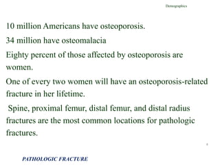
1) Pathologic fractures occur in abnormal bone that has been weakened, often by conditions like cancer or osteoporosis, and these fractures can occur during normal activity or minor trauma.
2) Evaluation of patients with possible pathologic fractures involves medical history, physical exam, laboratory tests, and imaging to determine the cause and extent of the bone abnormality.
3) Plain x-rays are usually the first imaging study and can provide clues about conditions like osteoporosis, fractures, or bone tumors, but other imaging may be needed to fully evaluate bone lesions or confirm a diagnosis.
The Indian Dental Academy is the Leader in continuing dental education , training dentists in all aspects of dentistry and
offering a wide range of dental certified courses in different formats.for more details please visit
www.indiandentalacademy.com
Imaging gives a clue on pattern recognition for differential diagnosis. Therefore imaging alone may not be sufficient for the diagnosis of MM.
Hematopathologic and Histopathologic correlation are important key for differentiation and definitive diagnosis.
amyloidosis(including history,physical and chemical properties, classification, variants, staining characteristics, lab diagnosis,morphological patterns according to organ involved ,), basically for undergraduates and residents in pathology
The vital role of oncology nurses in the care of patients with MM necessitates the awareness of the latest treatment advances and best practices for side-effect management. This CE-certified activity will provide updates in first-line, maintenance, and relapsed/refractory settings. Expert faculty will articulate the diagnosis, cytogenetics, and staging of the disease, as well as promising novel agents and evidence-based best practices for the management of side effects. To provide insight to attendees of the impact of evolving data on a personal level, a patient with MM will share personal perspectives on the journey from diagnosis, treatment, and overall patient experience.
Downloadable slide decks are a great tool for self study and teaching purposes. They are non-certified resources available to enhance your knowledge.
Review a downloadable slide deck by Beth Faiman, PhD(c), RN, APRN, BC, AOCN®, covering the most clinically relevant new data reported from Practical Navigation of a Changing Landscape: Keeping Current on Multiple Myeloma Treatments.
Target Audience
This activity has been designed to meet the educational needs of oncology nurses involved in the care of patients with multiple myeloma (MM).
Slide Deck Disclaimer
This slide deck in its original and unaltered format is for educational purposes and is current as of May 2012. All materials contained herein reflect the views of the faculty, and not those of IMER, the CE provider, or the commercial supporter. These materials may discuss therapeutic products that have not been approved by the US Food and Drug Administration and off-label uses of approved products. Readers should not rely on this information as a substitute for professional medical advice, diagnosis, or treatment. The use of any information provided is solely at your own risk, and readers should verify the prescribing information and all data before treating patients or employing any therapeutic products described in this educational activity.
For more information click here:
http://imeronline.com/gxpsites/hgxpp001.aspx?11,52,304,O,E,0,,743;561;8612
Download these clinical tools and resources to improve outcomes in care for patients with multiple myeloma:
http://imeronline.com/gxpsites/hgxpp001.aspx?11,52,304,O,E,0,,743;561;8613
Multiple myeloma is mostly a disease of the elderly. It is a form of haematological cancers that affects the Lymphocytes, and causes abnormal proliferation of plasma cells within the bone marrow, thus replacing the marrow, and is associated with multiple organ dysfunction.
This presentation is an introduction to the disease. It however leaves out the specific haematological treatment, because by that point, patient should have been referred to haematology.
By Dr. Usama Ragab, Zagazig Faculty of Medicine
PSC incidence ranges from 0.5 to 1.25 cases/100 000.
The prevalence of the disease ranges between six and 20 cases/100 000.
Men are more likely to be affected (70%).
Prevalence of PSC may be increased in first degree relatives of PSC patients
multiple myloma
By: Nader Amir Al-assadi
Supervised by : Dr/ Ghazi Alariqe
taiz university
Multiple myeloma (MM) is a plasma cell malignancy in which monoclonal plasma cells proliferate in bone marrow, resulting in an over abundance of monoclonal para protein (M protein), destruction of bone, and displacement of other hematopoietic cell lines.
The precise etiology of MM has not yet been established.
Roles have been suggested for a variety of factors, including genetic causes, environmental or occupational causes,radiation, chronic inflammation, and infection .
This presentation talks about Gastrointestinal carcinoid tumors specifically a review article published by certain authors (scientists) for further studies.
Evaluation of postpartum depression and relationship its with spiritual healt...Mohammad Mahdi Shater
Evaluation of postpartum depression and relationship its with spiritual health level and perceived social support in selected therapeutic centers in qom province
گزارش کارآموزی بهداشت روستای خورآباد قم سال 1395
برای دریافت اطلاعات تکمیلی با اکانت مکاتبه فرمایید.
دکتر محمد مهدی شاطر
Dr. Mohammad Mahdi Shater
Khorabad village, Qom, Iran Islamic Republic
Metabolic risk factors in children with asymptomatic hematuria
Francisco Rodolfo Spivacow, Elisa Elena del Valle, Paula Gabriela Rey
Dr.shater, Dr.razavi
Knee anatomy and clinical tests 2024.pdfvimalpl1234
This includes all relevant anatomy and clinical tests compiled from standard textbooks, Campbell,netter etc..It is comprehensive and best suited for orthopaedicians and orthopaedic residents.
Ozempic: Preoperative Management of Patients on GLP-1 Receptor Agonists Saeid Safari
Preoperative Management of Patients on GLP-1 Receptor Agonists like Ozempic and Semiglutide
ASA GUIDELINE
NYSORA Guideline
2 Case Reports of Gastric Ultrasound
Basavarajeeyam is an important text for ayurvedic physician belonging to andhra pradehs. It is a popular compendium in various parts of our country as well as in andhra pradesh. The content of the text was presented in sanskrit and telugu language (Bilingual). One of the most famous book in ayurvedic pharmaceutics and therapeutics. This book contains 25 chapters called as prakaranas. Many rasaoushadis were explained, pioneer of dhatu druti, nadi pareeksha, mutra pareeksha etc. Belongs to the period of 15-16 century. New diseases like upadamsha, phiranga rogas are explained.
micro teaching on communication m.sc nursing.pdfAnurag Sharma
Microteaching is a unique model of practice teaching. It is a viable instrument for the. desired change in the teaching behavior or the behavior potential which, in specified types of real. classroom situations, tends to facilitate the achievement of specified types of objectives.
Local Advanced Lung Cancer: Artificial Intelligence, Synergetics, Complex Sys...Oleg Kshivets
Overall life span (LS) was 1671.7±1721.6 days and cumulative 5YS reached 62.4%, 10 years – 50.4%, 20 years – 44.6%. 94 LCP lived more than 5 years without cancer (LS=2958.6±1723.6 days), 22 – more than 10 years (LS=5571±1841.8 days). 67 LCP died because of LC (LS=471.9±344 days). AT significantly improved 5YS (68% vs. 53.7%) (P=0.028 by log-rank test). Cox modeling displayed that 5YS of LCP significantly depended on: N0-N12, T3-4, blood cell circuit, cell ratio factors (ratio between cancer cells-CC and blood cells subpopulations), LC cell dynamics, recalcification time, heparin tolerance, prothrombin index, protein, AT, procedure type (P=0.000-0.031). Neural networks, genetic algorithm selection and bootstrap simulation revealed relationships between 5YS and N0-12 (rank=1), thrombocytes/CC (rank=2), segmented neutrophils/CC (3), eosinophils/CC (4), erythrocytes/CC (5), healthy cells/CC (6), lymphocytes/CC (7), stick neutrophils/CC (8), leucocytes/CC (9), monocytes/CC (10). Correct prediction of 5YS was 100% by neural networks computing (error=0.000; area under ROC curve=1.0).
These lecture slides, by Dr Sidra Arshad, offer a quick overview of the physiological basis of a normal electrocardiogram.
Learning objectives:
1. Define an electrocardiogram (ECG) and electrocardiography
2. Describe how dipoles generated by the heart produce the waveforms of the ECG
3. Describe the components of a normal electrocardiogram of a typical bipolar lead (limb II)
4. Differentiate between intervals and segments
5. Enlist some common indications for obtaining an ECG
6. Describe the flow of current around the heart during the cardiac cycle
7. Discuss the placement and polarity of the leads of electrocardiograph
8. Describe the normal electrocardiograms recorded from the limb leads and explain the physiological basis of the different records that are obtained
9. Define mean electrical vector (axis) of the heart and give the normal range
10. Define the mean QRS vector
11. Describe the axes of leads (hexagonal reference system)
12. Comprehend the vectorial analysis of the normal ECG
13. Determine the mean electrical axis of the ventricular QRS and appreciate the mean axis deviation
14. Explain the concepts of current of injury, J point, and their significance
Study Resources:
1. Chapter 11, Guyton and Hall Textbook of Medical Physiology, 14th edition
2. Chapter 9, Human Physiology - From Cells to Systems, Lauralee Sherwood, 9th edition
3. Chapter 29, Ganong’s Review of Medical Physiology, 26th edition
4. Electrocardiogram, StatPearls - https://www.ncbi.nlm.nih.gov/books/NBK549803/
5. ECG in Medical Practice by ABM Abdullah, 4th edition
6. Chapter 3, Cardiology Explained, https://www.ncbi.nlm.nih.gov/books/NBK2214/
7. ECG Basics, http://www.nataliescasebook.com/tag/e-c-g-basics
Title: Sense of Taste
Presenter: Dr. Faiza, Assistant Professor of Physiology
Qualifications:
MBBS (Best Graduate, AIMC Lahore)
FCPS Physiology
ICMT, CHPE, DHPE (STMU)
MPH (GC University, Faisalabad)
MBA (Virtual University of Pakistan)
Learning Objectives:
Describe the structure and function of taste buds.
Describe the relationship between the taste threshold and taste index of common substances.
Explain the chemical basis and signal transduction of taste perception for each type of primary taste sensation.
Recognize different abnormalities of taste perception and their causes.
Key Topics:
Significance of Taste Sensation:
Differentiation between pleasant and harmful food
Influence on behavior
Selection of food based on metabolic needs
Receptors of Taste:
Taste buds on the tongue
Influence of sense of smell, texture of food, and pain stimulation (e.g., by pepper)
Primary and Secondary Taste Sensations:
Primary taste sensations: Sweet, Sour, Salty, Bitter, Umami
Chemical basis and signal transduction mechanisms for each taste
Taste Threshold and Index:
Taste threshold values for Sweet (sucrose), Salty (NaCl), Sour (HCl), and Bitter (Quinine)
Taste index relationship: Inversely proportional to taste threshold
Taste Blindness:
Inability to taste certain substances, particularly thiourea compounds
Example: Phenylthiocarbamide
Structure and Function of Taste Buds:
Composition: Epithelial cells, Sustentacular/Supporting cells, Taste cells, Basal cells
Features: Taste pores, Taste hairs/microvilli, and Taste nerve fibers
Location of Taste Buds:
Found in papillae of the tongue (Fungiform, Circumvallate, Foliate)
Also present on the palate, tonsillar pillars, epiglottis, and proximal esophagus
Mechanism of Taste Stimulation:
Interaction of taste substances with receptors on microvilli
Signal transduction pathways for Umami, Sweet, Bitter, Sour, and Salty tastes
Taste Sensitivity and Adaptation:
Decrease in sensitivity with age
Rapid adaptation of taste sensation
Role of Saliva in Taste:
Dissolution of tastants to reach receptors
Washing away the stimulus
Taste Preferences and Aversions:
Mechanisms behind taste preference and aversion
Influence of receptors and neural pathways
Impact of Sensory Nerve Damage:
Degeneration of taste buds if the sensory nerve fiber is cut
Abnormalities of Taste Detection:
Conditions: Ageusia, Hypogeusia, Dysgeusia (parageusia)
Causes: Nerve damage, neurological disorders, infections, poor oral hygiene, adverse drug effects, deficiencies, aging, tobacco use, altered neurotransmitter levels
Neurotransmitters and Taste Threshold:
Effects of serotonin (5-HT) and norepinephrine (NE) on taste sensitivity
Supertasters:
25% of the population with heightened sensitivity to taste, especially bitterness
Increased number of fungiform papillae
NVBDCP.pptx Nation vector borne disease control programSapna Thakur
NVBDCP was launched in 2003-2004 . Vector-Borne Disease: Disease that results from an infection transmitted to humans and other animals by blood-feeding arthropods, such as mosquitoes, ticks, and fleas. Examples of vector-borne diseases include Dengue fever, West Nile Virus, Lyme disease, and malaria.
ABDOMINAL TRAUMA in pediatrics part one.drhasanrajab
Abdominal trauma in pediatrics refers to injuries or damage to the abdominal organs in children. It can occur due to various causes such as falls, motor vehicle accidents, sports-related injuries, and physical abuse. Children are more vulnerable to abdominal trauma due to their unique anatomical and physiological characteristics. Signs and symptoms include abdominal pain, tenderness, distension, vomiting, and signs of shock. Diagnosis involves physical examination, imaging studies, and laboratory tests. Management depends on the severity and may involve conservative treatment or surgical intervention. Prevention is crucial in reducing the incidence of abdominal trauma in children.
1. Section one: General Principles
Chapter 22:
PathologicFractures
INTRODUCTION PATHOLOGIC FRACTURE 1
Dr. Mohammad Mahdi Shater
Baqiyatallah University of Medical Sciences
2. Pathologic fractures occur in abnormal bone. Weakened
bone predisposes the patient for failure during normal
activity or after minor trauma.
Non-neoplastic skeletal disease separate into correctable
and uncorrectable conditions.
Correctable conditions include renal osteodystrophy,
hyperparathyroidism, osteomalacia, and disuse
osteoporosis.
Uncorrectable conditions include osteogenesis imperfecta,
polyostotic fibrous dysplasia, postmenopausal
osteoporosis, Paget disease, and osteopetrosis.
INTRODUCTION
PATHOLOGIC FRACTURE
2
3. -Fracture callus may not form normally, and healing often
occurs slowly.
-Many patients have additional fractures, delayed union,
and nonunion.
-In the management of patients with systemic skeletal
disease, it is important to prevent disuse osteoporosis,
which may lead to additional pathologic fractures.
- Osteoporosis is the most common condition associated
with pathologic fractures.
INTRODUCTION
PATHOLOGIC FRACTURE
3
4. 10 million Americans have osteoporosis.
34 million have osteomalacia
Eighty percent of those affected by osteoporosis are
women.
One of every two women will have an osteoporosis-related
fracture in her lifetime.
Spine, proximal femur, distal femur, and distal radius
fractures are the most common locations for pathologic
fractures.
Demographics
PATHOLOGIC FRACTURE
4
5. A comprehensive evaluation of a patient with a lytic bone lesion or
pathologic fracture is essential .
Certain symptoms should alert the orthopedic surgeon:
Pain is the most common presenting symptom before fracture, ranging
from a dull constant ache to an intense pain exacerbated by weight bearing.
Patients must be asked about previously diagnosed or treated cancer;
otherwise
Breast cancer can have a long latent period until bony metastases present.
A history of radiation is important.
Standard review of systems such as recent weight loss, fevers, night
sweats, and fatigue are important.
Questions about relevant risk factors such as smoking, dietary habits, and
toxic exposures should be asked.
EVALUATION
PATHOLOGIC FRACTURE
5
8. The physical examination should include a thorough
evaluation
Palpation of a mass, identi- fication of an obvious
deformity, and a detailed neurovascular examination.
All extremities and the entire spine evaluated for
additional lesions or lymphadenopathy
Multiple sites of involvement with bone metastasis,
lymphoma, multiple myeloma, or osteoporosis.
Evaluation of all possible primary sites (breast, prostate,
lung, thyroid) and a stool test for occult blood.
EVALUATION
PATHOLOGIC FRACTURE
8
9. Laboratory tests will not often make the diagnosis, especially in cases of
cancer.
A baseline laboratory profile should include
– CBC diff , ESR , serum chemistries, blood urea nitrogen (BUN),
serum glucose, liver function tests, protein, albumin, calcium,
phosphorus, and alkaline phosphatase.
Patients with widespread bone metastasis may exhibit anemia of chronic
disease, hypercalcemia, and increased alkaline phosphatase.
The hemoglobin is also often low in patients with multiple myeloma.
A standard urinalysis is necessary to look for microscopic hematuria,
which suggests renal cell carcinoma (RCC)
24-hour urine collection is necessary for a complete metabolic evaluation.
Serum and urine protein electrophoreses are important to exclude multiple
myeloma.
EVALUATION
PATHOLOGIC FRACTURE
9
10. Thyroid function tests, carcinoembryonic antigen (CEA),
CA125, and prostate specific antigen (PSA) are serum
markers for specific tumors that can be useful for
particular individuals.
N-telopeptide and C-telopeptide are new biomechanical
markers of bone collagen breakdown that can be measured
in the serum and urine.
These markers are used to confirm increased destruction
caused by bone metastasis, measure the overall extent of
bone involvement, and assess the response of the bone to
bisphosphonate treatment.
EVALUATION
PATHOLOGIC FRACTURE
10
12. Osteoporosis have normal values .
Osteomalacia have low serum calcium and phosphorus,
high serum ALP , high urinary phosphorus, and high
urinary hydroxyproline values (Table 22-3).
Primary hyperparathyroidism have high serum calcium,
alkaline phos- phatase, and parathyroid hormone levels
with low serum phosphorus. They also have high urinary
calcium, phosphorus, and hydroxyproline levels.
Renal osteodystrophy have low serum calcium with high
serum phosphorus, alkaline phos- phatase, and BUN
levels. When secondary hyperparathyroidism
EVALUATION
PATHOLOGIC FRACTURE
12
13. When secondary hyperparathyroidism develops in these patients, the
serum calcium increases to normal or elevated values with elevated
parathyroid hormone levels.
Urine values are difficult to assess in patients with secondary
hyperparathyroidism caused by abnormal glomerular filtration.
Patients with Paget disease have normal values for serum calcium and
phosphorus, but markedly elevated levels of alkaline phosphatase and
urinary hydroxyproline.
PSA is a sensitive measurement of prostate cancer.
A value less than 10 ng/mL essentially excludes the presence of bone
metastasis.
Serum calcium is a measurement of unbound calcium in the serum and,
therefore, determination of serum protein is necessary to interpret the cal-
cium level. If the serum protein is lower than normal, the normal range of
serum calcium is lowered.
EVALUATION
PATHOLOGIC FRACTURE
13
14. Patients often have marked pain or pathologic fractures that
leave them unable to ambulate or perform their activities of
daily living (adls).
Spinal fractures may develop neurologic deficits that lead to
paralysis.
Impending or actual extremity fractures may be forced to
remain in bed for prolonged periods of time, predisposing them
to hypercalcemia.
Anemia is a common hematologic abnormality .
The most tragic concern is the general loss in their quality of
life.
Associated Medical Problems
PATHOLOGIC FRACTURE
14
15. Hypercalcemia of malignancy, most commonly associated
with cancers of the lung, breast, kidney, genitourinary
tract, and multiple myeloma.
Much of the remainder is caused by primary hyper-
parathyroidism. Rarely, the two causes occur
simultaneously.
Hypercalcemia can be lethal if untreated (table 22-4).
Hypercalcemia portends a poor prognosis for the patient.
60% of patients with hypercalcemia will survive less than 3
months, and only 20% will be alive at 1 year.
Associated Medical Problems
PATHOLOGIC FRACTURE
15
17. Vigorous volume repletion is a temporizing measure, so
treatment must focus on reducing the degree of bone
resorption.
This can be accomplished by treating the primary tumor
directly or by using bisphosphonates to reduce osteoclastic
activity.
Correction of any electrolyte imbalance or hypercalcemia
should be done before surgery
Associated Medical Problems
PATHOLOGIC FRACTURE
17
18. The first and most important imaging study is a plain radiograph in two orthogonal planes.
Should be examined for diagnostic clues such as generalized osteopenia, periosteal
reaction, cortical thinning, Looser lines, and abnormal soft tissue shadows.
Osteopenia is the radiographic term used to indicate inadequate bone (osteoporosis) or
inadequately mineralized bone (osteomalacia).
Looser lines (compression-side radiolucent lines), calcification of small vessels, and
phalangeal periosteal reaction are features of osteomalacia or hyperparathyroidism.
Thin cortices and loss of the normal trabecular pattern without other abnormalities are more
suggestive of osteoporosis.
When an osteolytic or osteoblastic lesion is noted in other- wise normal bone, the process is
most likely neoplastic.
It is important to determine whether the lesion is inactive, active, or aggressive.
Small osteolytic lesions surrounded by a rim of reactive bone without endosteal or
periosteal reaction are usually inactive or minimally active benign bone tumors.
Lesions that erode the cortex but are contained by periosteum are usually active benign or
low-grade malignant bone tumors.
Radiographic Investigations – PLAIN Radiography
PATHOLOGIC FRACTURE
18
19. Large lesions that destroy the cortex are usually aggressive,
malignant lesions that can be primary or metastatic.
A permeative or “moth-eaten” pattern of cortical destruction is
highly suggestive of malignancy.
Most destructive bone lesions in patients older than 40 years of
age are caused by metastatic carcinoma followed in order of
incidence by multiple myeloma and lymphoma
A solitary bone lesion should be fully evaluated to rule out a
primary bone tumor such as a chondrosarcoma, malignant
fibrous histiocytoma, or osteosarcoma.
Radiographic Investigations – PLAIN Radiography
PATHOLOGIC FRACTURE
19
21. The radiographic appearance of bone metastasis can be
osteolytic, osteoblastic, or mixed.
Osteolytic destruction is most common and occurs in
metastases from cancers of the lung, thyroid, kidney, and
colon.
An osteoblastic appearance with sclerosis of the bone is
common in metastatic prostate cancer.
Metastatic breast cancer often has a mixed osteolytic and
osteoblastic appearance in the bone.
Radiographic Investigations – PLAIN Radiography
PATHOLOGIC FRACTURE
21
24. An isolated avulsion of the lesser trochanter is almost
always pathologic, and this specific injury should arouse
suspicion of occult metastatic disease or lymphoma and an
imminent femoral neck fracture.
A cortical lesion in an adult is usually a metastasis, most
commonly from lung cancer.
Radiographic Investigations – PLAIN Radiography
PATHOLOGIC FRACTURE
24
26. When a bone metastasis is diagnosed or suspected, the
remainder of the skeleton should be evaluated for additional
bony sites of disease.
Technetium bone scintigraphy is helpful in determining the
extent of metastatic disease to the skeleton, as it detects
osteoblastic activity and is quite sensitive.
Multiple myeloma is falsely negative on a bone scan as are
occasional cases of metastatic RCC because of the decreased
osteoblastic response to the tumor.
PET/computed tomography (CT) scanning had higher sensi-
tivity and specificity than PET scanning alone for detection of
malignant bone lesions.
Radiographic Investigations - Nuclear Medicine Nuclear
PATHOLOGIC FRACTURE
26
27. The recommended radiographic staging study is a CT scan of the
chest, abdomen, and pelvis with oral and intravenous contrast.
A mammogram should also be done if breast cancer is suspected,
MRI can also be used for early detection.
If multiple myeloma is considered, a skeletal survey including skull
films is recommended.
Magnetic resonance imaging (MRI) is useful in the evaluation of
patients with spinal metastasis
A standard angiogram is still useful when embolizing feeding tumor
vessels in vascular lesions such as metastatic RCC or multiple
myeloma as definitive treatment or before surgery.
Radiographic Investigations - Nuclear Medicine Nuclear
PATHOLOGIC FRACTURE
27
28. When and How to Perform a Biopsy?
A solitary bone lesion in a patient with or without a history of cancer
should be biopsied to obtain an accurate diagnosis.
A needle biopsy is usually definitive when differen- tiating a
carcinoma from a sarcoma.
If a needle biopsy is nondiagnostic or unable to be done, a careful
incisional biopsy should be performed using oncologic principles so
as not to preclude subsequent definitive surgical treatment.
When possible, the tissue should be obtained from a site near but
unaffected by the fracture.
The biopsy should be as small as possible, in a longitudinal fashion
in line with the extremity, and performed with excellent hemostasis.
Radiographic Investigations- Biopsy
PATHOLOGIC FRACTURE
28
29. Tissues contaminated by a postbiopsy hematoma must be
considered contaminated by tumor cells.
Cultures should always be sent at the time of biopsy to rule out
infection, which can be confused radiographically with a tumor.
If a definitive diagnosis of metastatic disease can be made on an
intraoperative frozen section, surgical treatment of the pathologic
fracture can be performed at the same operative setting.
If the frozen section is not diagnostic, it is best to wait for the
permanent sections before definitively treating the tumor and
fracture.
INTRODUCTION PATHOLOGIC FRACTURE 29
30. Bone metastases are painful even without an associated
fracture.
Treatment skeletal metastasis include
(a) prophylactic surgical stabilization before radiation
therapy or
(b) radiation and/or chemotherapy
Impending Pathologic Fractures
PATHOLOGIC FRACTURE
30
32. The goals of surgical treatment in a patient with an
impending pathologic fracture are to alleviate pain, reduce
narcotic utilization, restore skeletal stability, and regain
functional independence.
• Factors included in the decision making are
– Life expectancy of the patient
– Patient comorbidities
– Extent of the disease
– Tumor histology
– Anticipated future oncologic treatments
– Degree of pain
Patients with a life expectancy of less than 6 weeks may
not gain significant benefit from majorreconstructive
surgery.
Impending Pathologic Fractures - Classification Systems
PATHOLOGIC FRACTURE
32
33. Treatment Options for Patients with Metastatic or Systemic Disease
General Considerations
The local bone lesion can be treated with nonsurgical management
(radiation, functional bracing, and bisphosphonates) or surgical
stabilization with or without resection.
Medical treatment with bisphosphonates has decreased the incidence of
pathologic fractures because of inhibition of osteoclast-mediated bone
destruction.
Patients with small bone lesions, especially in nonweight-bearing
bones,are often candidates for radiation therapy rather than surgical
stabilization.
Surgical intervention is usually employed for large lytic lesions at risk
for fracture or for existing pathologic fractures. Postoperatively,
external beam radiation is used as an adjuvant local treatment for the
entire operative field and implant unless the metastatic lesion is
completely resected
PATHOLOGIC FRACTURE 33
34. Nonoperative Treatment
Nonsurgical candidates are those with limited life expectancies, severe
comorbidities,small lesions, or radiosensitive tumors.
The use of a fracture brace works well for lesions in the upper
extremity. A braced lesion may heal with or without radiation therapy.
Patients with proximal humeral lesions can be treated with a sling, and
those with distal humeral lesions can be immobilized in a posterior
elbow splint with or without a hinge.
The factors that influence whether healing will occur include location
of the lesion, extent of bony destruction, tumor histology, type of
treatment, and length of patient
survival.
The most important factor affecting union was length of patient
survival(greater than 6 months).
Fractures secondary to multiple myeloma were most likely to heal.
Nonoperative Treatment PATHOLOGIC FRACTURE 34
35. Operative Treatment
An intramedullary device or modular prosthesis provides more
definitive stability.
Polymethyl methacrylate(PMMA) is often used to increase the
strength of the fixation,but it should not be used alone to replace
a segment of bone.
In metastatic bone disease:
-Reconstruction requires metal and PMMA.
-When a prosthesis is used to replace a joint affected by a
metastatic lesion or a pathologic fracture, it should be cemented
into the host bone. The goal is to have the patient weight bearing
as tolerated after the surgical procedure.
Operative Treatment PATHOLOGIC FRACTURE 35
36. Metastatic RCC is the most likely lesion to cause excessive blood
loss, but metastatic thyroid cancer and multiple myeloma are also
hypervascular.
-tourniquet
-embolization
-evaluation of their renal status should be performed before
injecting nephrotoxic dye for angiography.
Operative Treatment
PATHOLOGIC FRACTURE 36
37. Upper-Extremity Fractures
Twenty percent of osseous metastases occur in the upper
extremity with approximately 50% occurring in the
humerus.
The benefits to quality of life should outweigh the risks
of potential surgery.
Gentle pendulum exercises can maintain motion in the
shoulder and,with appropriate precautions against using
torsion, are safe for most proximal and midhumeral
impending fractures.
Upper-Extremity Fractures PATHOLOGIC FRACTURE 37
38. Scapula/Clavicle
Nonoperatively with shoulder immobilization,
radiation,and/or medical management.
Occasionally a large, destructive metastasis will
occur in the inferior body or articular portion
(glenoid) of the scapula.As pain dictates, these
areas of the scapula can be resected.
Upper-Extremity Fractures
PATHOLOGIC FRACTURE 38
39. Proximal Humerus
Humeral head or neck: proximal humeral replacement
or intramedullary fixation.
When there is extensive destruction of the proximal
humerus or a fracture leaving minimal bone for
adequate fixation, resection of the lesion and
reconstruction with a cemented proximal humeral
endoprosthesis are indicated.
In the face of distal disease progression, it can be
modified to a total humeral prosthesis.
Upper-Extremity Fractures PATHOLOGIC FRACTURE 39
41. Humeral Diaphysis
locked intramedullary fixation or an intercalary
metal spacer.
Intercalary spacers:
Used in segmental defects and cases of
failed fixation caused by progressive disease.
Plate fixation:need for extensive exposure/With
disease progression, there is risk of hardware
failure.
PATHOLOGIC FRACTURE 41
42. Distal Humerus:flexible intramedullary nails,
bicondylar plate fixation, or resection with modular
distal humeral reconstruction
Forearm:Metastases distal to the elbow are unusual, and
the most common are from the lung, breast, and
kidney.Metastatic lesionsto the radius and ulna can be
treated with flexible rods or rigid plate fixation.
Pathologic fractures of the radial head can be
treated with resection.
Hand:curettage,internal fixation,and cementation
Distal or extensive: Amputation
PATHOLOGIC FRACTURE 42
43. Pelvic/Acetabular Fractures:Many bone metastasis or pathologic
fractures in the bony pelvis do not affect weight-bearing
functions; consequently, they do not require surgical intervention.
(iliac wing,superior/inferior pubic rami, or sacroiliac region)
PATHOLOGIC FRACTURE 43
44. Lower-Extremity Fractures:
The femur is the most common long bone to be affected by
metastasis.The proximal third is involved in 50% of
cases,with the intertrochanteric region accounting for 20%
of cases.
Metastatic disease to the femur is the most painful of the
bone metastasis, likely because of the high weight-bearing
stresses through the proximal region.
Painful destructive lesions in the proximal femur should
be prophylactically stabilized whenever possible.
Femoral Neck: Pathologic fractures of the femoral head
and neck rarely heal, and the neoplastic process tends to
progress.
The procedure of choice:Cemented replacement prosthesis
(hemiarthroplasty versus a total hip replacement depends on
the presence of acetabular involvement)
PATHOLOGIC FRACTURE 44
46. Intertrochanteric Region:screw and side-plate
fixation has a high rate of failure when used in the
setting of metastatic bone disease, even when
supplemented with adjuvant PMMA and
postoperative radiation. The standard of care is
IMN or prosthetic replacement.
SubtrochantericRegion:Statically locked
intramedullary fixation with or without PMMA
will stabilize the area and provide weight-bearing
support.
Femoral Diaphysis:statically locked
cephalomedullary nail, with or without PMMA.
PATHOLOGIC FRACTURE 46
48. Supracondylar Femur:lateral locking plate
fixation supplemented with PMMA or a modular
distal femoral prosthesis.
Tibia:A locking plate with PMMA after thorough
curettage of the lesion
Extensive lesions:modular proximal tibial
Prosthesis
Diaphyseal lesions: locked intramedullary device
Distal lesion: internal fixation + PMMA
PATHOLOGIC FRACTURE 48
49. Spinal Fractures:
The metastases most commonly involve the vertebral
body rather than the posterior elements.
The lesions are often discovered incidentally on a
bone scan during a routine metastatic workup in a
patient with known cancer.
If apatient treated for an osteoporotic compression
fracture does not respond to the treatment or if there
is progressive destruction of bone, a biopsy should be
performed.
The classic plain radiographic finding in metastatic
involvement of the spine is loss of a pedicle on an
anteroposterior view.
PATHOLOGIC FRACTURE 49
50. MRI:complete replacement of the vertebral
segment, multiple vertebral body lesions,
pedicle involvement, and an intact intervertebral
disk, metastatic disease is most likely.
Treatment options for patients with symptomatic
metastatic disease to the spine include
nonoperative management with radiation,
corticosteroids, and/or bracing; minimally
invasive techniques such as kyphoplasty and
vertebroplasty; and surgical treatment with
adjuvant radiation.
PATHOLOGIC FRACTURE 50
51. -If the patient has pain but no neurologic compromise or
risk of impending fracture, radiation treatment is indicated.
-Radiation is also used for radiation-sensitive tumors such
as lymphoma or myeloma even when they present with
neurologic compromise.
-When there is minimal or no bone destruction but cord
compression is caused by tumor extension,emergent
radiation is recommended+short course of high-dose
corticosteroids.
Other indications for radiation:
- patients with medical comorbidities precluding surgery
- patients with 6 weeks or less to live
- those with multilevel disease.
PATHOLOGIC FRACTURE 51
52. Benign Bone Tumors:
Indications for surgical treatment of the fracture
-include unacceptable deformity in a cast
-open fracture
-fracturenonunion,
-active or aggressive lesions such as giant cell
tumor (GCT) or aneurysmal bone cyst (ABC).
Benign Bone Tumors PATHOLOGIC FRACTURE 52
53. Unicameral Bone Cyst:
A pathologic fracture is the presenting complaint in two-thirds
of patients with a unicameral bone cyst.
lytic lesions are located in the proximal humerus or proximal
Femur
A humeral fracture should be allowed to heal in a satisfactory
position as the fracture occasionally stimulates healing of the
cyst.
If the cyst does not heal spontaneously after the fracture callus
remodels, corticosteroid injection with bone graft or bone
marrow aspirate into the cyst is recommended.
A displaced fracture through a proximal femoral UBC
in a child usually requires open reduction, bone grafting of the
cyst, and internal fixation due to weight-bearing requirements.
PATHOLOGIC FRACTURE 53
54. Aneurysmal Bone Cyst:
-An active benign lesion that can grow rapidly in
the metaphysis of a young patient, simulating a
malignancy.
-15% to 20% of lesions occur in the posterior
elements of the spine and can cause neurologic
compromise.
-The standard treatment of an ABC with or
without a fracture is intralesional curettage and
bone grafting +- internal fixation
PATHOLOGIC FRACTURE 54
55. Eosinophilic Granuloma:
-Is a solitary lesion in the spectrum of disease
known as Langerhans cell histiocytosis.
-Can cause collapse of a vertebral body (vertebra
plana)and neurologic symptoms.
Patients with symptomatic vertebra plana are
braced, and eventually the vertebral height is
restored without surgery.
-corticosteroid injection
-Open curettage
PATHOLOGIC FRACTURE 55
56. Nonossifying Fibroma:
-Nonossifying fibromas are extremely common
lytic lesions in young patients. They
spontaneously resolve after skeletal maturity.
-They are asymptomatic, but large lesions can
fracture.
-The distal tibia, distal femur, and proximal tibia.
-Closed reduction and cast immobilization
-Curettage and bone grafting
-Internal fixation.
PATHOLOGIC FRACTURE 56
57. Giant Cell Tumor:
-An aggressive benign bone tumor that occurs in
young adults.
-10% present with a pathologic fracture.
-Curettage and bone grafting or cementation.
-Internal fixation is often necessary after a
pathologic fracture as there is usually extensive
bone loss and deformity.
-Adjuvant treatment with phenol or cryosurgery
Primary wide resection and reconstruction is
only necessary when the associated joint is
beyond salvage.
PATHOLOGIC FRACTURE 57
58. Malignant Bone Tumors
Primary malignant bone tumors are treated with a
combination of surgery, chemotherapy, and/or
radiation.
Pathologic fractures in patients with myeloma,
lymphoma, and metastatic carcinoma can be
treated with fixation through the tumor
+chemotherapy and radiation.
Primary malignant bone tumors such as
osteosarcoma, Ewing sarcoma, and chondrosarcoma
are treated much differently than systemic neoplastic
disease.
Local control of the primary lesion is achieved by
complete surgical resection.
Malignant Bone Tumors PATHOLOGIC FRACTURE 58
59. A pathologic fracture through the lesion
theoretically decreases the chance of local
control, because tumor cells spread throughout
the hematoma.
Amputation should be discussed as a potential
surgical option for patients with a pathologic
fracture through a primary malignant bone
tumor.
Before initiating treatment for a patient with a
pathologic fracture through a presumed primary
bone sarcoma, the patient should be staged and a
biopsy performed.
Malignant Bone Tumors PATHOLOGIC FRACTURE 59
60. Osteosarcoma/Ewing Sarcoma:
-These are the two most common primary malignant bone
tumors in children.
- Approximately 10% of patients present with a pathologic
fracture.
-Closed treatment of the fracture in a cast is indicated
after a needle or open biopsy is performed.
-When staging is complete, preoperative chemotherapy is
used for patients with osteosarcoma or Ewing sarcoma.
After 3 to 4 months of systemic therapy, a decision is
made about local control of the primary tumor.
-For patients with osteosarcoma,surgical resection is
indicated.
-Local control in Ewing sarcoma can be achieved with
surgical resection, radiation, or both.
Malignant Bone Tumors PATHOLOGIC FRACTURE 60