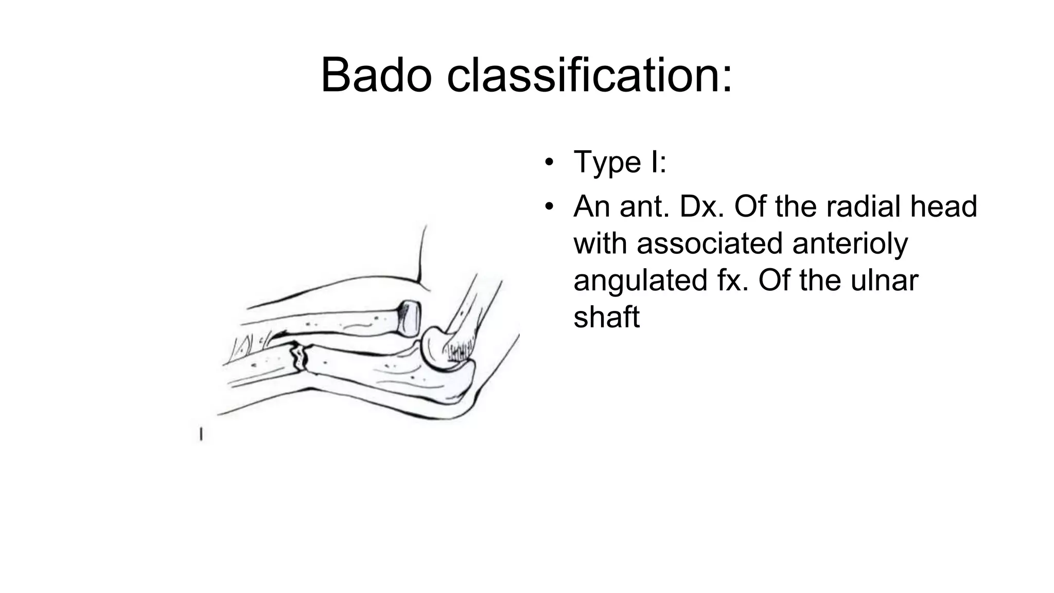
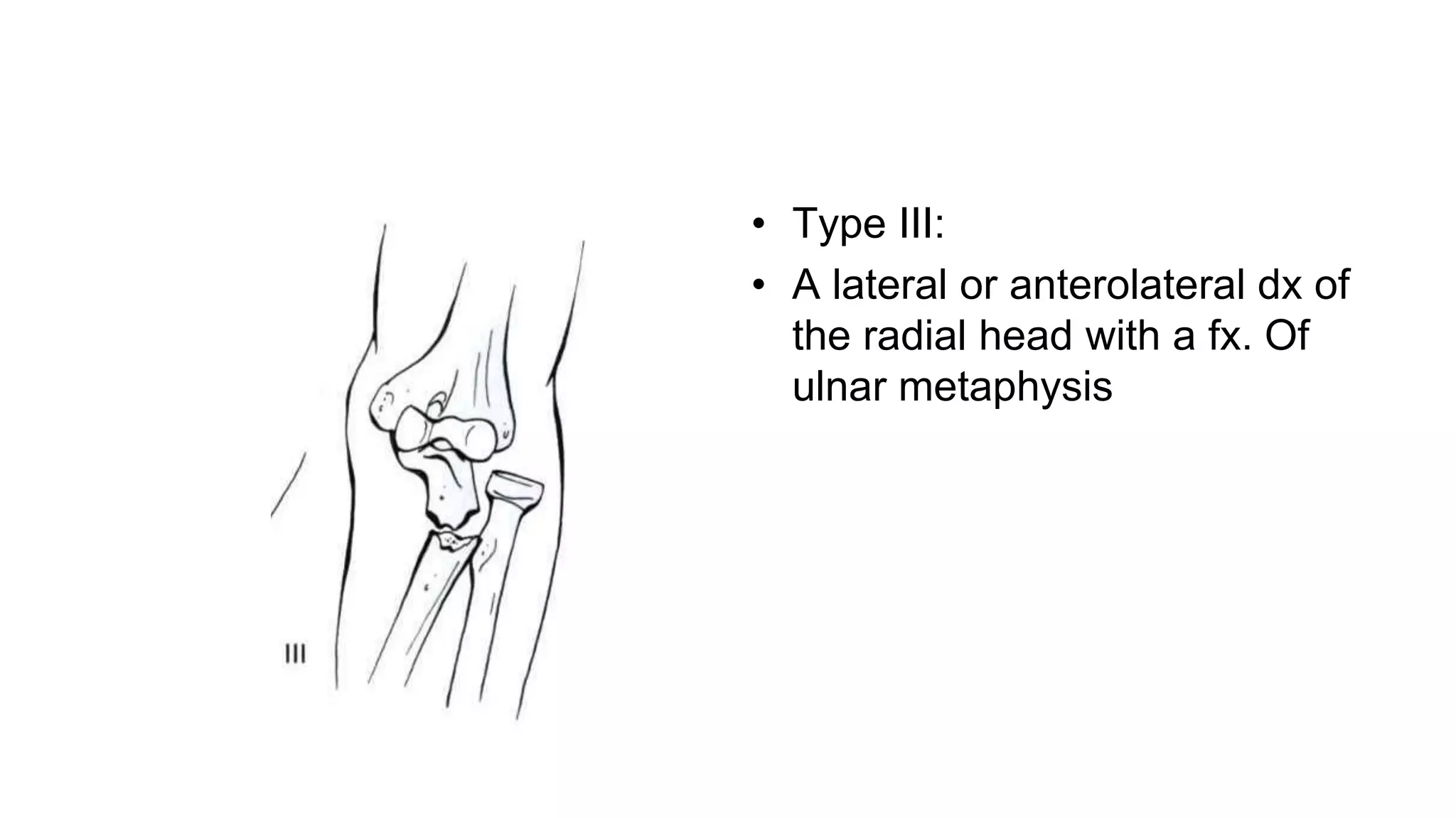
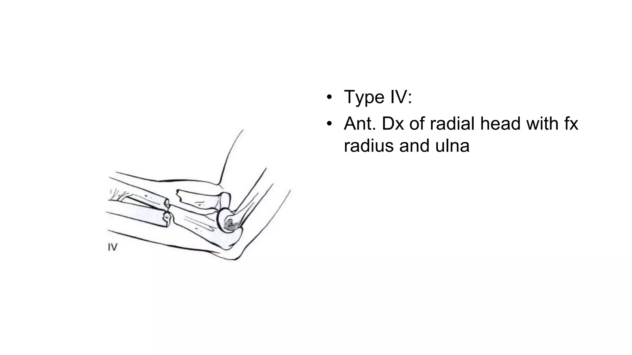
SlideShare a Scribd company logo
FRACTURES OF THE SHAFTS OF
THE RADIUS AND ULNA
Dr. Mohammad Mahdi Shater
Orthopedic Surgery Resident
Baqiyatallah University of Medical Sciences
‫الرحیم‬ ‫الرحمن‬ ‫اهلل‬ ‫بسم‬
Rockwood and Green's Fractures in Adults
Introduction
• Role
• Radial shaft fractures are defined as those occurring between the radial neck
proximally and the junction of the metaphysis and diaphysis distally,
approximately 3 cm proximal to the distal articular surface.
• Ulnar shaft fractures are defined as those occurring between the distal
aspect of the coronoid proximally and the ulnar neck distally.
Epidemiology
• Forearm fx are more common in men than women
because of higher incidence in men of MCA ,falls,sports
participation,…
• The ratio of open fx to closed fx is higher for the foream
than for any other bone except tibia
Mechanism of Injury
• Direct blow is the most common.
-road traffic accidents
-fights in which the defender uses the forearm for protection.
• Less frequently may result from falls and sports injuries.
Night stick
• direct blows occur in fights in which the defender uses the
forearm for protection a direct blow under these
circumstances may cause an isolated ulnar fracture, often
called a "night stick“ fracture, or a Monteggia fracture
dislocation
Signs and Symptoms
• Pain
• Swelling
• Loss of function of the forearm and hand
• Local tenderness
• Deformity
• In closed fractures, a tense swelling and pain on passive
stretch of the forearm may suggest compartment
syndrome.
Associated Musculoskeletal Injuries
• Muscle
• Radioulnar joint disruption(Monteggia , Galeazzi)
Open Diaphyseal Fractures
• The frequency of open fractures ranges from less than 10% in
isolated radial shaft fractures to 43% of both-bone forearm
fractures
Compartment Syndrome
• Swelling ,Pain
• Pain out of proportion with passive extension of the digits is the
most sensitive test to clinically diagnose compartment syndrome
• Compartmental pressures within 30 mm Hg
• absolute compartment pressure is an unreliable indicator
of the requirement for fasciotomy and that a pressure
differential of less than 30 mm Hg between the diastolic
blood pressure, and the ompartment pressure indicates
that immediate fasciotomy is required
Associated Nonmusculoskeletal Injuries
• Head injury
• Nerve injuries
• Artery disruption
• Abdominal and pelvic trauma
Imaging and Other Diagnostic Studies
• Anterior–posterior (AP) and lateral radiographs of the forearm
• Tuberosity view
• Computed tomography (CT)
• Magnetic resonance imaging diagnose injuries to the DRUJ and
associated TFCC
• Ultrasound
Forearm fractures
Classification
• What bone(s) is (are) fractured?
• In what location is the fracture (proximal, middle, distal
third)
• What is the fracture pattern (simple transverse, simple
oblique, comminuted)?
• Is there instability at the distal or proximal radioulnar
joint?
• Is the fracture open or closed?
• Is a previous implant present?
• Is a previous deformity present?
• Is the bone stock normal?
OTA
Classification
Type A fractures
• are simple fractures of the ulna, radius, or both bones. In Al fractures there is
a simple fracture of the ulna and the radius is intact. In A2 fractures there is a
simple fractures of the radius and the ulna is intact. In both these groups, .1
refers to an oblique fractures, .2 to a transverse fracture, and .3 to a fracture
associated with the dislocation. The A1.3 fracture represents a Monteggia
fracture dislocation and the A2.3 fracture represents a Galeazzi fracture
dislocation.
• In the A3 group, .1 refers to a radial fracture in the proximal third of the bone,
.2 to a radial fracture in the middle third, and .3 to a radial fracture in the
distal third.
Type B fractures
• are wedge fractures of either the ulna (Bl), the radius (B2), or both
the radius and ulna (B3). In groups Bl and B2, .1 refers to an
intact wedge, .2 to a fragmented wedge, and .3 to an associated
dislocation with B1.3 fractures being Monteggia fracture
dislocations and B2.3 fractures being Galeazzi fracture
dislocations. In the B3 group, B3.1 fractures have an ulnar wedge
and a simple fracture of the radius, B3.2 fractures have a radial
wedge and a simple fracture of the ulna, and B3.3 fractures have
radial and ulnar wedges
Type C fractures
• are complex fractures. Cl fractures are complex fractures of the ulna, C2
fractures involve the radius, and C3 fractures involve both the radius and
ulna. In Cl.l fractures there is a bifocal fracture of the ulna with an intact
radius, in Cl.2 fractures there is a bifocal fracture of the ulna with a radial
fracture, and in Cl.3 fractures the ulnar fracture is irregular. In C2.1 fractures
the radial fracture is bifocal and the ulna is intact. In C2.2 fractures the radial
fracture is bifocal but the ulna is fractured, and in C2.3 fractures the radial
fracture is irregular. In C3.1 fractures both bones show a bifocal fracture and
in C3.2 fractures there is a bifocal fracture of one bone with an irregular
fracture of the other. In the rare C3.3 fracture, both fractures are irregular or
comminuted.
Monteggia Fracture
• Fractures between the proximal third of the ulna
accompanied by radial head dx.
• Bado divided Monteggia lesions into four types with the
classification depending on the direction of the radial head
Bado classification:
• Type I:
• An ant. Dx. Of the radial head
with associated anterioly
angulated fx. Of the ulnar
shaft
• Type II:post. Dx. Radial head
with a poteriorly angulated fx
of ulna
• Type III:
• A lateral or anterolateral dx of
the radial head with a fx. Of
ulnar metaphysis
• Type IV:
• Ant. Dx of radial head with fx
radius and ulna
Galeazzi Fracture
• Dislocation or subluxation of the DRUJ in association with
a solitary fx. of the radius at the junction of the middle and
distal third is called Galeazzi fx.
• Injuries to the DRUj are generally subdivided into stable,
partially unstable (subluxation), and unstable.
• Open reduction and internal fixation of the radius depends
on DRUj stability.
• Most authorities indicate that :
• about 60% of Monteggia fracture dislocations are type I
• 15% are type II
• 10% are type III (virtually only occur in children)
• 10% are type IV
Anatomy
Forearm fractures can be regarded as articular fractures as
slight deviations in orientation of the radius and ulna will
significantly decrease the forearm's rotation and impair
the positioning and function of the hand.
Consist of :
• Ulna : Which is straight
• Radius: Bowed
• Interrosseous membrane occupied the space between
them
• The central band is approx, 3.5 cm wide running
obliquelyfrom proximal origin on the radius to its distal
insertion on the ulna
Anatomy
• Radius
• Ulna
• Interosseous Space
• Triangular Fibrocartilage Complex
• Proximal Radioulnar Joint
• Muscles
• Nerves
• Arteries
CURRENT TREATMENT OPTIONS
• Nonoperative Treatment
• Operative Treatment
• External fixator
• Intramedullary Nailing
• Plating
Surgical Approach
• Ulna
• Radius
• Volar Approach of Henry
• Posterior (Thompson) approach
Operative Treatment
External fixator:
• There are only a few indications for the use of external
fixation in forearm fx:
1- Initial management of open fractures of the radius
and ulna associated with extensive soft tissue damage
• 2-Maintaining the length in fx with severe bone loss
2- multiply-injured patient
Intramedullary Nailing
• intramedullary nails are not as strong and do not maintain
forearm reduction as well as plate osteosynthesis
• Children vs adult
• Type of Nails
• Indications/Contraindications
Plating
• Internal fixation with plates allows excellent control of the
fracture
fragments and therefore permits accurate restoration of the
anatomy, which remains the key principle in treating
forearm
fractures as it preserves maximal forearm function
• The plates most widely used for internal fixation of
forearm fractures are the 3.5 (DCP) and the 3.5 limited
contact-dynamic compression plate (LC-DCP)
• In comparison to the DCP, the contact
area between the bone and the LC-OCP is reduced by
about 50%
• The concept of limited contact between the plate and the
bone has lead to the development of the point contact-
fixator (PC-Fix) in which the contact area between the
plate and the underlying bone is reduced to two contact
points every 16 mm .Fixation achived between the screws
and the bone, like an external fixator.
AUTHORS' PREFERRED TREATMENT
Nonoperative treatment:
-undisplaced fractures
-in patients with significant comorbidities
Operative treatment:
-For the treatment of forearm diaphyseal fractures the best
device is plate osteosynthesis(3.5-mm LC-DCP).
Pearls and Pitfalls
Pearls
• There is a high incidence of associated musculoskeletal injury.
• Tense swelling in the forearm suggests compartment syndrome.
• In type II Monteggia fracture dislocations, an anterior triangular or
quadrilateral anterior fragment must be looked for on x-rays.
• The results of using the 3.5-mm LC-DCPhave not been bettered
by more modern plates.
Pitfalls
• Subluxation of dislocation of the DRUJ may develop slowly.
• Failure to restore the radial bow will restrict forearm rotation.
• Intramedullary nailing frequently does not restore the radial bow.
• In initially undisplaced fractures, nonoperative management can be
• associated with poor results.
• Minimally invasive techniques are not recommended.
• Early plate removal may cause refracture.
• Narrowing of the interosseous space may cause radio-ulnar synostosis.
COMPLICATIONS
• Compartment Syndrome
• Malunion
• Nonunion
• Infection
• Plate Removal and Refracture
• Radio-Ulnar Synostosis
• Nerve palsy(FPL)
Compatment syndrome
• One pathway is tissue hypoxia followed by swelling which further
reduces the perfusion pressure at the capillary level, eventually
leading to ischemic muscle and myonecrosis.
• Another more common pathway is direct or indirect muscle
damage leading to muscle swelling followed by increased
intracompartmental pressure
• pressure will lead to reduced capillary blood flow, muscle
ischemia, and myonecrosis
• The forearm is the most common site for compartment syndrome
in the upper extremity.
The three compartments of the forearm include the anterior (or flexor
compartment), the posterior
(or extensor compartment), and the mobile wad (including
• brachioradialis and extensor carpi radialis longus and brevis).
• Flexor digitorum profundus and flexor pollicis longus are the most severely
affected muscles because of their deep location.
Malunion
• It seems therefore that early correction yields better
results than late correction if osteotomy is required.
Nonunion
• In general, fracture nonunion results from unstable
fixation or a compromised blood supply secondary to the
severity of the injury or poor surgical technique.
Infection
• If infection does occur, surgical treatment with an
adequate debridement is recommended.
• Implant removal should not be under taken
• appropriate antibiotic treatment depending on the bacteria
involved in the infection.
Plate removal
• It has been suggested that remodeling takes up to 12-18
months to complete
• 4-6 w brace or splint
• We do not advocate routine plate removal in the
asymptomatic patient
General
• Open fractures
• Infection
• Multiply-injuredpatients with head trauma
• Delayed internal fixation
Surgical (less common)
• Narrowing the interosseous space by nonanatomic reduction
• Use of too long screws
• Bone grafting
 Indomethasin and Radiotherapy
Radio-Ulnar Synostosis
Assessment of peripheral n. function:
• Making a fist (median and ulnar n.)
• Wrist and finger extension (radial n.)
• Abd. of extended fingers (ulnar n. when radial n.is intact)
• Extension in the IP joint of the thumb (posterior interosseous n.)
• Flexion in the IP joint of the thumb (anterior interosseous n.)
Assessment of Blood Supply
• Lesion of the medial collateral ligament complex of the
elbow?
• Lesion of the lateral collateral ligament complex of the
elbow?
• Lesion of radio-carpal ligaments?
• Pending or established compartment syndrome?
• Collateral circulation of the forearm in the presence of either isolated radial or
ulnar arterial damage is usually sufficient to maintain viability of the hand and
forearm.
• Viability may even be maintained if both the radial and ulnar arteries are
damaged because the longitudinally orientated collateral vessels may still
provide sufficient blood supply
• If one major artery is intact and there is adequate perfusion to the hand, the
damaged vessel does not have to be repaired.
• However, in combination with nerve injuries, it has been argued that recovery
of the associated nerve lesion will be improved by an enhanced blood supply
and vascular repair is therefore advocated.
Assessment of Bony Injury
Proximal radio-ulnar joint
• Dislocation or abnormally wide joint space?
• Direction of the dislocation?
• Is the annular ligament probably torn or has the radial
head just slipped out of the intact annular ligament?
Interosseous membrane
• Is the membrane torn? If so, proximally or distally?
Distal radio-ulnar joint
• Dislocation
• Abnormal separation
• Shortening of the radius relative to the ulnar head
• Nerve injuries in closed forearm fractures are relatively
uncommon.
• posterior interosseous n. most commonly
• Most nerve injuries are neuropraxias
• Collateral circulation of the forearm in the presence of
either isolated radial or ulnar arterial damage is usually
sufficient to maintain viability of the hand and forearm.
Viability may even be maintained if both the radial and
ulnar arteries are damaged because the longitudinally
orientated collateral vessels may still provide sufficient
blood supply

More Related Content

What's hot

Phalangeal fractures of hand
Phalangeal fractures of handPhalangeal fractures of hand
Phalangeal fractures of hand
Darshan K Swamy
 
Distal Humerus Fractures.pptx
Distal Humerus Fractures.pptxDistal Humerus Fractures.pptx
Distal Humerus Fractures.pptx
SethiNet presentations
 
Subtrochanteric
SubtrochantericSubtrochanteric
Subtrochanteric
Orthosurg2016
 
External fixator
External fixatorExternal fixator
External fixator
Dr. Ditesh Jain
 
Distal radioulnar joint
Distal radioulnar joint Distal radioulnar joint
Distal radioulnar joint orthoprince
 
Ankle fractures
Ankle fracturesAnkle fractures
Ankle fractures
Dr.Anshu Sharma
 
Distal radius fractures
Distal radius fracturesDistal radius fractures
Distal radius fractures
Asi-oqua Bassey
 
Fracture IT Femur
Fracture IT FemurFracture IT Femur
Fracture IT Femur
AbhishekPathak218
 
Proximal humerus fracture Management
Proximal humerus  fracture ManagementProximal humerus  fracture Management
Proximal humerus fracture Managementvaruntandra
 
Biomechanics and biology of relative stability
Biomechanics and biology of relative stabilityBiomechanics and biology of relative stability
Biomechanics and biology of relative stability
Orthosurg2016
 
Surgical Approaches to distal humerus fractures - DR.S.SENTHIL SAILESH, M.S...
 Surgical Approaches to distal  humerus fractures - DR.S.SENTHIL SAILESH, M.S... Surgical Approaches to distal  humerus fractures - DR.S.SENTHIL SAILESH, M.S...
Surgical Approaches to distal humerus fractures - DR.S.SENTHIL SAILESH, M.S...
Senthil sailesh
 
Pilon Fractures
Pilon FracturesPilon Fractures
Pilon Fracturesjfreshour
 
Henry versus thompson approach for fixation of proximal third radial shaft fr...
Henry versus thompson approach for fixation of proximal third radial shaft fr...Henry versus thompson approach for fixation of proximal third radial shaft fr...
Henry versus thompson approach for fixation of proximal third radial shaft fr...
BipulBorthakur
 
Acetabulum fractures
Acetabulum fractures  Acetabulum fractures
Acetabulum fractures orthoprince
 
AO Principles of Fracture treatment & Different Implants.
AO Principles of Fracture treatment & Different Implants.AO Principles of Fracture treatment & Different Implants.
AO Principles of Fracture treatment & Different Implants.
Dr.Anshu Sharma
 
Approaches to Acetabulum- Dr Sharan
Approaches to Acetabulum- Dr SharanApproaches to Acetabulum- Dr Sharan
Approaches to Acetabulum- Dr Sharan
T Sharan Achar
 
Trochanteric fractures
Trochanteric fracturesTrochanteric fractures
Trochanteric fractures
Ahmad Sulong
 
Tips, tricks and pitfalls of proximal femoral nailing (PFN)
Tips, tricks and pitfalls of proximal femoral nailing (PFN)Tips, tricks and pitfalls of proximal femoral nailing (PFN)
Tips, tricks and pitfalls of proximal femoral nailing (PFN)
Puneeth Pai
 

What's hot (20)

Phalangeal fractures of hand
Phalangeal fractures of handPhalangeal fractures of hand
Phalangeal fractures of hand
 
Distal Humerus Fractures.pptx
Distal Humerus Fractures.pptxDistal Humerus Fractures.pptx
Distal Humerus Fractures.pptx
 
Subtrochanteric
SubtrochantericSubtrochanteric
Subtrochanteric
 
External fixator
External fixatorExternal fixator
External fixator
 
Distal radioulnar joint
Distal radioulnar joint Distal radioulnar joint
Distal radioulnar joint
 
Ankle fractures
Ankle fracturesAnkle fractures
Ankle fractures
 
Distal radius fractures
Distal radius fracturesDistal radius fractures
Distal radius fractures
 
Fracture IT Femur
Fracture IT FemurFracture IT Femur
Fracture IT Femur
 
Proximal humerus fracture Management
Proximal humerus  fracture ManagementProximal humerus  fracture Management
Proximal humerus fracture Management
 
Biomechanics and biology of relative stability
Biomechanics and biology of relative stabilityBiomechanics and biology of relative stability
Biomechanics and biology of relative stability
 
Surgical Approaches to distal humerus fractures - DR.S.SENTHIL SAILESH, M.S...
 Surgical Approaches to distal  humerus fractures - DR.S.SENTHIL SAILESH, M.S... Surgical Approaches to distal  humerus fractures - DR.S.SENTHIL SAILESH, M.S...
Surgical Approaches to distal humerus fractures - DR.S.SENTHIL SAILESH, M.S...
 
Suzukiframepaper
SuzukiframepaperSuzukiframepaper
Suzukiframepaper
 
Pilon Fractures
Pilon FracturesPilon Fractures
Pilon Fractures
 
Henry versus thompson approach for fixation of proximal third radial shaft fr...
Henry versus thompson approach for fixation of proximal third radial shaft fr...Henry versus thompson approach for fixation of proximal third radial shaft fr...
Henry versus thompson approach for fixation of proximal third radial shaft fr...
 
Acetabulum fractures
Acetabulum fractures  Acetabulum fractures
Acetabulum fractures
 
AO Principles of Fracture treatment & Different Implants.
AO Principles of Fracture treatment & Different Implants.AO Principles of Fracture treatment & Different Implants.
AO Principles of Fracture treatment & Different Implants.
 
Approaches to Acetabulum- Dr Sharan
Approaches to Acetabulum- Dr SharanApproaches to Acetabulum- Dr Sharan
Approaches to Acetabulum- Dr Sharan
 
Phalanx Fracture
Phalanx FracturePhalanx Fracture
Phalanx Fracture
 
Trochanteric fractures
Trochanteric fracturesTrochanteric fractures
Trochanteric fractures
 
Tips, tricks and pitfalls of proximal femoral nailing (PFN)
Tips, tricks and pitfalls of proximal femoral nailing (PFN)Tips, tricks and pitfalls of proximal femoral nailing (PFN)
Tips, tricks and pitfalls of proximal femoral nailing (PFN)
 

Similar to Radius and ulnar shaft fx

Intertrochanteric fractures and its management with DHS or PFN or Arthroplast...
Intertrochanteric fractures and its management with DHS or PFN or Arthroplast...Intertrochanteric fractures and its management with DHS or PFN or Arthroplast...
Intertrochanteric fractures and its management with DHS or PFN or Arthroplast...
ASRAM Medical College, Eluru, Andhra Pradesh
 
Cervical trauma
Cervical traumaCervical trauma
Cervical trauma
Ali Jiwani
 
Cervical spine injuries.pptx
Cervical spine injuries.pptxCervical spine injuries.pptx
Cervical spine injuries.pptx
Sairamakrishnan Sivadasan
 
Distal Radioulnar Joint (DRUJ) Injuries
Distal Radioulnar Joint (DRUJ) InjuriesDistal Radioulnar Joint (DRUJ) Injuries
Distal Radioulnar Joint (DRUJ) Injuries
Kaushik Velugoori
 
Upper extremity fracture principles
Upper extremity fracture principlesUpper extremity fracture principles
Upper extremity fracture principles
YudiNug1
 
neck x ray.pptx
neck x ray.pptxneck x ray.pptx
neck x ray.pptx
FatimaAmirlou
 
Radius and Ulna Shaft Fracture
Radius and Ulna Shaft  FractureRadius and Ulna Shaft  Fracture
Radius and Ulna Shaft Fracture
Dr Sandip Biswas
 
Comparative study of ORIF with philos plate vs CRIF with k wiring of Neers 2p...
Comparative study of ORIF with philos plate vs CRIF with k wiring of Neers 2p...Comparative study of ORIF with philos plate vs CRIF with k wiring of Neers 2p...
Comparative study of ORIF with philos plate vs CRIF with k wiring of Neers 2p...
BalagangadharaC
 
Upper and Lower Extremity fractures
Upper and Lower Extremity fracturesUpper and Lower Extremity fractures
Upper and Lower Extremity fractures
Aubreyvale Sagun
 
Femoral fracture
Femoral fractureFemoral fracture
Femoral fracture
aya tya
 
Imaging of atlanto occipital and atlantoaxial traumatic injuries
Imaging of atlanto occipital and atlantoaxial traumatic injuriesImaging of atlanto occipital and atlantoaxial traumatic injuries
Imaging of atlanto occipital and atlantoaxial traumatic injuries
Sumiya Arshad
 
distal radius # ppt
 distal radius # ppt distal radius # ppt
distal radius # ppt
rangaraya medical college
 
femoral neck fracture .pptx
femoral neck fracture .pptxfemoral neck fracture .pptx
femoral neck fracture .pptx
BiehasAlwakaf1
 
C2 (Axis) cervical Spine Fracture Presentation
C2 (Axis) cervical Spine Fracture PresentationC2 (Axis) cervical Spine Fracture Presentation
C2 (Axis) cervical Spine Fracture Presentation
Anas Ahmed
 
fractures around Elbow.pdf
fractures around Elbow.pdffractures around Elbow.pdf
fractures around Elbow.pdf
FuadNaji1
 
Terrible triad of the Elbow
Terrible triad of the ElbowTerrible triad of the Elbow
Terrible triad of the Elbow
Libin Thomas
 
Forearm Fractures of Adults
Forearm Fractures of AdultsForearm Fractures of Adults
Forearm Fractures of Adults
Pulasthi Kanchana
 
fracture and dislocation ppt . Almas khan. khorfakkhan hospital dubai
fracture and dislocation ppt . Almas khan. khorfakkhan hospital dubaifracture and dislocation ppt . Almas khan. khorfakkhan hospital dubai
fracture and dislocation ppt . Almas khan. khorfakkhan hospital dubai
almasmkm
 
Clavicle fracture
Clavicle fractureClavicle fracture
Clavicle fracture
Mohammad Mahdi Shater
 
Nailing it hip fractures short versus long; locked versus non locked
Nailing it hip fractures short versus long; locked versus non lockedNailing it hip fractures short versus long; locked versus non locked
Nailing it hip fractures short versus long; locked versus non locked
Love2jaipal
 

Similar to Radius and ulnar shaft fx (20)

Intertrochanteric fractures and its management with DHS or PFN or Arthroplast...
Intertrochanteric fractures and its management with DHS or PFN or Arthroplast...Intertrochanteric fractures and its management with DHS or PFN or Arthroplast...
Intertrochanteric fractures and its management with DHS or PFN or Arthroplast...
 
Cervical trauma
Cervical traumaCervical trauma
Cervical trauma
 
Cervical spine injuries.pptx
Cervical spine injuries.pptxCervical spine injuries.pptx
Cervical spine injuries.pptx
 
Distal Radioulnar Joint (DRUJ) Injuries
Distal Radioulnar Joint (DRUJ) InjuriesDistal Radioulnar Joint (DRUJ) Injuries
Distal Radioulnar Joint (DRUJ) Injuries
 
Upper extremity fracture principles
Upper extremity fracture principlesUpper extremity fracture principles
Upper extremity fracture principles
 
neck x ray.pptx
neck x ray.pptxneck x ray.pptx
neck x ray.pptx
 
Radius and Ulna Shaft Fracture
Radius and Ulna Shaft  FractureRadius and Ulna Shaft  Fracture
Radius and Ulna Shaft Fracture
 
Comparative study of ORIF with philos plate vs CRIF with k wiring of Neers 2p...
Comparative study of ORIF with philos plate vs CRIF with k wiring of Neers 2p...Comparative study of ORIF with philos plate vs CRIF with k wiring of Neers 2p...
Comparative study of ORIF with philos plate vs CRIF with k wiring of Neers 2p...
 
Upper and Lower Extremity fractures
Upper and Lower Extremity fracturesUpper and Lower Extremity fractures
Upper and Lower Extremity fractures
 
Femoral fracture
Femoral fractureFemoral fracture
Femoral fracture
 
Imaging of atlanto occipital and atlantoaxial traumatic injuries
Imaging of atlanto occipital and atlantoaxial traumatic injuriesImaging of atlanto occipital and atlantoaxial traumatic injuries
Imaging of atlanto occipital and atlantoaxial traumatic injuries
 
distal radius # ppt
 distal radius # ppt distal radius # ppt
distal radius # ppt
 
femoral neck fracture .pptx
femoral neck fracture .pptxfemoral neck fracture .pptx
femoral neck fracture .pptx
 
C2 (Axis) cervical Spine Fracture Presentation
C2 (Axis) cervical Spine Fracture PresentationC2 (Axis) cervical Spine Fracture Presentation
C2 (Axis) cervical Spine Fracture Presentation
 
fractures around Elbow.pdf
fractures around Elbow.pdffractures around Elbow.pdf
fractures around Elbow.pdf
 
Terrible triad of the Elbow
Terrible triad of the ElbowTerrible triad of the Elbow
Terrible triad of the Elbow
 
Forearm Fractures of Adults
Forearm Fractures of AdultsForearm Fractures of Adults
Forearm Fractures of Adults
 
fracture and dislocation ppt . Almas khan. khorfakkhan hospital dubai
fracture and dislocation ppt . Almas khan. khorfakkhan hospital dubaifracture and dislocation ppt . Almas khan. khorfakkhan hospital dubai
fracture and dislocation ppt . Almas khan. khorfakkhan hospital dubai
 
Clavicle fracture
Clavicle fractureClavicle fracture
Clavicle fracture
 
Nailing it hip fractures short versus long; locked versus non locked
Nailing it hip fractures short versus long; locked versus non lockedNailing it hip fractures short versus long; locked versus non locked
Nailing it hip fractures short versus long; locked versus non locked
 

More from Mohammad Mahdi Shater

Lateral condylar fractures of the distal humerus
Lateral condylar fractures of the distal humerusLateral condylar fractures of the distal humerus
Lateral condylar fractures of the distal humerus
Mohammad Mahdi Shater
 
Surgical techniques
Surgical techniques Surgical techniques
Surgical techniques
Mohammad Mahdi Shater
 
Pelvic and hip anatomy
Pelvic and hip anatomyPelvic and hip anatomy
Pelvic and hip anatomy
Mohammad Mahdi Shater
 
Ankle fracture
Ankle fractureAnkle fracture
Ankle fracture
Mohammad Mahdi Shater
 
Elbow dislocations and terrible triad
Elbow dislocations and terrible triadElbow dislocations and terrible triad
Elbow dislocations and terrible triad
Mohammad Mahdi Shater
 
Postoperative care of femoral shaft and supracondylar fx
Postoperative care of femoral shaft and supracondylar fxPostoperative care of femoral shaft and supracondylar fx
Postoperative care of femoral shaft and supracondylar fx
Mohammad Mahdi Shater
 
Injuries to the sternoclavicular joint
Injuries to the sternoclavicular jointInjuries to the sternoclavicular joint
Injuries to the sternoclavicular joint
Mohammad Mahdi Shater
 
Femoral neck fracture
Femoral neck fractureFemoral neck fracture
Femoral neck fracture
Mohammad Mahdi Shater
 
Pathologic fractures
Pathologic fracturesPathologic fractures
Pathologic fractures
Mohammad Mahdi Shater
 
Evaluation of postpartum depression and relationship its with spiritual healt...
Evaluation of postpartum depression and relationship its with spiritual healt...Evaluation of postpartum depression and relationship its with spiritual healt...
Evaluation of postpartum depression and relationship its with spiritual healt...
Mohammad Mahdi Shater
 
Neonatal hypopituitarism approaches to diagnosis and treatment
Neonatal hypopituitarism approaches to diagnosis and treatmentNeonatal hypopituitarism approaches to diagnosis and treatment
Neonatal hypopituitarism approaches to diagnosis and treatment
Mohammad Mahdi Shater
 
TCA
TCATCA
Chest trauma
Chest traumaChest trauma
Chest trauma
Mohammad Mahdi Shater
 
Chart & structure
Chart & structureChart & structure
Chart & structure
Mohammad Mahdi Shater
 
Khorabad
KhorabadKhorabad
Metabolic risk factors in children with asymptomatic hematuria
Metabolic risk factors in children with asymptomatic hematuriaMetabolic risk factors in children with asymptomatic hematuria
Metabolic risk factors in children with asymptomatic hematuria
Mohammad Mahdi Shater
 
Pharmacology
PharmacologyPharmacology
Pharmacology
Mohammad Mahdi Shater
 
Intravenous accsess
Intravenous accsessIntravenous accsess
Intravenous accsess
Mohammad Mahdi Shater
 

More from Mohammad Mahdi Shater (20)

Lateral condylar fractures of the distal humerus
Lateral condylar fractures of the distal humerusLateral condylar fractures of the distal humerus
Lateral condylar fractures of the distal humerus
 
Surgical techniques
Surgical techniques Surgical techniques
Surgical techniques
 
Pelvic and hip anatomy
Pelvic and hip anatomyPelvic and hip anatomy
Pelvic and hip anatomy
 
Ankle fracture
Ankle fractureAnkle fracture
Ankle fracture
 
Elbow dislocations and terrible triad
Elbow dislocations and terrible triadElbow dislocations and terrible triad
Elbow dislocations and terrible triad
 
Postoperative care of femoral shaft and supracondylar fx
Postoperative care of femoral shaft and supracondylar fxPostoperative care of femoral shaft and supracondylar fx
Postoperative care of femoral shaft and supracondylar fx
 
Injuries to the sternoclavicular joint
Injuries to the sternoclavicular jointInjuries to the sternoclavicular joint
Injuries to the sternoclavicular joint
 
Femoral neck fracture
Femoral neck fractureFemoral neck fracture
Femoral neck fracture
 
Pathologic fractures
Pathologic fracturesPathologic fractures
Pathologic fractures
 
Ankle
AnkleAnkle
Ankle
 
Evaluation of postpartum depression and relationship its with spiritual healt...
Evaluation of postpartum depression and relationship its with spiritual healt...Evaluation of postpartum depression and relationship its with spiritual healt...
Evaluation of postpartum depression and relationship its with spiritual healt...
 
Neonatal hypopituitarism approaches to diagnosis and treatment
Neonatal hypopituitarism approaches to diagnosis and treatmentNeonatal hypopituitarism approaches to diagnosis and treatment
Neonatal hypopituitarism approaches to diagnosis and treatment
 
Sepsis
SepsisSepsis
Sepsis
 
TCA
TCATCA
TCA
 
Chest trauma
Chest traumaChest trauma
Chest trauma
 
Chart & structure
Chart & structureChart & structure
Chart & structure
 
Khorabad
KhorabadKhorabad
Khorabad
 
Metabolic risk factors in children with asymptomatic hematuria
Metabolic risk factors in children with asymptomatic hematuriaMetabolic risk factors in children with asymptomatic hematuria
Metabolic risk factors in children with asymptomatic hematuria
 
Pharmacology
PharmacologyPharmacology
Pharmacology
 
Intravenous accsess
Intravenous accsessIntravenous accsess
Intravenous accsess
 

Recently uploaded

Are There Any Natural Remedies To Treat Syphilis.pdf
Are There Any Natural Remedies To Treat Syphilis.pdfAre There Any Natural Remedies To Treat Syphilis.pdf
Are There Any Natural Remedies To Treat Syphilis.pdf
Little Cross Family Clinic
 
MANAGEMENT OF ATRIOVENTRICULAR CONDUCTION BLOCK.pdf
MANAGEMENT OF ATRIOVENTRICULAR CONDUCTION BLOCK.pdfMANAGEMENT OF ATRIOVENTRICULAR CONDUCTION BLOCK.pdf
MANAGEMENT OF ATRIOVENTRICULAR CONDUCTION BLOCK.pdf
Jim Jacob Roy
 
Report Back from SGO 2024: What’s the Latest in Cervical Cancer?
Report Back from SGO 2024: What’s the Latest in Cervical Cancer?Report Back from SGO 2024: What’s the Latest in Cervical Cancer?
Report Back from SGO 2024: What’s the Latest in Cervical Cancer?
bkling
 
ARTIFICIAL INTELLIGENCE IN HEALTHCARE.pdf
ARTIFICIAL INTELLIGENCE IN  HEALTHCARE.pdfARTIFICIAL INTELLIGENCE IN  HEALTHCARE.pdf
ARTIFICIAL INTELLIGENCE IN HEALTHCARE.pdf
Anujkumaranit
 
The hemodynamic and autonomic determinants of elevated blood pressure in obes...
The hemodynamic and autonomic determinants of elevated blood pressure in obes...The hemodynamic and autonomic determinants of elevated blood pressure in obes...
The hemodynamic and autonomic determinants of elevated blood pressure in obes...
Catherine Liao
 
Factory Supply Best Quality Pmk Oil CAS 28578–16–7 PMK Powder in Stock
Factory Supply Best Quality Pmk Oil CAS 28578–16–7 PMK Powder in StockFactory Supply Best Quality Pmk Oil CAS 28578–16–7 PMK Powder in Stock
Factory Supply Best Quality Pmk Oil CAS 28578–16–7 PMK Powder in Stock
rebeccabio
 
Physiology of Special Chemical Sensation of Taste
Physiology of Special Chemical Sensation of TastePhysiology of Special Chemical Sensation of Taste
Physiology of Special Chemical Sensation of Taste
MedicoseAcademics
 
Physiology of Chemical Sensation of smell.pdf
Physiology of Chemical Sensation of smell.pdfPhysiology of Chemical Sensation of smell.pdf
Physiology of Chemical Sensation of smell.pdf
MedicoseAcademics
 
Cervical & Brachial Plexus By Dr. RIG.pptx
Cervical & Brachial Plexus By Dr. RIG.pptxCervical & Brachial Plexus By Dr. RIG.pptx
Cervical & Brachial Plexus By Dr. RIG.pptx
Dr. Rabia Inam Gandapore
 
Flu Vaccine Alert in Bangalore Karnataka
Flu Vaccine Alert in Bangalore KarnatakaFlu Vaccine Alert in Bangalore Karnataka
Flu Vaccine Alert in Bangalore Karnataka
addon Scans
 
The POPPY STUDY (Preconception to post-partum cardiovascular function in prim...
The POPPY STUDY (Preconception to post-partum cardiovascular function in prim...The POPPY STUDY (Preconception to post-partum cardiovascular function in prim...
The POPPY STUDY (Preconception to post-partum cardiovascular function in prim...
Catherine Liao
 
How to Give Better Lectures: Some Tips for Doctors
How to Give Better Lectures: Some Tips for DoctorsHow to Give Better Lectures: Some Tips for Doctors
How to Give Better Lectures: Some Tips for Doctors
LanceCatedral
 
Surgical Site Infections, pathophysiology, and prevention.pptx
Surgical Site Infections, pathophysiology, and prevention.pptxSurgical Site Infections, pathophysiology, and prevention.pptx
Surgical Site Infections, pathophysiology, and prevention.pptx
jval Landero
 
Pulmonary Thromboembolism - etilogy, types, medical- Surgical and nursing man...
Pulmonary Thromboembolism - etilogy, types, medical- Surgical and nursing man...Pulmonary Thromboembolism - etilogy, types, medical- Surgical and nursing man...
Pulmonary Thromboembolism - etilogy, types, medical- Surgical and nursing man...
VarunMahajani
 
Non-respiratory Functions of the Lungs.pdf
Non-respiratory Functions of the Lungs.pdfNon-respiratory Functions of the Lungs.pdf
Non-respiratory Functions of the Lungs.pdf
MedicoseAcademics
 
Lung Cancer: Artificial Intelligence, Synergetics, Complex System Analysis, S...
Lung Cancer: Artificial Intelligence, Synergetics, Complex System Analysis, S...Lung Cancer: Artificial Intelligence, Synergetics, Complex System Analysis, S...
Lung Cancer: Artificial Intelligence, Synergetics, Complex System Analysis, S...
Oleg Kshivets
 
Couples presenting to the infertility clinic- Do they really have infertility...
Couples presenting to the infertility clinic- Do they really have infertility...Couples presenting to the infertility clinic- Do they really have infertility...
Couples presenting to the infertility clinic- Do they really have infertility...
Sujoy Dasgupta
 
Antiulcer drugs Advance Pharmacology .pptx
Antiulcer drugs Advance Pharmacology .pptxAntiulcer drugs Advance Pharmacology .pptx
Antiulcer drugs Advance Pharmacology .pptx
Rohit chaurpagar
 
basicmodesofventilation2022-220313203758.pdf
basicmodesofventilation2022-220313203758.pdfbasicmodesofventilation2022-220313203758.pdf
basicmodesofventilation2022-220313203758.pdf
aljamhori teaching hospital
 
Hemodialysis: Chapter 3, Dialysis Water Unit - Dr.Gawad
Hemodialysis: Chapter 3, Dialysis Water Unit - Dr.GawadHemodialysis: Chapter 3, Dialysis Water Unit - Dr.Gawad
Hemodialysis: Chapter 3, Dialysis Water Unit - Dr.Gawad
NephroTube - Dr.Gawad
 

Recently uploaded (20)

Are There Any Natural Remedies To Treat Syphilis.pdf
Are There Any Natural Remedies To Treat Syphilis.pdfAre There Any Natural Remedies To Treat Syphilis.pdf
Are There Any Natural Remedies To Treat Syphilis.pdf
 
MANAGEMENT OF ATRIOVENTRICULAR CONDUCTION BLOCK.pdf
MANAGEMENT OF ATRIOVENTRICULAR CONDUCTION BLOCK.pdfMANAGEMENT OF ATRIOVENTRICULAR CONDUCTION BLOCK.pdf
MANAGEMENT OF ATRIOVENTRICULAR CONDUCTION BLOCK.pdf
 
Report Back from SGO 2024: What’s the Latest in Cervical Cancer?
Report Back from SGO 2024: What’s the Latest in Cervical Cancer?Report Back from SGO 2024: What’s the Latest in Cervical Cancer?
Report Back from SGO 2024: What’s the Latest in Cervical Cancer?
 
ARTIFICIAL INTELLIGENCE IN HEALTHCARE.pdf
ARTIFICIAL INTELLIGENCE IN  HEALTHCARE.pdfARTIFICIAL INTELLIGENCE IN  HEALTHCARE.pdf
ARTIFICIAL INTELLIGENCE IN HEALTHCARE.pdf
 
The hemodynamic and autonomic determinants of elevated blood pressure in obes...
The hemodynamic and autonomic determinants of elevated blood pressure in obes...The hemodynamic and autonomic determinants of elevated blood pressure in obes...
The hemodynamic and autonomic determinants of elevated blood pressure in obes...
 
Factory Supply Best Quality Pmk Oil CAS 28578–16–7 PMK Powder in Stock
Factory Supply Best Quality Pmk Oil CAS 28578–16–7 PMK Powder in StockFactory Supply Best Quality Pmk Oil CAS 28578–16–7 PMK Powder in Stock
Factory Supply Best Quality Pmk Oil CAS 28578–16–7 PMK Powder in Stock
 
Physiology of Special Chemical Sensation of Taste
Physiology of Special Chemical Sensation of TastePhysiology of Special Chemical Sensation of Taste
Physiology of Special Chemical Sensation of Taste
 
Physiology of Chemical Sensation of smell.pdf
Physiology of Chemical Sensation of smell.pdfPhysiology of Chemical Sensation of smell.pdf
Physiology of Chemical Sensation of smell.pdf
 
Cervical & Brachial Plexus By Dr. RIG.pptx
Cervical & Brachial Plexus By Dr. RIG.pptxCervical & Brachial Plexus By Dr. RIG.pptx
Cervical & Brachial Plexus By Dr. RIG.pptx
 
Flu Vaccine Alert in Bangalore Karnataka
Flu Vaccine Alert in Bangalore KarnatakaFlu Vaccine Alert in Bangalore Karnataka
Flu Vaccine Alert in Bangalore Karnataka
 
The POPPY STUDY (Preconception to post-partum cardiovascular function in prim...
The POPPY STUDY (Preconception to post-partum cardiovascular function in prim...The POPPY STUDY (Preconception to post-partum cardiovascular function in prim...
The POPPY STUDY (Preconception to post-partum cardiovascular function in prim...
 
How to Give Better Lectures: Some Tips for Doctors
How to Give Better Lectures: Some Tips for DoctorsHow to Give Better Lectures: Some Tips for Doctors
How to Give Better Lectures: Some Tips for Doctors
 
Surgical Site Infections, pathophysiology, and prevention.pptx
Surgical Site Infections, pathophysiology, and prevention.pptxSurgical Site Infections, pathophysiology, and prevention.pptx
Surgical Site Infections, pathophysiology, and prevention.pptx
 
Pulmonary Thromboembolism - etilogy, types, medical- Surgical and nursing man...
Pulmonary Thromboembolism - etilogy, types, medical- Surgical and nursing man...Pulmonary Thromboembolism - etilogy, types, medical- Surgical and nursing man...
Pulmonary Thromboembolism - etilogy, types, medical- Surgical and nursing man...
 
Non-respiratory Functions of the Lungs.pdf
Non-respiratory Functions of the Lungs.pdfNon-respiratory Functions of the Lungs.pdf
Non-respiratory Functions of the Lungs.pdf
 
Lung Cancer: Artificial Intelligence, Synergetics, Complex System Analysis, S...
Lung Cancer: Artificial Intelligence, Synergetics, Complex System Analysis, S...Lung Cancer: Artificial Intelligence, Synergetics, Complex System Analysis, S...
Lung Cancer: Artificial Intelligence, Synergetics, Complex System Analysis, S...
 
Couples presenting to the infertility clinic- Do they really have infertility...
Couples presenting to the infertility clinic- Do they really have infertility...Couples presenting to the infertility clinic- Do they really have infertility...
Couples presenting to the infertility clinic- Do they really have infertility...
 
Antiulcer drugs Advance Pharmacology .pptx
Antiulcer drugs Advance Pharmacology .pptxAntiulcer drugs Advance Pharmacology .pptx
Antiulcer drugs Advance Pharmacology .pptx
 
basicmodesofventilation2022-220313203758.pdf
basicmodesofventilation2022-220313203758.pdfbasicmodesofventilation2022-220313203758.pdf
basicmodesofventilation2022-220313203758.pdf
 
Hemodialysis: Chapter 3, Dialysis Water Unit - Dr.Gawad
Hemodialysis: Chapter 3, Dialysis Water Unit - Dr.GawadHemodialysis: Chapter 3, Dialysis Water Unit - Dr.Gawad
Hemodialysis: Chapter 3, Dialysis Water Unit - Dr.Gawad
 

Radius and ulnar shaft fx

  • 1. FRACTURES OF THE SHAFTS OF THE RADIUS AND ULNA Dr. Mohammad Mahdi Shater Orthopedic Surgery Resident Baqiyatallah University of Medical Sciences ‫الرحیم‬ ‫الرحمن‬ ‫اهلل‬ ‫بسم‬ Rockwood and Green's Fractures in Adults
  • 2. Introduction • Role • Radial shaft fractures are defined as those occurring between the radial neck proximally and the junction of the metaphysis and diaphysis distally, approximately 3 cm proximal to the distal articular surface. • Ulnar shaft fractures are defined as those occurring between the distal aspect of the coronoid proximally and the ulnar neck distally.
  • 3. Epidemiology • Forearm fx are more common in men than women because of higher incidence in men of MCA ,falls,sports participation,… • The ratio of open fx to closed fx is higher for the foream than for any other bone except tibia
  • 4. Mechanism of Injury • Direct blow is the most common. -road traffic accidents -fights in which the defender uses the forearm for protection. • Less frequently may result from falls and sports injuries.
  • 5. Night stick • direct blows occur in fights in which the defender uses the forearm for protection a direct blow under these circumstances may cause an isolated ulnar fracture, often called a "night stick“ fracture, or a Monteggia fracture dislocation
  • 6.
  • 7. Signs and Symptoms • Pain • Swelling • Loss of function of the forearm and hand • Local tenderness • Deformity • In closed fractures, a tense swelling and pain on passive stretch of the forearm may suggest compartment syndrome.
  • 8. Associated Musculoskeletal Injuries • Muscle • Radioulnar joint disruption(Monteggia , Galeazzi)
  • 9. Open Diaphyseal Fractures • The frequency of open fractures ranges from less than 10% in isolated radial shaft fractures to 43% of both-bone forearm fractures
  • 10. Compartment Syndrome • Swelling ,Pain • Pain out of proportion with passive extension of the digits is the most sensitive test to clinically diagnose compartment syndrome • Compartmental pressures within 30 mm Hg
  • 11. • absolute compartment pressure is an unreliable indicator of the requirement for fasciotomy and that a pressure differential of less than 30 mm Hg between the diastolic blood pressure, and the ompartment pressure indicates that immediate fasciotomy is required
  • 12. Associated Nonmusculoskeletal Injuries • Head injury • Nerve injuries • Artery disruption • Abdominal and pelvic trauma
  • 13. Imaging and Other Diagnostic Studies • Anterior–posterior (AP) and lateral radiographs of the forearm • Tuberosity view • Computed tomography (CT) • Magnetic resonance imaging diagnose injuries to the DRUJ and associated TFCC • Ultrasound
  • 14.
  • 15. Forearm fractures Classification • What bone(s) is (are) fractured? • In what location is the fracture (proximal, middle, distal third) • What is the fracture pattern (simple transverse, simple oblique, comminuted)? • Is there instability at the distal or proximal radioulnar joint? • Is the fracture open or closed? • Is a previous implant present? • Is a previous deformity present? • Is the bone stock normal?
  • 16. OTA Classification Type A fractures • are simple fractures of the ulna, radius, or both bones. In Al fractures there is a simple fracture of the ulna and the radius is intact. In A2 fractures there is a simple fractures of the radius and the ulna is intact. In both these groups, .1 refers to an oblique fractures, .2 to a transverse fracture, and .3 to a fracture associated with the dislocation. The A1.3 fracture represents a Monteggia fracture dislocation and the A2.3 fracture represents a Galeazzi fracture dislocation. • In the A3 group, .1 refers to a radial fracture in the proximal third of the bone, .2 to a radial fracture in the middle third, and .3 to a radial fracture in the distal third.
  • 17. Type B fractures • are wedge fractures of either the ulna (Bl), the radius (B2), or both the radius and ulna (B3). In groups Bl and B2, .1 refers to an intact wedge, .2 to a fragmented wedge, and .3 to an associated dislocation with B1.3 fractures being Monteggia fracture dislocations and B2.3 fractures being Galeazzi fracture dislocations. In the B3 group, B3.1 fractures have an ulnar wedge and a simple fracture of the radius, B3.2 fractures have a radial wedge and a simple fracture of the ulna, and B3.3 fractures have radial and ulnar wedges
  • 18. Type C fractures • are complex fractures. Cl fractures are complex fractures of the ulna, C2 fractures involve the radius, and C3 fractures involve both the radius and ulna. In Cl.l fractures there is a bifocal fracture of the ulna with an intact radius, in Cl.2 fractures there is a bifocal fracture of the ulna with a radial fracture, and in Cl.3 fractures the ulnar fracture is irregular. In C2.1 fractures the radial fracture is bifocal and the ulna is intact. In C2.2 fractures the radial fracture is bifocal but the ulna is fractured, and in C2.3 fractures the radial fracture is irregular. In C3.1 fractures both bones show a bifocal fracture and in C3.2 fractures there is a bifocal fracture of one bone with an irregular fracture of the other. In the rare C3.3 fracture, both fractures are irregular or comminuted.
  • 19.
  • 20. Monteggia Fracture • Fractures between the proximal third of the ulna accompanied by radial head dx. • Bado divided Monteggia lesions into four types with the classification depending on the direction of the radial head
  • 21. Bado classification: • Type I: • An ant. Dx. Of the radial head with associated anterioly angulated fx. Of the ulnar shaft
  • 22. • Type II:post. Dx. Radial head with a poteriorly angulated fx of ulna
  • 23. • Type III: • A lateral or anterolateral dx of the radial head with a fx. Of ulnar metaphysis
  • 24. • Type IV: • Ant. Dx of radial head with fx radius and ulna
  • 25. Galeazzi Fracture • Dislocation or subluxation of the DRUJ in association with a solitary fx. of the radius at the junction of the middle and distal third is called Galeazzi fx. • Injuries to the DRUj are generally subdivided into stable, partially unstable (subluxation), and unstable. • Open reduction and internal fixation of the radius depends on DRUj stability.
  • 26. • Most authorities indicate that : • about 60% of Monteggia fracture dislocations are type I • 15% are type II • 10% are type III (virtually only occur in children) • 10% are type IV
  • 27. Anatomy Forearm fractures can be regarded as articular fractures as slight deviations in orientation of the radius and ulna will significantly decrease the forearm's rotation and impair the positioning and function of the hand.
  • 28. Consist of : • Ulna : Which is straight • Radius: Bowed • Interrosseous membrane occupied the space between them • The central band is approx, 3.5 cm wide running obliquelyfrom proximal origin on the radius to its distal insertion on the ulna
  • 29. Anatomy • Radius • Ulna • Interosseous Space • Triangular Fibrocartilage Complex • Proximal Radioulnar Joint • Muscles • Nerves • Arteries
  • 30. CURRENT TREATMENT OPTIONS • Nonoperative Treatment • Operative Treatment • External fixator • Intramedullary Nailing • Plating
  • 31.
  • 32. Surgical Approach • Ulna • Radius • Volar Approach of Henry • Posterior (Thompson) approach
  • 33. Operative Treatment External fixator: • There are only a few indications for the use of external fixation in forearm fx: 1- Initial management of open fractures of the radius and ulna associated with extensive soft tissue damage • 2-Maintaining the length in fx with severe bone loss 2- multiply-injured patient
  • 34. Intramedullary Nailing • intramedullary nails are not as strong and do not maintain forearm reduction as well as plate osteosynthesis • Children vs adult • Type of Nails • Indications/Contraindications
  • 35. Plating • Internal fixation with plates allows excellent control of the fracture fragments and therefore permits accurate restoration of the anatomy, which remains the key principle in treating forearm fractures as it preserves maximal forearm function
  • 36. • The plates most widely used for internal fixation of forearm fractures are the 3.5 (DCP) and the 3.5 limited contact-dynamic compression plate (LC-DCP) • In comparison to the DCP, the contact area between the bone and the LC-OCP is reduced by about 50%
  • 37. • The concept of limited contact between the plate and the bone has lead to the development of the point contact- fixator (PC-Fix) in which the contact area between the plate and the underlying bone is reduced to two contact points every 16 mm .Fixation achived between the screws and the bone, like an external fixator.
  • 38. AUTHORS' PREFERRED TREATMENT Nonoperative treatment: -undisplaced fractures -in patients with significant comorbidities Operative treatment: -For the treatment of forearm diaphyseal fractures the best device is plate osteosynthesis(3.5-mm LC-DCP).
  • 39. Pearls and Pitfalls Pearls • There is a high incidence of associated musculoskeletal injury. • Tense swelling in the forearm suggests compartment syndrome. • In type II Monteggia fracture dislocations, an anterior triangular or quadrilateral anterior fragment must be looked for on x-rays. • The results of using the 3.5-mm LC-DCPhave not been bettered by more modern plates.
  • 40. Pitfalls • Subluxation of dislocation of the DRUJ may develop slowly. • Failure to restore the radial bow will restrict forearm rotation. • Intramedullary nailing frequently does not restore the radial bow. • In initially undisplaced fractures, nonoperative management can be • associated with poor results. • Minimally invasive techniques are not recommended. • Early plate removal may cause refracture. • Narrowing of the interosseous space may cause radio-ulnar synostosis.
  • 41. COMPLICATIONS • Compartment Syndrome • Malunion • Nonunion • Infection • Plate Removal and Refracture • Radio-Ulnar Synostosis • Nerve palsy(FPL)
  • 42. Compatment syndrome • One pathway is tissue hypoxia followed by swelling which further reduces the perfusion pressure at the capillary level, eventually leading to ischemic muscle and myonecrosis. • Another more common pathway is direct or indirect muscle damage leading to muscle swelling followed by increased intracompartmental pressure • pressure will lead to reduced capillary blood flow, muscle ischemia, and myonecrosis
  • 43. • The forearm is the most common site for compartment syndrome in the upper extremity. The three compartments of the forearm include the anterior (or flexor compartment), the posterior (or extensor compartment), and the mobile wad (including • brachioradialis and extensor carpi radialis longus and brevis). • Flexor digitorum profundus and flexor pollicis longus are the most severely affected muscles because of their deep location.
  • 44. Malunion • It seems therefore that early correction yields better results than late correction if osteotomy is required.
  • 45. Nonunion • In general, fracture nonunion results from unstable fixation or a compromised blood supply secondary to the severity of the injury or poor surgical technique.
  • 46. Infection • If infection does occur, surgical treatment with an adequate debridement is recommended. • Implant removal should not be under taken • appropriate antibiotic treatment depending on the bacteria involved in the infection.
  • 47. Plate removal • It has been suggested that remodeling takes up to 12-18 months to complete • 4-6 w brace or splint • We do not advocate routine plate removal in the asymptomatic patient
  • 48. General • Open fractures • Infection • Multiply-injuredpatients with head trauma • Delayed internal fixation Surgical (less common) • Narrowing the interosseous space by nonanatomic reduction • Use of too long screws • Bone grafting  Indomethasin and Radiotherapy Radio-Ulnar Synostosis
  • 49. Assessment of peripheral n. function: • Making a fist (median and ulnar n.) • Wrist and finger extension (radial n.) • Abd. of extended fingers (ulnar n. when radial n.is intact) • Extension in the IP joint of the thumb (posterior interosseous n.) • Flexion in the IP joint of the thumb (anterior interosseous n.)
  • 50. Assessment of Blood Supply • Lesion of the medial collateral ligament complex of the elbow? • Lesion of the lateral collateral ligament complex of the elbow? • Lesion of radio-carpal ligaments? • Pending or established compartment syndrome?
  • 51. • Collateral circulation of the forearm in the presence of either isolated radial or ulnar arterial damage is usually sufficient to maintain viability of the hand and forearm. • Viability may even be maintained if both the radial and ulnar arteries are damaged because the longitudinally orientated collateral vessels may still provide sufficient blood supply • If one major artery is intact and there is adequate perfusion to the hand, the damaged vessel does not have to be repaired. • However, in combination with nerve injuries, it has been argued that recovery of the associated nerve lesion will be improved by an enhanced blood supply and vascular repair is therefore advocated.
  • 52. Assessment of Bony Injury Proximal radio-ulnar joint • Dislocation or abnormally wide joint space? • Direction of the dislocation? • Is the annular ligament probably torn or has the radial head just slipped out of the intact annular ligament?
  • 53. Interosseous membrane • Is the membrane torn? If so, proximally or distally? Distal radio-ulnar joint • Dislocation • Abnormal separation • Shortening of the radius relative to the ulnar head
  • 54. • Nerve injuries in closed forearm fractures are relatively uncommon. • posterior interosseous n. most commonly • Most nerve injuries are neuropraxias
  • 55. • Collateral circulation of the forearm in the presence of either isolated radial or ulnar arterial damage is usually sufficient to maintain viability of the hand and forearm. Viability may even be maintained if both the radial and ulnar arteries are damaged because the longitudinally orientated collateral vessels may still provide sufficient blood supply