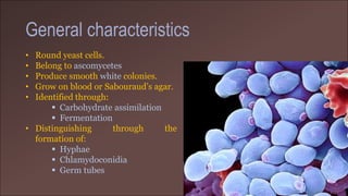
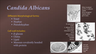
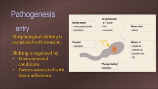
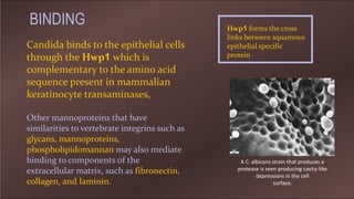
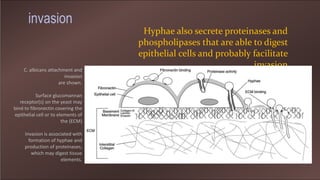
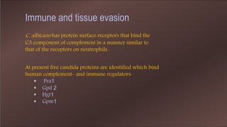
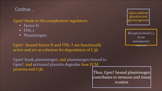
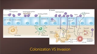
Pathogenesis of Candida Albicans
Madiha Siddiqui
M.Phil-1st
Molecular Mechanism of
Pathogenesis
General characteristics
• Round yeast cells.
• Belong to ascomycetes
• Produce smooth white colonies.
• Grow on blood or Sabouraud’s agar.
• Identified through:
 Carbohydrate assimilation
 Fermentation
• Distinguishing through the
formation of:
 Hyphae
 Chlamydoconidia
 Germ tubes
Candida Albicans
Different Morphological forms:
 Yeast
 Hyphae
 Pseudohyphae
When incubated
at 37°C, C. albicans
rapidly
forms elongated
hyphae called
germ tubes.
On specialized
media,
C. albicans forms
thick walled
chlamydoconidia,
which
differentiate it
from other
Candida
species.
Cell wall includes:
 β-glucan
 Chitin
 Lipid
 Mannan covalently bonded
with protein Cell wall
Structure
observed
through
TEM
Pathogenesis
Morphological shifting is
associated with invasion.
Shifting is regulated by:
• Environmental
conditions
• Factors associated with
tissue adherence.
entry
Candida binds to the epithelial cells
through the Hwp1 which is
complementary to the amino acid
sequence present in mammalian
keratinocyte transaminases,
Hwp1 forms the cross
links between squamous
epithelial specific
protein
Other mannoproteins that have
similarities to vertebrate integrins such as
glycans, mannoproteins,
phospholipidomannan may also mediate
binding to components of the
extracellular matrix, such as fibronectin,
collagen, and laminin.
A C. albicans strain that produces a
protease is seen producing cavity-like
depressions in the cell
surface.
BINDING
Hyphae also secrete proteinases and
phospholipases that are able to digest
epithelial cells and probably facilitate
invasionC. albicans attachment and
invasion
are shown.
Surface glucomannan
receptor(s) on the yeast may
bind to fibronectin covering the
epithelial cell or to elements of
the (ECM)
Invasion is associated with
formation of hyphae and
production of proteinases,
which may digest tissue
elements.
invasion
C. albicans has protein surface receptors that bind the
C3 component of complement in a manner similar to
that of the receptors on neutrophils.
At present five candida proteins are identified which bind
human complement- and immune regulators:
 Pra1
 Gpd 2
 Hgt1
 Gpm1
Immune and tissue evasion
Gpm1 binds to the complement regulators:
 Factor H
 FHL-1
 Plasminogen.
Gpm1 -bound Factor H and FHL-1 are functionally
active and act as cofactors for degradation of C3b.
Gpm1 binds plasminogen, and plasminogen bound to
Gpm1, and activated plasmin degrades host ECM
proteins and C3b.
Gpm1 catalyzes
glycolysis and
glyconeogenesis.
Thus, Gpm1 bound plasminogen
contributes to immune and tissue
evasion
Receptors bind C3
in an
antiopsonic
manner
Continue…
Colonization VS Invasion
Antimicrobics and immunosuppression
increase risk local and invasive infection
 Antibacterial therapy
 Leukopenia or corticosteroid therapy
 Acquired immunodeficiency
syndrome
Mechanical disruptions may provide access to
ECM
 Indwelling devices
 Cancer chemotherapy
Vulnerability
Diabetes mellitus also predisposes to C. albicans
infection,
 Greater production of the surface
mannoproteins
in the presence of high glucose
 Neutrophils are the primary first-line
defense.
 Candida mannan may downregulate CMI
responses.
 Modulation of tryptophan metabolism
by C. albicans hyphae can down-
modulate TH17-type responses
IMMUNITY
 C. albicans produces a white, cheesy plaque that is loosely adherent to
the mucosal surface.
 The lesion is usually painless,
 Oral lesions, called thrush, occur on the tongue, palate, and other
mucosal surfaces as single or multiple, ragged white patches.
 C.albicans skin infection occurs on the moist places.
 Chronic mucocutaneous candidiasis is associated with specific T-cell
cell defects
Clinical manifestation
diagnosis
A direct aspirate, biopsy, or bronchoalveolar lavage is often required
to establish the diagnosis.
Exudate or epithelial scrapings examined by potassium hydroxide
(KOH) preparations
treatment
C. albicans is usually susceptible to amphotericin B,
nystatin, flucytosine, and the azoles
Thank
you

Pathogenesis of candida

  • 1.
    Pathogenesis of CandidaAlbicans Madiha Siddiqui M.Phil-1st Molecular Mechanism of Pathogenesis
  • 2.
    General characteristics • Roundyeast cells. • Belong to ascomycetes • Produce smooth white colonies. • Grow on blood or Sabouraud’s agar. • Identified through:  Carbohydrate assimilation  Fermentation • Distinguishing through the formation of:  Hyphae  Chlamydoconidia  Germ tubes
  • 3.
    Candida Albicans Different Morphologicalforms:  Yeast  Hyphae  Pseudohyphae When incubated at 37°C, C. albicans rapidly forms elongated hyphae called germ tubes. On specialized media, C. albicans forms thick walled chlamydoconidia, which differentiate it from other Candida species. Cell wall includes:  β-glucan  Chitin  Lipid  Mannan covalently bonded with protein Cell wall Structure observed through TEM
  • 4.
    Pathogenesis Morphological shifting is associatedwith invasion. Shifting is regulated by: • Environmental conditions • Factors associated with tissue adherence. entry
  • 5.
    Candida binds tothe epithelial cells through the Hwp1 which is complementary to the amino acid sequence present in mammalian keratinocyte transaminases, Hwp1 forms the cross links between squamous epithelial specific protein Other mannoproteins that have similarities to vertebrate integrins such as glycans, mannoproteins, phospholipidomannan may also mediate binding to components of the extracellular matrix, such as fibronectin, collagen, and laminin. A C. albicans strain that produces a protease is seen producing cavity-like depressions in the cell surface. BINDING
  • 6.
    Hyphae also secreteproteinases and phospholipases that are able to digest epithelial cells and probably facilitate invasionC. albicans attachment and invasion are shown. Surface glucomannan receptor(s) on the yeast may bind to fibronectin covering the epithelial cell or to elements of the (ECM) Invasion is associated with formation of hyphae and production of proteinases, which may digest tissue elements. invasion
  • 7.
    C. albicans hasprotein surface receptors that bind the C3 component of complement in a manner similar to that of the receptors on neutrophils. At present five candida proteins are identified which bind human complement- and immune regulators:  Pra1  Gpd 2  Hgt1  Gpm1 Immune and tissue evasion
  • 8.
    Gpm1 binds tothe complement regulators:  Factor H  FHL-1  Plasminogen. Gpm1 -bound Factor H and FHL-1 are functionally active and act as cofactors for degradation of C3b. Gpm1 binds plasminogen, and plasminogen bound to Gpm1, and activated plasmin degrades host ECM proteins and C3b. Gpm1 catalyzes glycolysis and glyconeogenesis. Thus, Gpm1 bound plasminogen contributes to immune and tissue evasion Receptors bind C3 in an antiopsonic manner Continue…
  • 9.
  • 10.
    Antimicrobics and immunosuppression increaserisk local and invasive infection  Antibacterial therapy  Leukopenia or corticosteroid therapy  Acquired immunodeficiency syndrome Mechanical disruptions may provide access to ECM  Indwelling devices  Cancer chemotherapy Vulnerability Diabetes mellitus also predisposes to C. albicans infection,  Greater production of the surface mannoproteins in the presence of high glucose
  • 11.
     Neutrophils arethe primary first-line defense.  Candida mannan may downregulate CMI responses.  Modulation of tryptophan metabolism by C. albicans hyphae can down- modulate TH17-type responses IMMUNITY
  • 12.
     C. albicansproduces a white, cheesy plaque that is loosely adherent to the mucosal surface.  The lesion is usually painless,  Oral lesions, called thrush, occur on the tongue, palate, and other mucosal surfaces as single or multiple, ragged white patches.  C.albicans skin infection occurs on the moist places.  Chronic mucocutaneous candidiasis is associated with specific T-cell cell defects Clinical manifestation
  • 13.
    diagnosis A direct aspirate,biopsy, or bronchoalveolar lavage is often required to establish the diagnosis. Exudate or epithelial scrapings examined by potassium hydroxide (KOH) preparations
  • 14.
    treatment C. albicans isusually susceptible to amphotericin B, nystatin, flucytosine, and the azoles
  • 15.