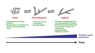
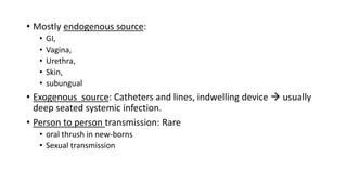
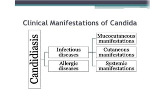
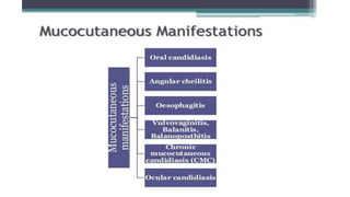
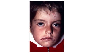
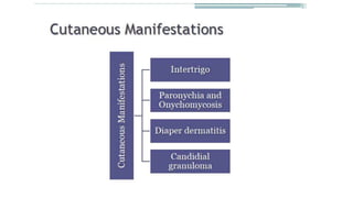
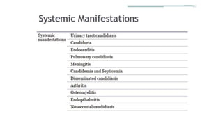
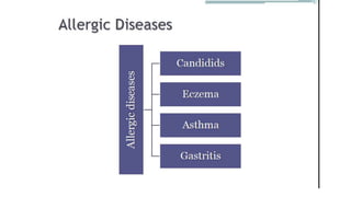
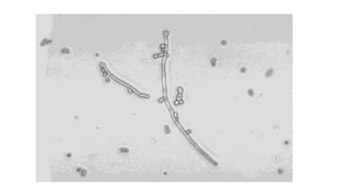
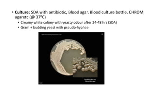
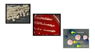
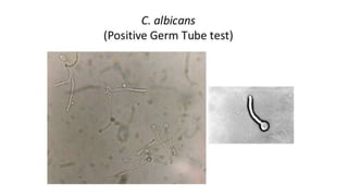
Candida
Pathology
Introduction
• 4th Century BC – ‘Epidemics’ (by Hippocrates) mentioned oral aphtha
(Thrush)
• 200 species  20 A/W human or animal diseases
• Major pathogen – Candida albicans
• Others: C. dubliniensis, C. glabrata, C. guilliermondii, C.kefyr, C.kruesi,
C. lusitaniae, C. parapsilosis, C.tropicalis etc.
• Budding cells, psudohyphae and true hyphae – all simultaneously
possible
Pathogenesis
• Most common fungal infection in human
• Increased risk of infection –
• Extremes of age,
• Pregnancy,
• HIV infected,
• Steroid, cytotoxic drug (or similar) therapy,
• Malignancy,
• post transplantation,
• Broad spectrum antibiotics,
• Diabetes,
• neutropenia etc.
• Virulence factors
• Adhesins
• Enzymes: proteinases, phospholipase
• Toxins: GP extracts pyrogenic properties
• Pseudohyphae : Active infection marker
• Candida albicans transforms frequently between 3 phenotypic forms
in tissues  Phenotypic switching (evasion of host defence)
• Mostly endogenous source:
• GI,
• Vagina,
• Urethra,
• Skin,
• subungual
• Exogenous source: Catheters and lines, indwelling device  usually
deep seated systemic infection.
• Person to person transmission: Rare
• oral thrush in new-borns
• Sexual transmission
Mucosal candidiasis
Oral thrush Chronic mucocutaneous candidiasis
Cutaneous candidiasis
Onychomycosis and paronychia Intertrigo
Lab diagnosis
• Specimen: Depending on site – swabs (mucosal patches, skin lesion),
nail scrapings, sputum, blood, urine
• Direct Microscopy:
• Gram positive, oval budding yeast (4-6um) or KOH mount
• Pseudohyphae
• Pseudohyphae V/S true hyphae
Feature Pseudohyphae True Hyphae
Septa Constricted No Constriction
Origin of branches Constricted and septate No Constriction & No
septum present
Grows by Budding Apical elongation
• Culture: SDA with antibiotic, Blood agar, Blood culture bottle, CHROM
agaretc (@ 370C)
• Creamy white colony with yeasty odour after 24-48 hrs (SDA)
• Gram + budding yeast with pseudo-hyphae
Species identification
• Germ tube test: Reynold Braude phenomenon
• Colonies mixed with serum & incubated for 2 hours
• Wet mount under microscope
• Long tube like projections extending from yeast cells no constriction at
origin
• Specific for C. albicans (C. dubliniensis)
• Cornmeal agar:
• Chlamydiospore is specific for Candida albicans
• CHROME Agar
• Growth at 450C : C. albicans & C. dublinensis
• Sugar assimilation tests
• PCR
• Serology
• Antibody : ELISA, Latex agglutination
• ANTIGEN DETECTION: Cell wall mannan & Cytoplasmic antigen by ELISA
• Enzyme assay : Enolase, Proteinase
• Metabolites – alpha1-3 glucan (G test)
Treatment
• Depending on type of candidiasis
• Azoles
• Amphotericin B

Candida pathogen.pptx

  • 1.
  • 2.
    Introduction • 4th CenturyBC – ‘Epidemics’ (by Hippocrates) mentioned oral aphtha (Thrush) • 200 species  20 A/W human or animal diseases • Major pathogen – Candida albicans • Others: C. dubliniensis, C. glabrata, C. guilliermondii, C.kefyr, C.kruesi, C. lusitaniae, C. parapsilosis, C.tropicalis etc. • Budding cells, psudohyphae and true hyphae – all simultaneously possible
  • 3.
    Pathogenesis • Most commonfungal infection in human • Increased risk of infection – • Extremes of age, • Pregnancy, • HIV infected, • Steroid, cytotoxic drug (or similar) therapy, • Malignancy, • post transplantation, • Broad spectrum antibiotics, • Diabetes, • neutropenia etc.
  • 4.
    • Virulence factors •Adhesins • Enzymes: proteinases, phospholipase • Toxins: GP extracts pyrogenic properties • Pseudohyphae : Active infection marker
  • 5.
    • Candida albicanstransforms frequently between 3 phenotypic forms in tissues  Phenotypic switching (evasion of host defence)
  • 7.
    • Mostly endogenoussource: • GI, • Vagina, • Urethra, • Skin, • subungual • Exogenous source: Catheters and lines, indwelling device  usually deep seated systemic infection. • Person to person transmission: Rare • oral thrush in new-borns • Sexual transmission
  • 10.
    Mucosal candidiasis Oral thrushChronic mucocutaneous candidiasis
  • 14.
  • 19.
    Lab diagnosis • Specimen:Depending on site – swabs (mucosal patches, skin lesion), nail scrapings, sputum, blood, urine
  • 20.
    • Direct Microscopy: •Gram positive, oval budding yeast (4-6um) or KOH mount • Pseudohyphae
  • 21.
    • Pseudohyphae V/Strue hyphae Feature Pseudohyphae True Hyphae Septa Constricted No Constriction Origin of branches Constricted and septate No Constriction & No septum present Grows by Budding Apical elongation
  • 23.
    • Culture: SDAwith antibiotic, Blood agar, Blood culture bottle, CHROM agaretc (@ 370C) • Creamy white colony with yeasty odour after 24-48 hrs (SDA) • Gram + budding yeast with pseudo-hyphae
  • 25.
    Species identification • Germtube test: Reynold Braude phenomenon • Colonies mixed with serum & incubated for 2 hours • Wet mount under microscope • Long tube like projections extending from yeast cells no constriction at origin • Specific for C. albicans (C. dubliniensis)
  • 27.
    • Cornmeal agar: •Chlamydiospore is specific for Candida albicans
  • 28.
    • CHROME Agar •Growth at 450C : C. albicans & C. dublinensis • Sugar assimilation tests • PCR
  • 29.
    • Serology • Antibody: ELISA, Latex agglutination • ANTIGEN DETECTION: Cell wall mannan & Cytoplasmic antigen by ELISA • Enzyme assay : Enolase, Proteinase • Metabolites – alpha1-3 glucan (G test)
  • 30.
    Treatment • Depending ontype of candidiasis • Azoles • Amphotericin B