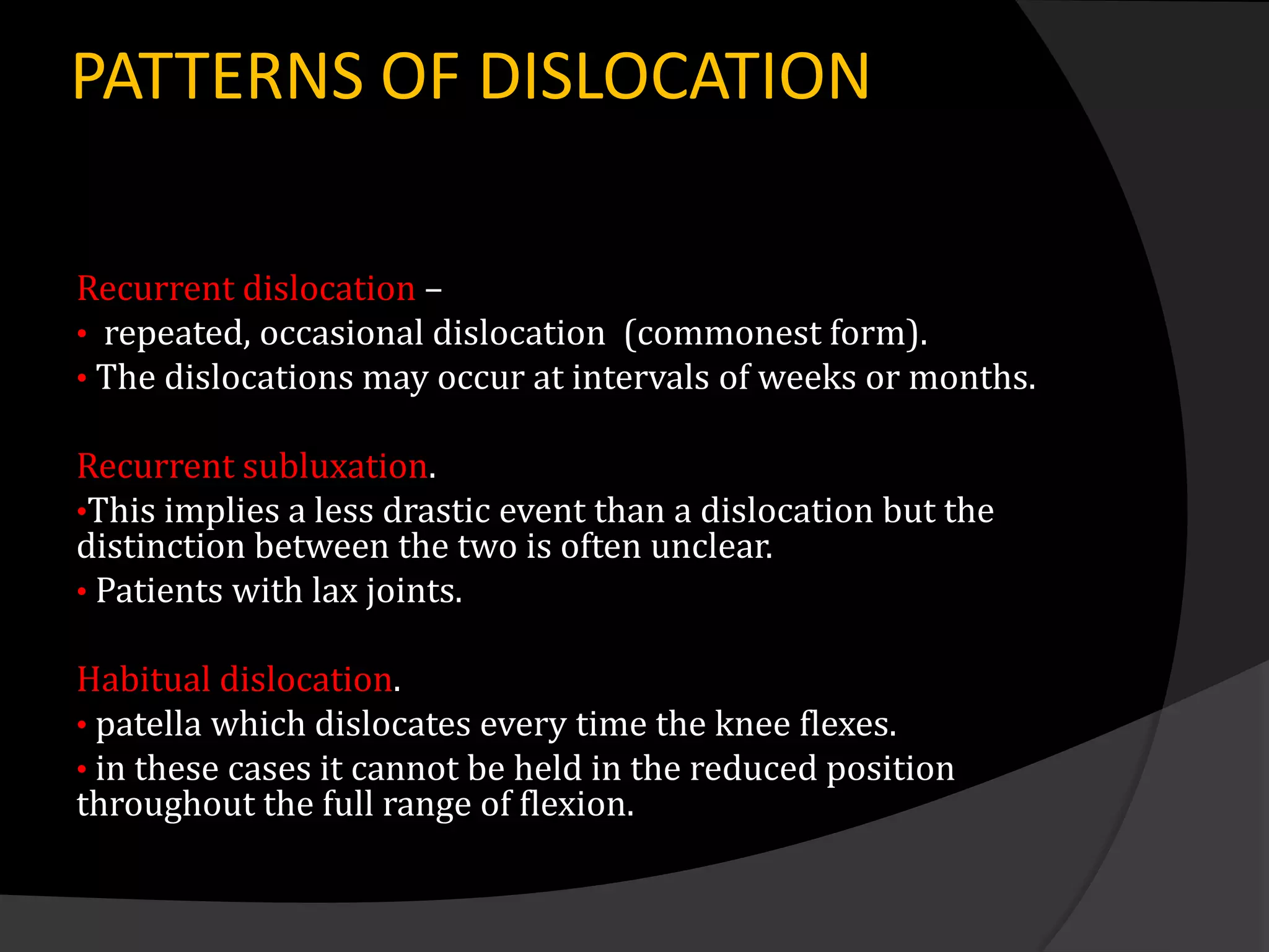
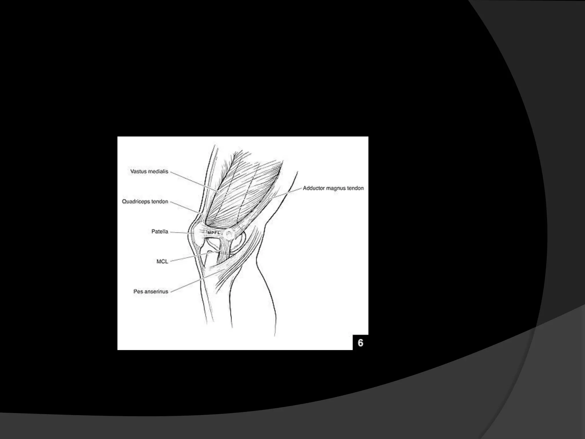
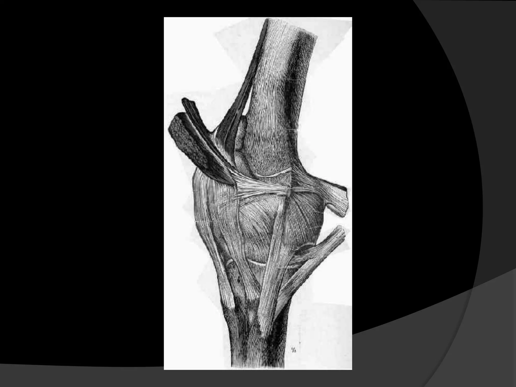
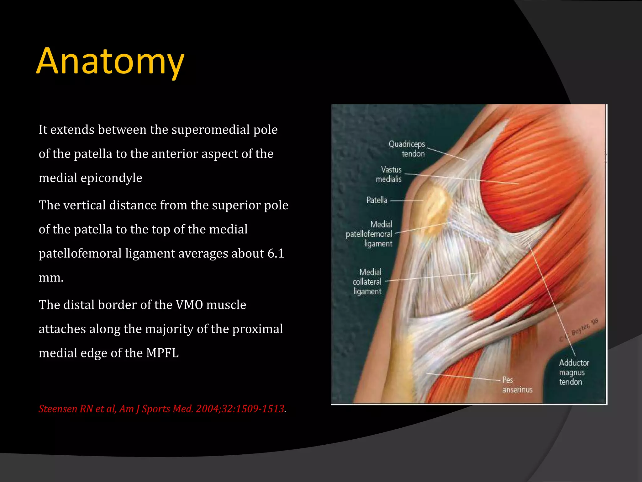
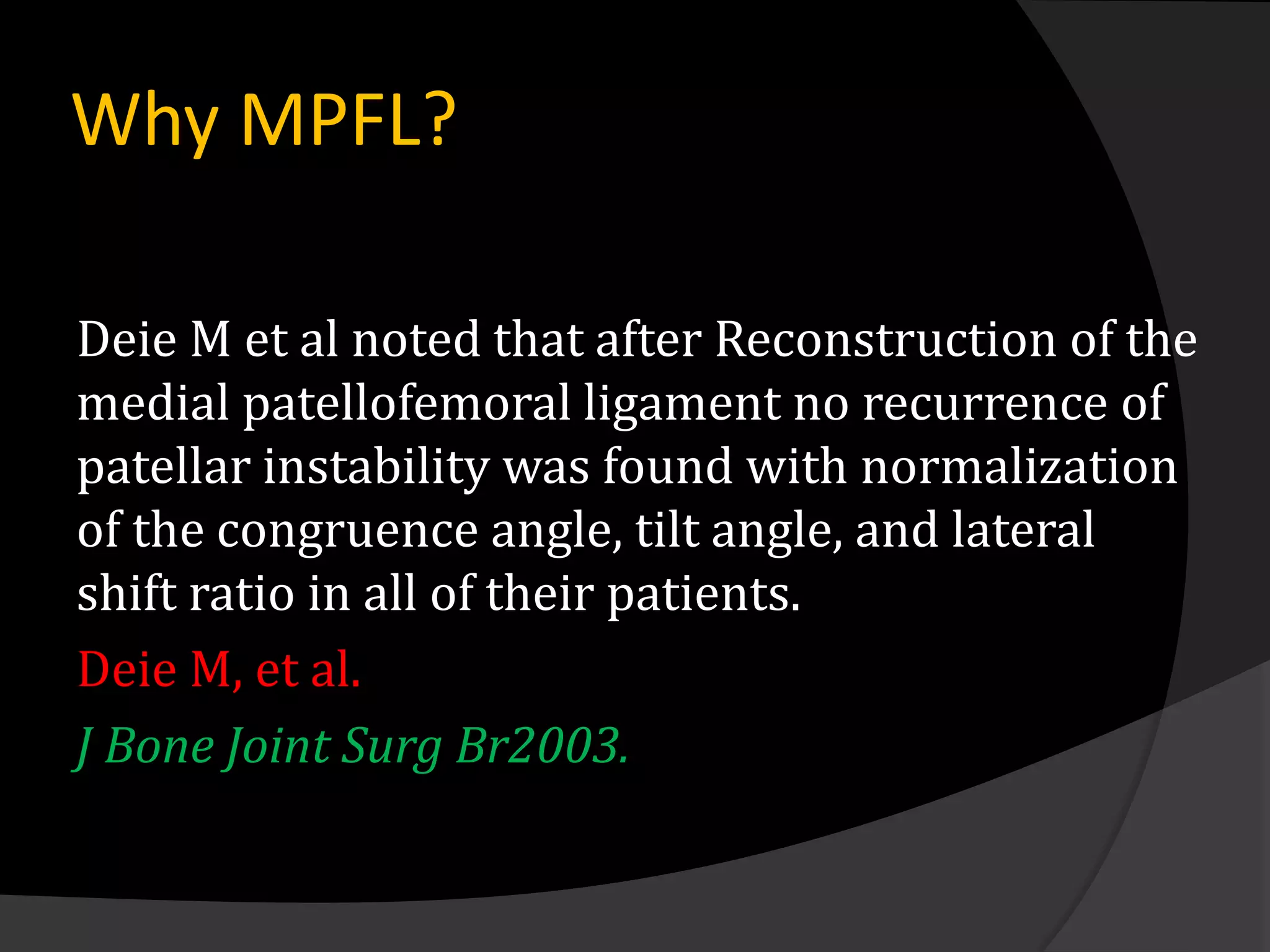
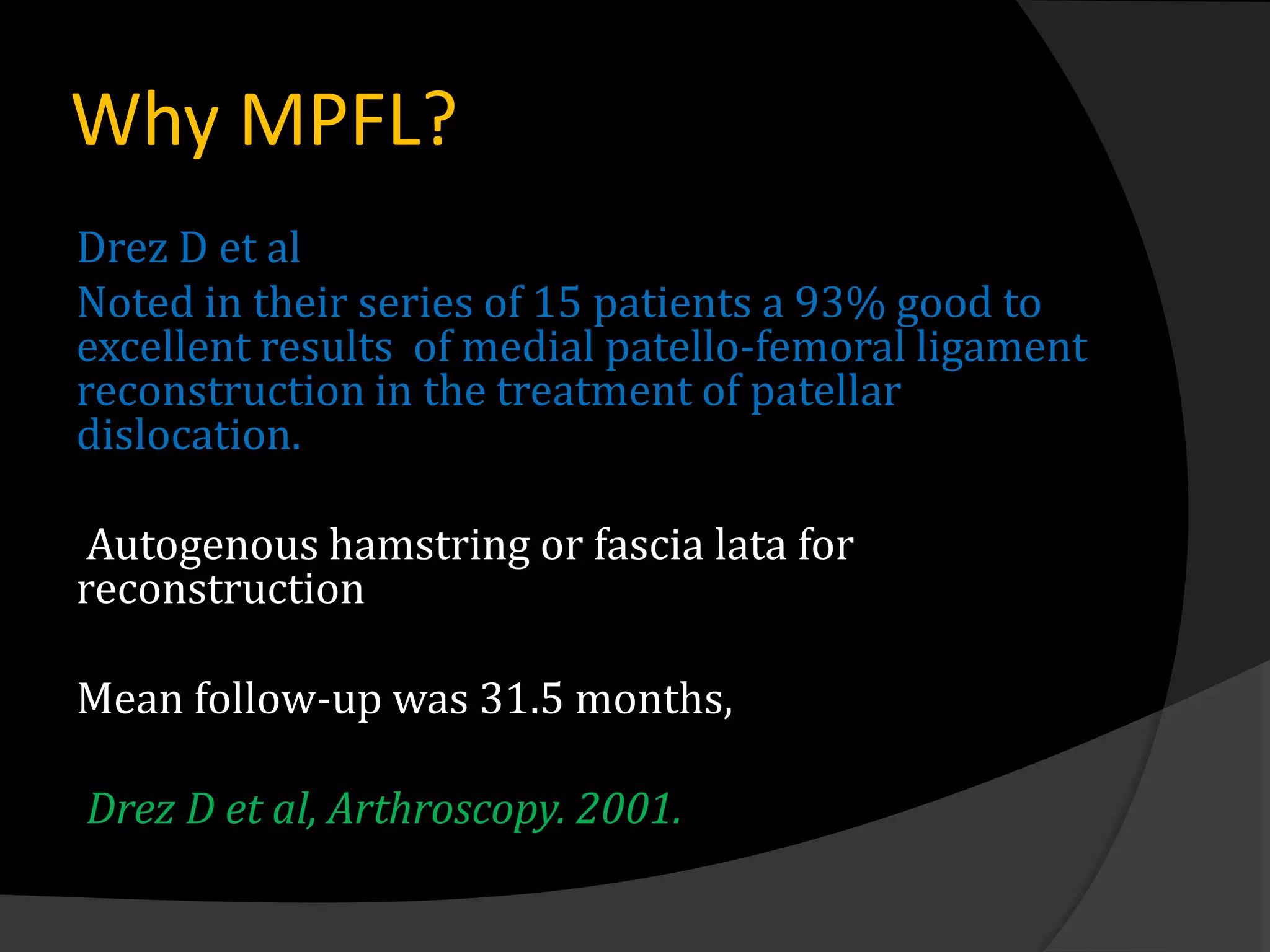
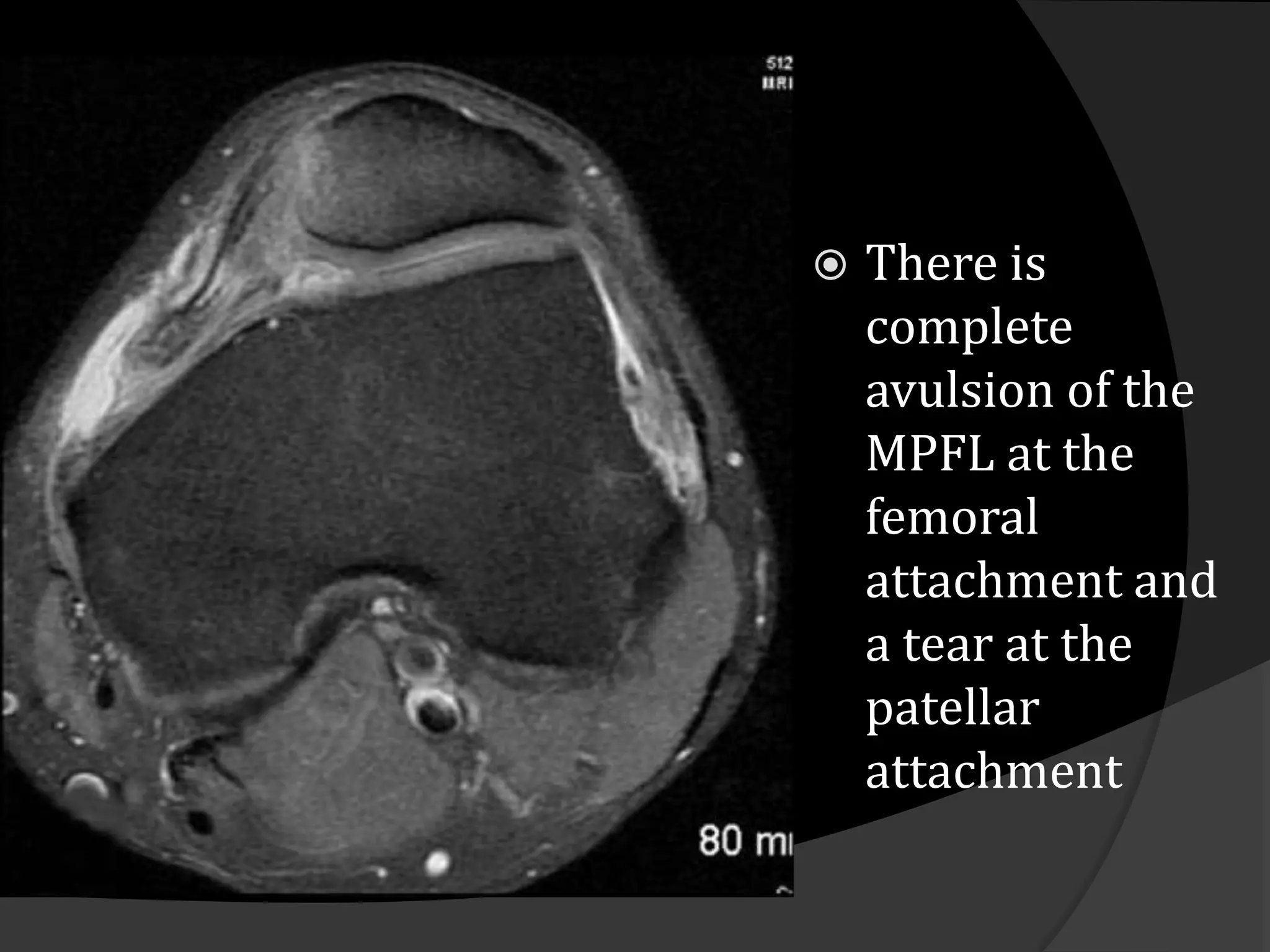
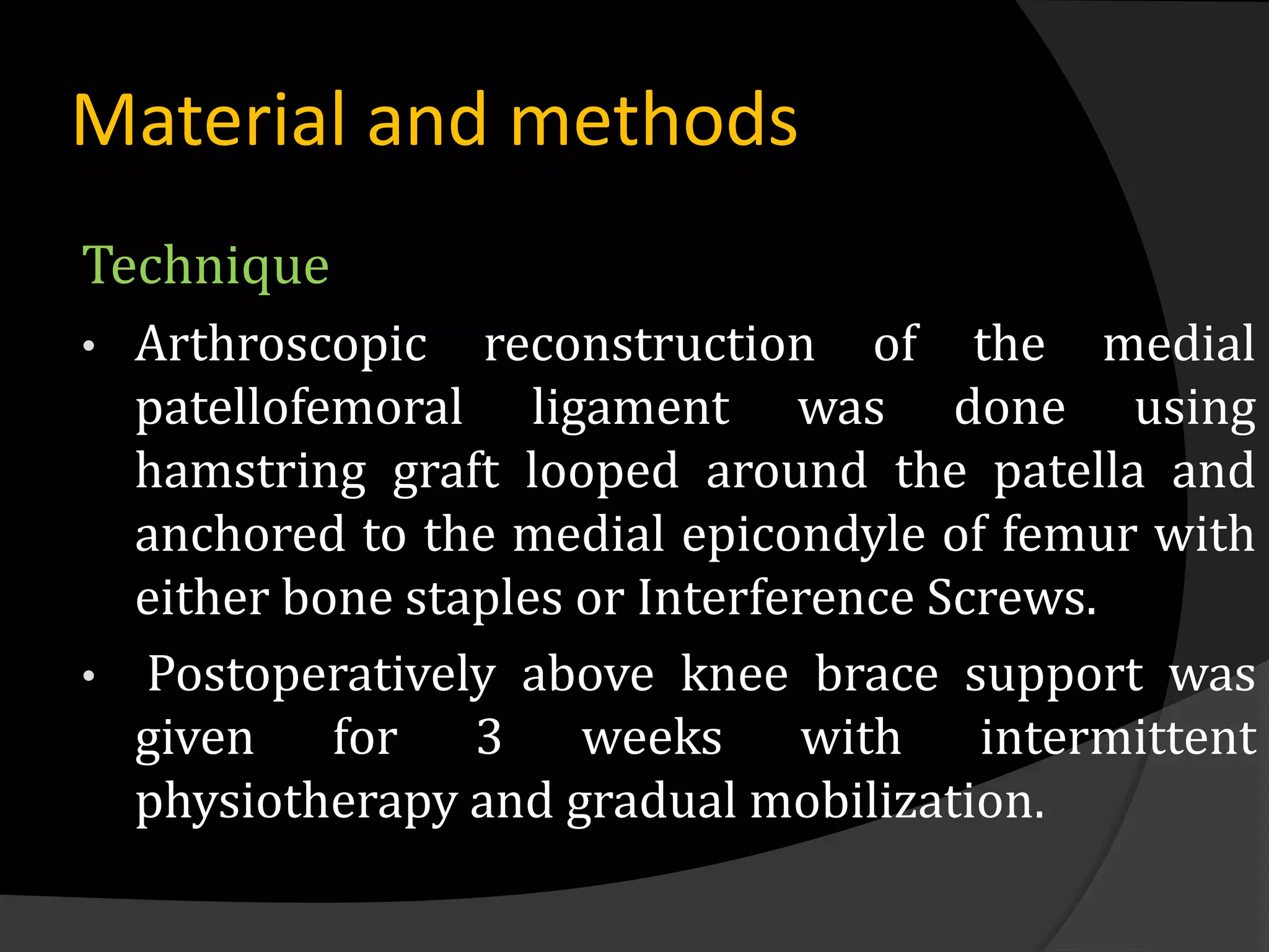
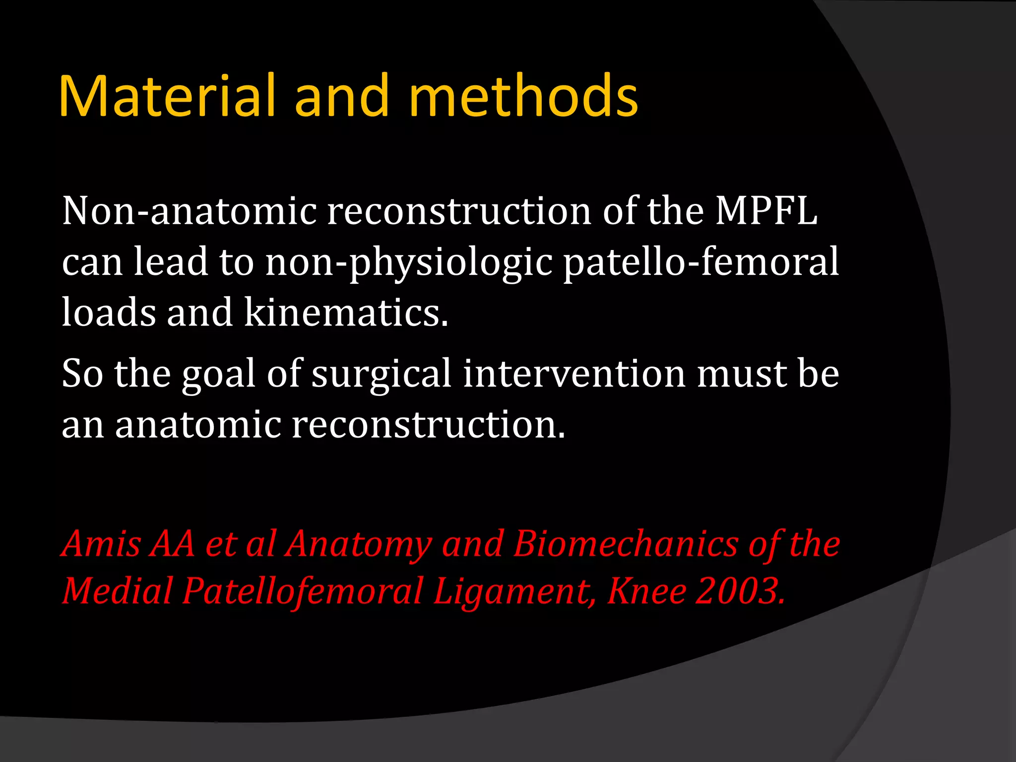
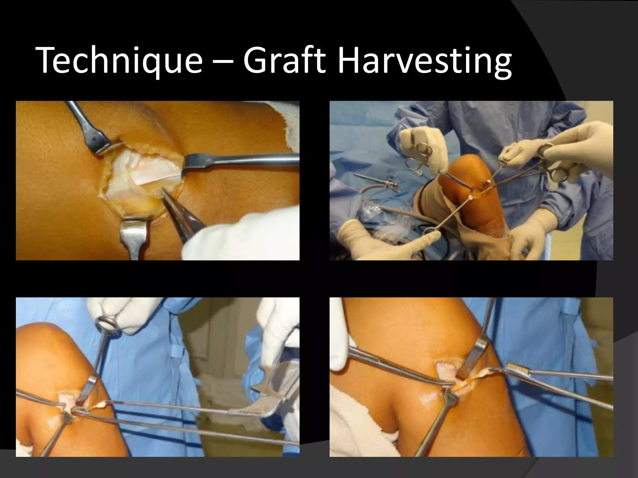
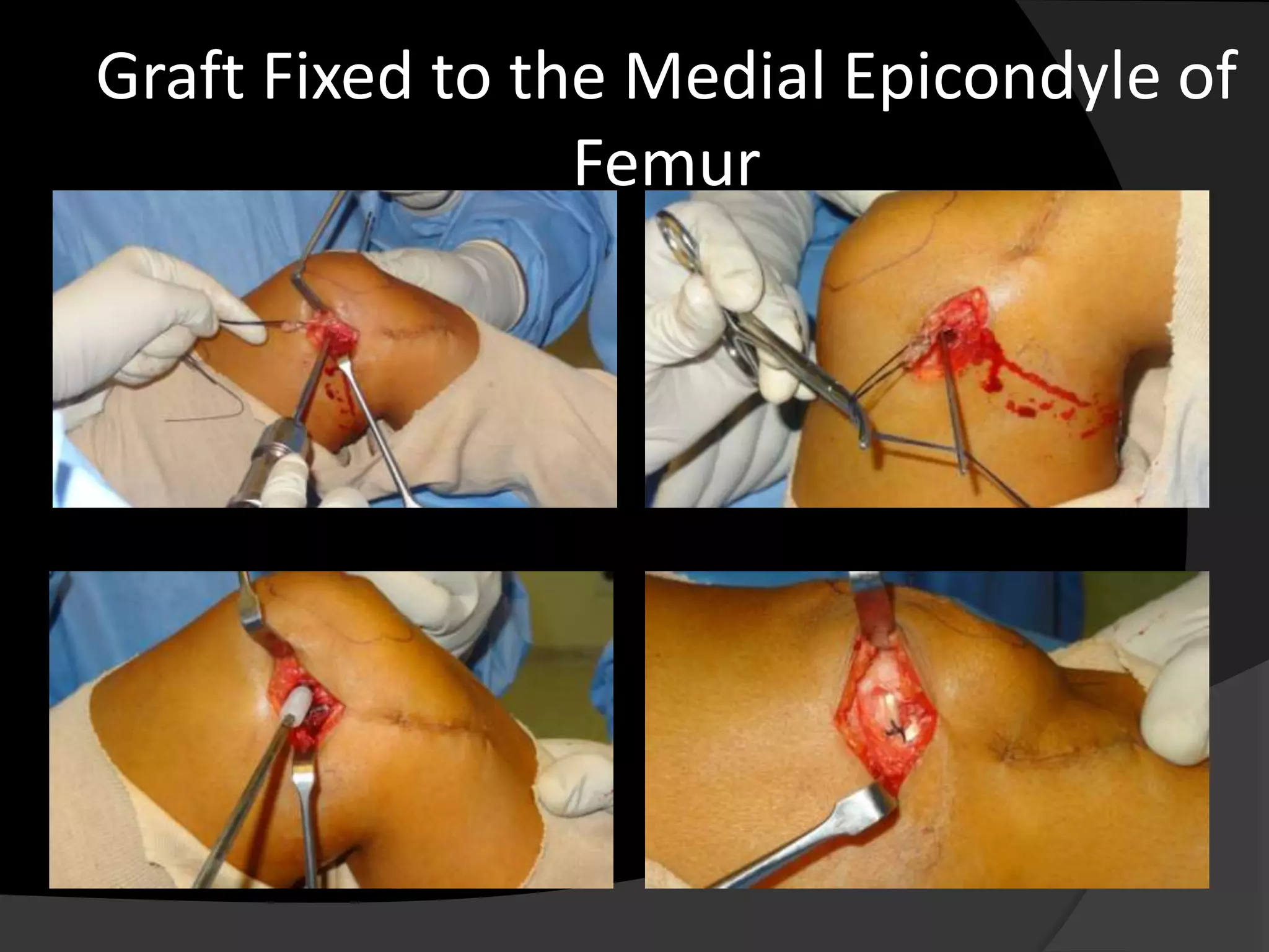
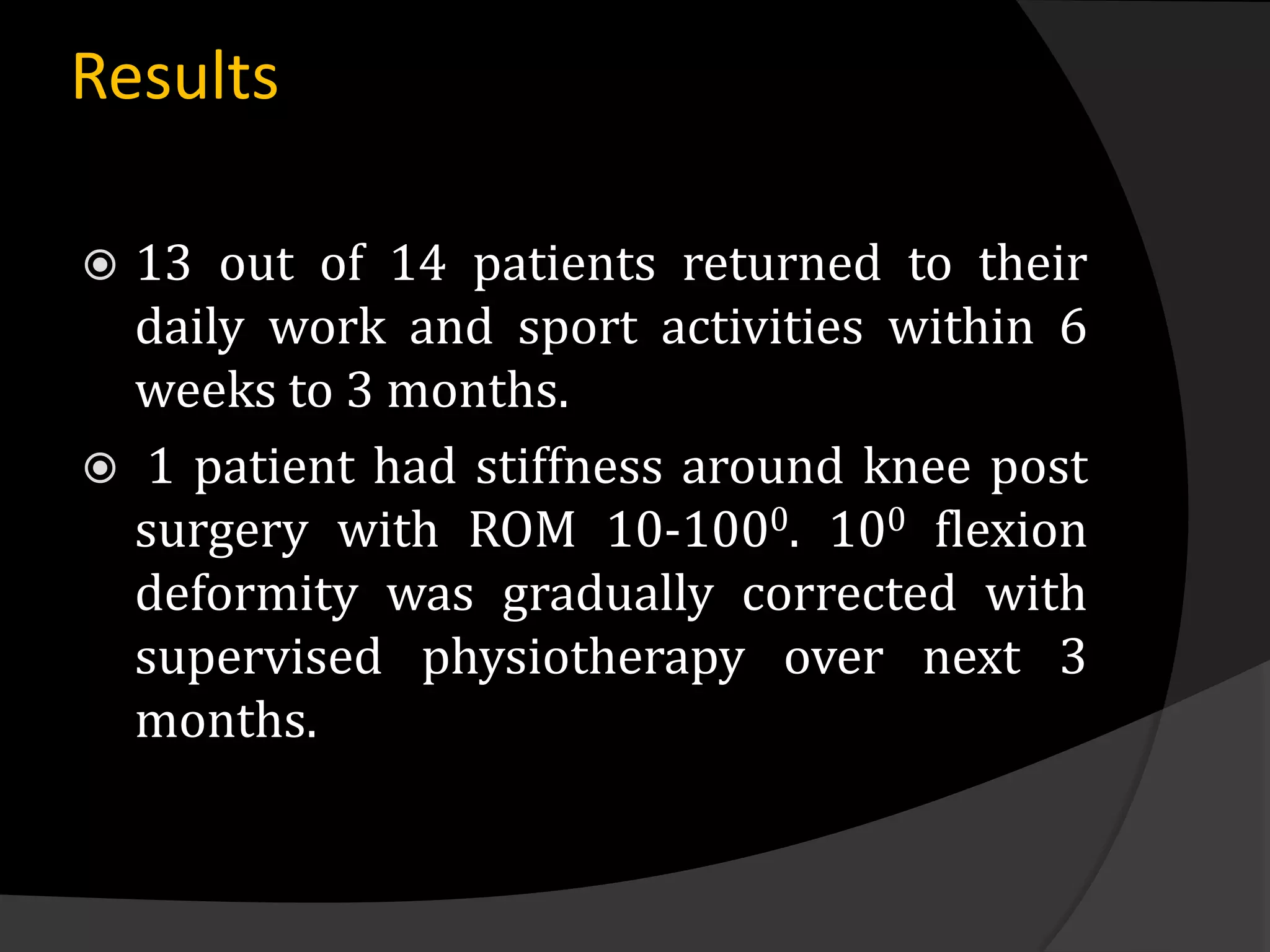
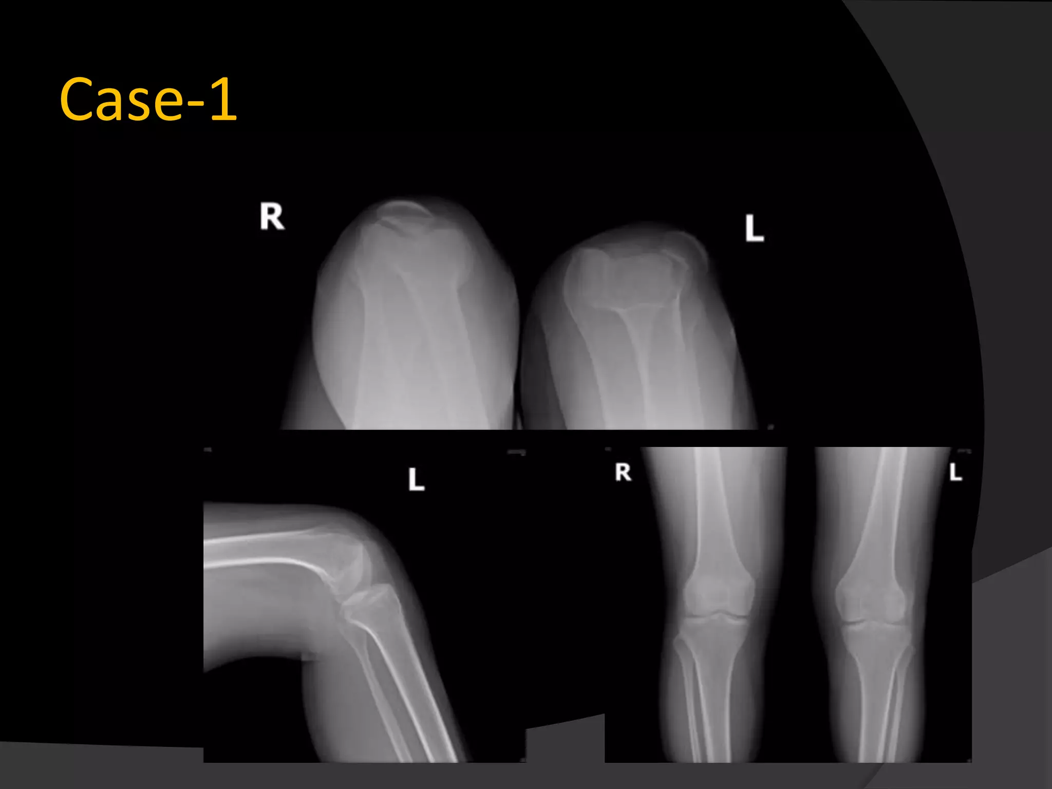
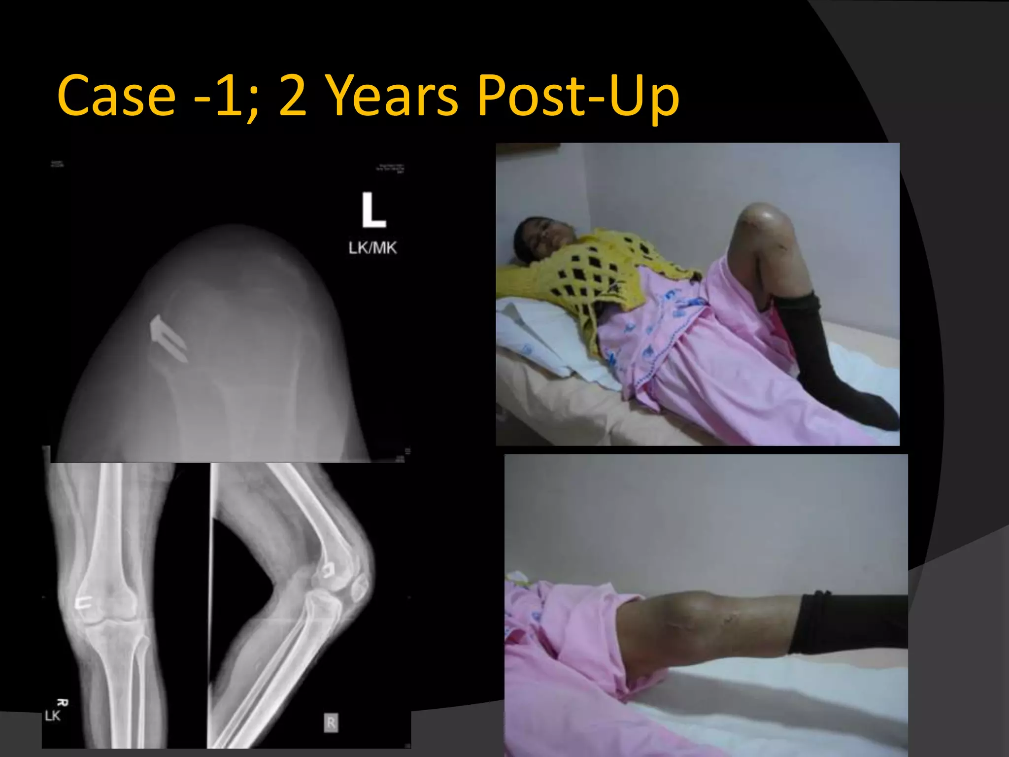
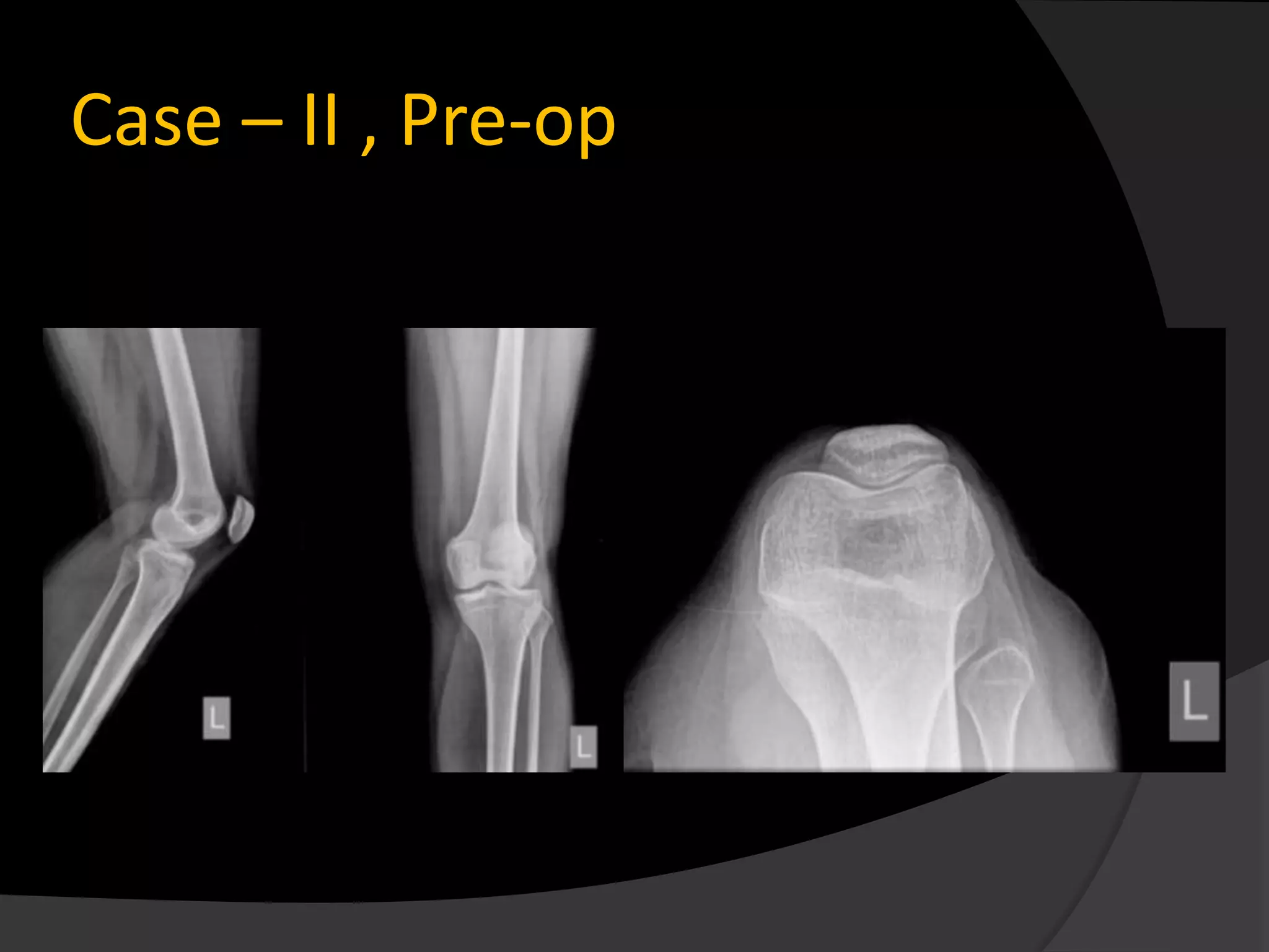
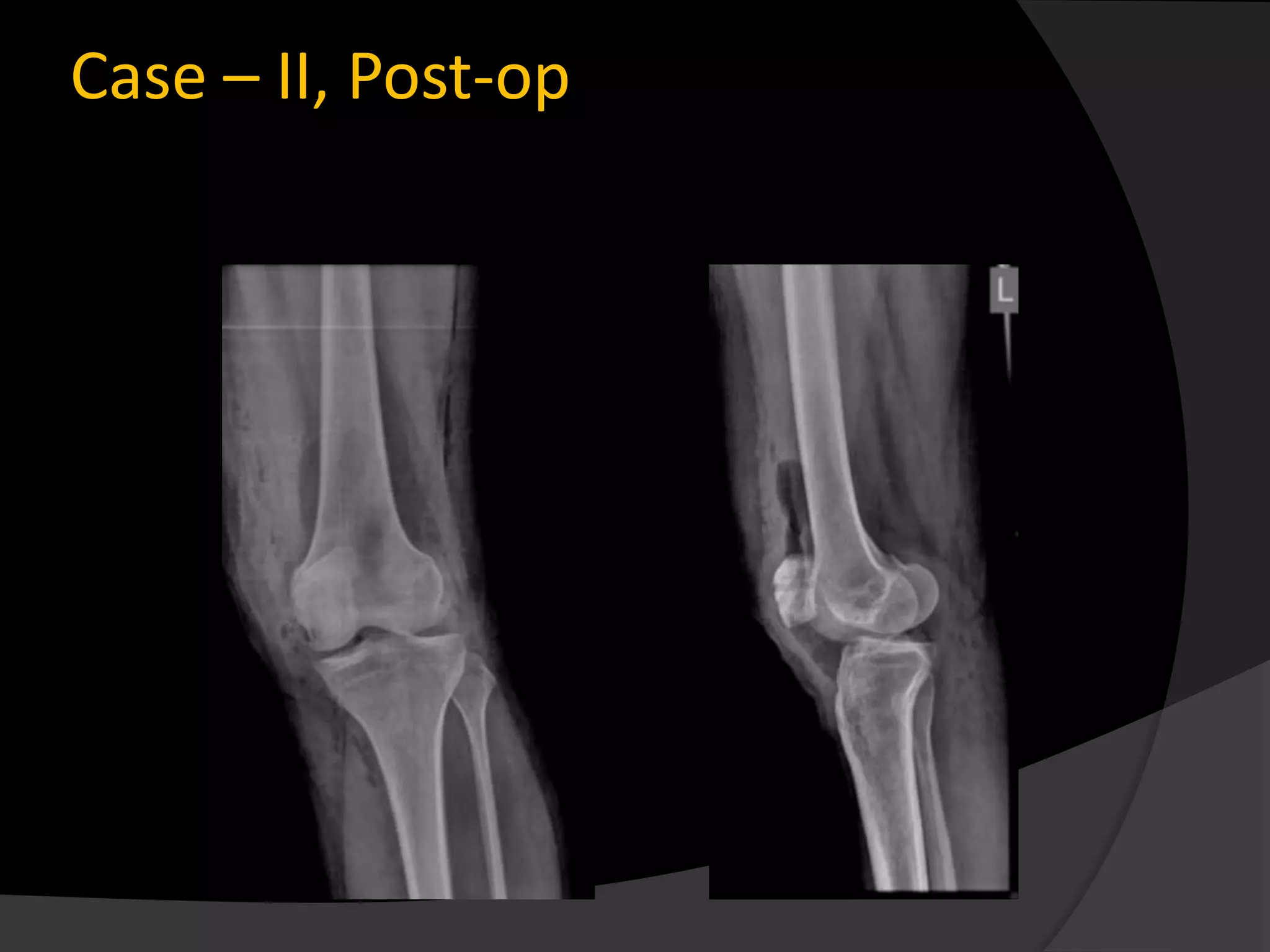
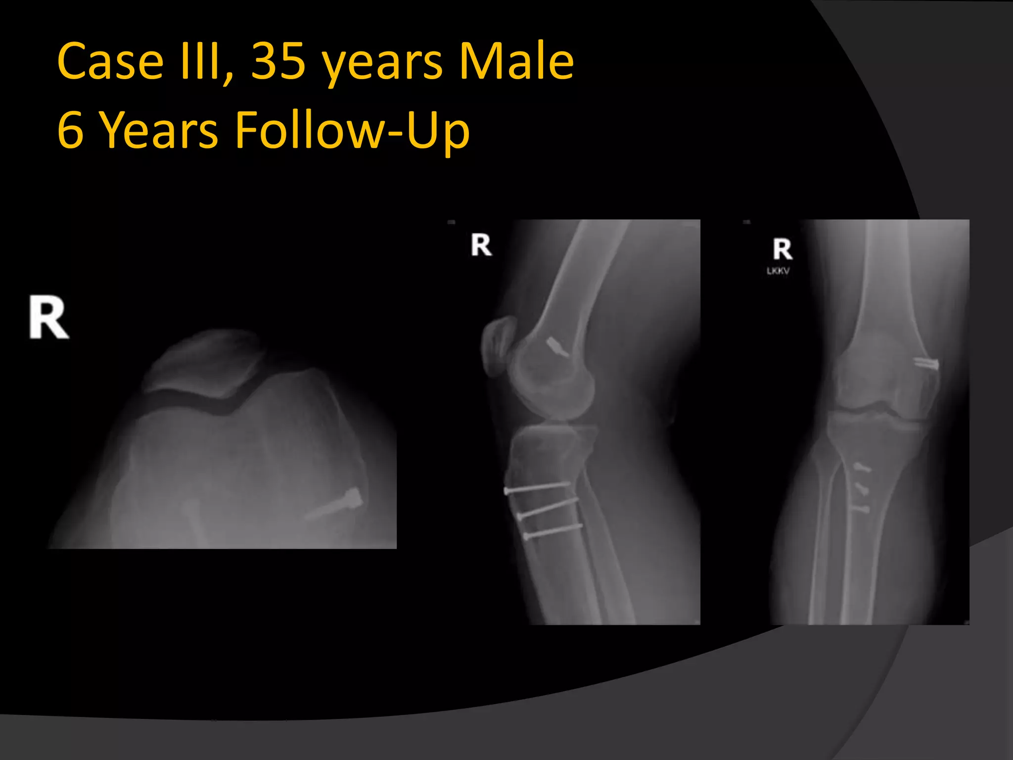
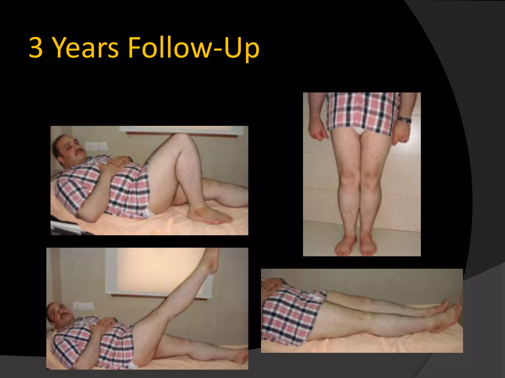
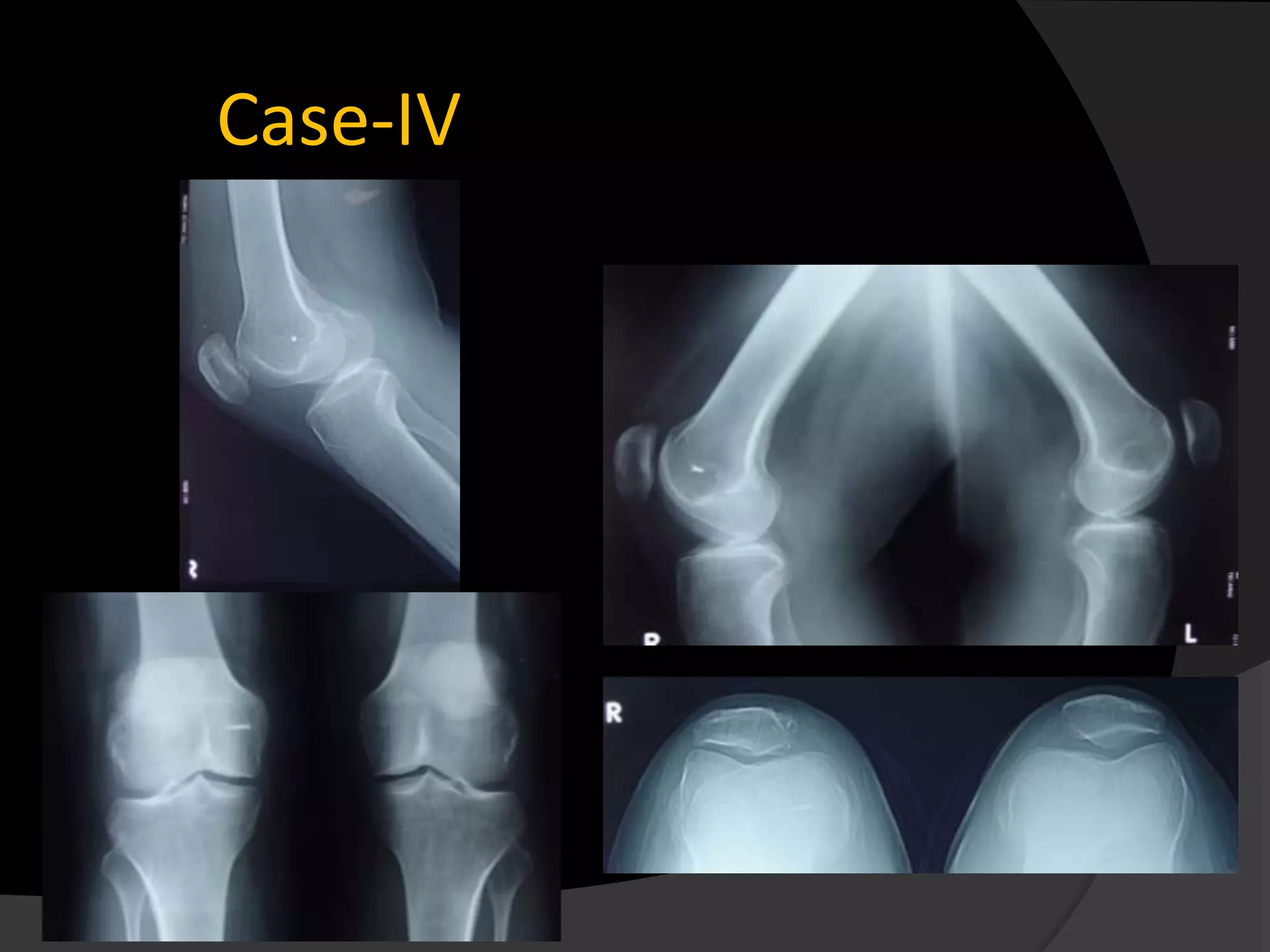
The document discusses patterns of patellar dislocation including recurrent dislocation, recurrent subluxation, and habitual dislocation. It then summarizes the anatomy and biomechanics of the medial patellofemoral ligament (MPFL), which is the primary soft tissue restraint preventing abnormal lateral displacement of the patella. Surgical reconstruction of the MPFL has gained popularity for treating recurrent patellar instability due to studies showing good postoperative outcomes with normalization of patellofemoral tracking and no recurrence of instability. The document presents the technique and results for MPFL reconstruction in 14 patients with patellar instability.
Medial patellofemoral ligament reconstruction ---- an update on techniques used. This lecture was taken by me at Trinity Arthroscopy Course, Chandigarh.
Mpfl tech - MPFL Reconstruction for Patellar InstabilityDelhiArthroscopy
MPFL Rec onstruction for Patellar Instability - By Dr Shekhar Srivastav .
Surgical Technique
- Diagnostic Arthroscopy
- Look for any Osteochondral fragment
(Loose body)
- Look for any Chondral damage
- Patellar tracking though Supero-lateral portal
Post-op Protocol
Ambulation with stick and Knee Brace- 3 wks
ROM exer – Next day upto 300 and progress
Review every 2 wks,6 wks,3 mnths,6 mnths and
yearly thereafter
Post-op assessment (Crosby-Insall criteria)
Excellent- No pain,normal activity
Good- Occasional pain,discomfort
Fair/Poor- Pain,loss of flexion,recurrent
dislocation/subluxation
Worse- Pain increased,displacement more
frequent
Caution
Must avoid overtightening-
Medial instability
Medial patellar arthritis
Patellar fractures
Preexisting Chondromalacia
Details @ http://www.delhiarthroscopy.com/
Medial patellofemoral ligament.
stabilizer of patella during initial 30 degree of flexion.
More than 18 techniques of reconstruction described.
Runs in layer 2 on medial aspect of the knee.
Origin- 1.9mm anterior/3.8 mm distal to adductor tubercle.(Laparade JBJS- Am.07)
Insertion – proximal 2/3 of patella
Along Distal edge of VMO
Broad insertion over 28.2+_ 5.6 mm over proximal 2/3 patella.
Average length 59.8mm +_ 4.8mm.
Passive- Trochlear constraints
Depth ,length(Height)
Patellar engagement
Capsular ligamentous tethers, especially MPFL
(Hautamaa, Fithian et al. 1998)
Dynamic elements
Simulated muscle tension has little effect on patellar mobility. Regardless of flexion angle
(Scnavongers, Farahmand et al 2003
This presentation goes in depth about Primary and Recurrent Patellar dislocation. Its cause, clinical and radiographic evaluation and various modalities of management with update from latest literature.
Posterolateral corner injuries of knee joint Samir Dwidmuthe
Missed posterolateral corner injuries of knee joint is a common cause for failure of ACL and PCL reconstruction only next to malpositioned tunnels.
Isolated PLC injuries are uncommon, making up <2% of all acute knee ligamentous injuries. Covey JBJS 2001
Incidence of PLC injuries associated with concomitant ACL and PCL disruptions are much more common (43% to 80%). Ranawat JAAOS 2008
A recent (MRI) analysis of surgical tibialplateau fractures demonstrated an incidence of PLC injuries in 68% of cases. Gardner JOT 2005
Take home message
PLC injuries to be ruled out in every case of ACL& PCL rupture.
Neurovascular integrity to be checked in every case.
Grade I & II can be managed conservatively.
Grade III Acute- Repair.
Grade III Chronic- Anatomic PLC recon.
Beware of varus knee alignment.
Surgical Versus Ponseti Approach for the Management of CTEV - Dr. CHINTAN N. ...DrChintan Patel
Surgical Versus Ponseti Approach for the Management of CTEV (congenital tallipes equino varus): A Comparative Study (J Pediatr Orthop Volume 33, Number 3, April/May 2013)
Patella dislocation is a common problem in the young. Recurrence of dislocation can be significant problem causing pain and discomfort. The assessment and guidelines towards non-surgical and surgical treatment options are discussed here.
Medial Patellofemoral Ligament Reconstruction with Patellar TendonDavid Sadigursky
Medial Patellofemoral Ligament Reconstruction with Patellar Tendon - ISAKOS NEWSLETTER
Review related to MPFL reconstruction and the use of the patellar tendon as a graft.
Revisão a respeito da reconstrução do Ligamento Patelofemoral Medial com o enxerto do tendão patelar.
Management of recurrent dislocation of patella by reconstructing2
1.
2. PATTERNS OF DISLOCATION
Recurrent dislocation –
• repeated, occasional dislocation (commonest form).
• The dislocations may occur at intervals of weeks or months.
Recurrent subluxation.
•This implies a less drastic event than a dislocation but the
distinction between the two is often unclear.
• Patients with lax joints.
Habitual dislocation.
• patella which dislocates every time the knee flexes.
• in these cases it cannot be held in the reduced position
throughout the full range of flexion.
3.
4.
5. Introduction
Instability of the patella is a
common problem.
The medial patellofemoral ligament
(MPFL) has been demonstrated to
be the major soft tissue stabilizer to
prevent abnormal lateral
displacement of the patella.
Desio SM et al
Soft Tissue Restraints to Lateral
Patellar Translation in the Human
Knee, Am J Sports Med
6. Anatomy
It extends between the superomedial pole
of the patella to the anterior aspect of the
medial epicondyle
The vertical distance from the superior pole
of the patella to the top of the medial
patellofemoral ligament averages about 6.1
mm.
The distal border of the VMO muscle
attaches along the majority of the proximal
medial edge of the MPFL
Steensen RN et al, Am J Sports Med. 2004;32:1509-1513.
7. Anatomy-Design fault?
The patellofemoral This apparent
joint is intrinsically ‘design fault’ is
unstable joint? minimised by the
Tibial tubercle lies resistance of the
lateral to the long axis lateral lip of trochlea
of the femur and the to lateral movement
quadriceps muscle,
and the patella is of the patella during
therefore subject to a flexion.
laterally directed
force.
8. Biomechanics
Biomechanically, the medial patello-femoral
ligament is considered the primary passive restraint
to patellar lateral displacement, with a mean tensile
strength of 208 N
Amis AA et al, Knee. 2003;10:215-220.
9. Biomechanics
From 0° to 30° of flexion- the median ridge of the
patella lies lateral to the centre of the trochlea.
30° and 60° of flexion-it moves medially to
become centred in the trochlear groove.
As flexion proceeds the patella is more deeply
engaged in the trochlea and is held firmly by soft-
tissue tension.
Beyond 90°-It tilts so that its medial facet
articulates with the medial femoral condyle.
10. Biomechanics
Lateral patellar displacement tests in vitro showed
that the patella subluxed most easily at 20 degrees
knee flexion.
The contribution of the MPFL to resisting patellar
lateral subluxation was greatest in the extended
knee
Amis AA et al, Knee. 2003,
Anatomy and biomechanics of the medial patello-femoral ligament.
11. Factors Leading To Patellar
Instability
Poor engagement Failure to stay in the trochlea
abnormally high •Defective lateral trochlear margin
•unusually shallow trochlear groove.
patella, patellar •Greater laterally directed force -excessive valgus.
dysplasia • excessively tight lateral structures -fibrosis of the
or a poorly vastus lateralis or
developed trochlea. •deficient medial structures -injury to the medial
retinaculum,
• stretching of medial structures after repeated
dislocations,
•severe wasting of the vastus medialis.
12. Management-Options are more
than 100
lateral release,
medial imbrication,
medial patellofemoral ligament repair,
and a number of distal realignment
procedures.
13. Management
Recently, medial patellofemoral ligament
reconstruction has gained popularity as a
treatment modality for recurrent patellar
instability.
14. Why MPFL?
Deie M et al noted that after Reconstruction of the
medial patellofemoral ligament no recurrence of
patellar instability was found with normalization
of the congruence angle, tilt angle, and lateral
shift ratio in all of their patients.
Deie M, et al.
J Bone Joint Surg Br2003.
15. Why MPFL?
Drez D et al
Noted in their series of 15 patients a 93% good to
excellent results of medial patello-femoral ligament
reconstruction in the treatment of patellar
dislocation.
Autogenous hamstring or fascia lata for
reconstruction
Mean follow-up was 31.5 months,
Drez D et al, Arthroscopy. 2001.
16. Why MPFL?
Ellera Gomes JL et al
showed in their series of 16 knees with Medial patello-
femoral ligament reconstruction with semitendinosus
autograft for chronic patellar instability
94% of patient outcomes were rated good or excellent
according to the Crosby-Insall criteria.
88% of the patients were satisfied with their surgery
15 knees showed a negative apprehension test at follow-
up.
Arthroscopy. 2004
17. Why MPFL
MPFL is injured in most cases with acute
patellar dsilocation and MPFL insufficiency
is present in all cases with recurrent patellar
dislocation.
…Now, the role of the MPFL has been almost
established in the Management of
Dislocations of patella.
18. Material and methods
14 Patients with patellar instability were
enrolled in this study from 2008-2012
5 habitual dislocation 9 Recurrent dislocation
Medial patellofemoral ligament pathology was confirmed
by both clinical and radiological examination.
Apprension test was positive in 12 patients
Average Q angle..
20. There is
complete
avulsion of the
MPFL at the
femoral
attachment and
a tear at the
patellar
attachment
21. Material and methods
Technique
• Arthroscopic reconstruction of the medial
patellofemoral ligament was done using
hamstring graft looped around the patella and
anchored to the medial epicondyle of femur with
either bone staples or Interference Screws.
• Postoperatively above knee brace support was
given for 3 weeks with intermittent
physiotherapy and gradual mobilization.
22. Material and methods
Non-anatomic reconstruction of the MPFL
can lead to non-physiologic patello-femoral
loads and kinematics.
So the goal of surgical intervention must be
an anatomic reconstruction.
Amis AA et al Anatomy and Biomechanics of the
Medial Patellofemoral Ligament, Knee 2003.
29. Results
13 out of 14 patients returned to their
daily work and sport activities within 6
weeks to 3 months.
1 patient had stiffness around knee post
surgery with ROM 10-1000. 100 flexion
deformity was gradually corrected with
supervised physiotherapy over next 3
months.
30. Results
Complications:
Anterior knee pain-3 Patients.
Numbness -5 patients experience mild
numbness on the shin, close to the
surgical scar.
No case of graft rupture and infection was
noted
38. Conclusion
The principal advantage of this procedure
is the ability to definitively reconstruct
the medial patellofemoral ligament on the
femur.
Allowing reasonable MPFL isometry
throughout the arc of knee motion.
A minimal invasive surgery.
With this procedure early rehabilitation
can be started and is a good technique for
sport persons with MPFL injuries.