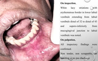
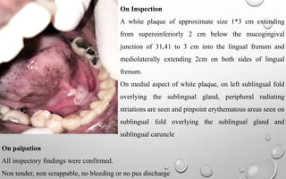
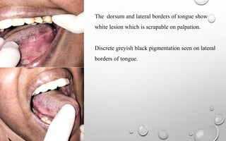
CASE PRESENTATION
Dr. Nitha Willy
First Year PG
Department Of Oral Medicine And Radiology
BIOGRAPHIC DATA
Name : Jaseela Shaji
Age :44 years
Sex : F
Occupation : Home maker
Address : Thanickamolath Methala
O P No :2971
Phone no: 9446972349
CHIEF COMPLAINT
Patient complaints of burning sensation in mouth since two months.
HISTORY OF PRESENTING ILLNESS
Patient complaints of burning sensation in mouth since 2 months while eating spicy food.
Patient complaints that mucosa gets ulcerated which is then painful.
Patient visited a dental clinic for the pain in lower mouth last day and was prescribed a
dologel which was not effective in reducing patient’s complaint.
Patient had consulted a gastroenterologist at Lisie hospital before one month relating
problem of mouth to be associated with gastric problem and she was informed that there was
no problems associated with GIT.
Patient has stress related to family issues.
Patient has not taken any special foods, patient has not changed toothpaste.
VAS score : 8/10
MEDICAL HISTORY
H/O Hypertension and hyperlipidemia under medication for 4 years
Patient doesn’t remember name of medications.
Patient had no recent hospitalization history or recent episodes of fever.
Drug Allergy
No known drug allergy
REVIEW OF SYSTEMS
CNS: No abnormalities reported
GIT: No abnormalities reported
Respiratory system: No abnormalities reported
CVS: No abnormalities reported
Endocrine system: No abnormalities reported
Genito urinary system: No abnormalities reported
PAST DENTAL VISIT
For similar complaint two day before in a private dental clinic.
PERSONAL HISTORY
Marital status: Married
Sleep and appetite: decreased sleep and loss of appetite.
Bowel and bladder movements: regular
Diet: mixed
Oral hygiene: brushes twice daily using tooth paste and toothbrush.
No deleterious and parafunctional habits noticed
FAMILY HISTORY
Parents and siblings are diabetic and hypertensive patients
GENERAL PHYSICAL EXAMINATION
Patient was conscious, co-operative and well oriented with person, place, time.
1. Gait: steady gait
2. Built: moderately built
3. Nourishment: moderately nourished
4. Temperature: 37 ͦ C
5. Pulse rate:72 beats/minute
6. Respiratory rate: 16 beats/minute
7. Blood pressure: 120/90 mm Hg
8. No signs of pallor, Icterus, Cyanosis/ Clubbing/ edema and no lymphadenopathy
9. Height: 5 ft 5 in
10. Weight: 70 kg
LOCAL EXAMINATION OF HEAD AND NECK
Extra oral examination
1) Head : Mesencephalic
2) Hair : No abnormalities detected
3) Face :Apparently symmetrical
4) Skin : no abnormalities detected
5) Eyes : no abnormalities detected
6) Ears : no abnormalities detected
7) Nose: no abnormalities detected
8) Lips :competent
9) TMJ : Mouth Opening Within Normal Limits, No Deviation, No Clicking/ Crepitus
10) Muscles of mastication : non-tender
11) Lymph nodes : not palpable
12) Cranial nerve examination: no abnormalities
Intra oral examination
Soft tissue examination
1. Labial mucosa: no abnormalities detected
2. Labial vestibule: white lacy striations with erythematous border in lower labial vestibule
extending distal aspect of 32 to distal aspect of 43.
3. Buccal mucosa: no abnormalities detected
4. Buccal vestibule: no abnormalities detected.
5. Lingual vestibule: A white plaque seen extending from lingual vestibule of 31,41 to
lingual frenum, of size 2*2 cm.
6. Gingiva : coral pink color, scalloped contours with knife edge margins, stippling present,
gingiva is firm and resilient.
7. Bleeding on probing: absent
8. Periodontal pocket: absent
9. Tongue: The dorsal and lateral borders of tongue had presence of white coating.
10. Floor of mouth: no abnormalities detected
11. Frenal attachment : mucosal frenal attachment detected
12. Palate: no abnormalities detected
13. Oropharynx: no abnormalities detected
14. Salivary gland orifices: no abnormalities detected
Hard tissue examination
1. Number of teeth : 16-27,36-47
2. Carious teeth :0
3. Missing teeth : 18,17,28,38,37,48
4. Root stump:0
5. Restored tooth: 47,46,35,34,36,14,24
6. Fractured tooth:0
7. Mobility :0
8. Attrition : generalised
Examination of lesion
On inspection
On inspection,
White lacy striations with
erythematous border in lower labial
vestibule extending from labial
vestibule distal of 32 to distal of 43
and supero-inferiorly from
mucogingival junction to labial
vestibule was noted.
On palpation,
All inspectory findings were
confirmed.
Non tender, non scrappable, no
bleeding or no pus discharge
On Inspection
A white plaque of approximate size 1*3 cm extending
from superoinferiorly 2 cm below the mucogingival
junction of 31,41 to 3 cm into the lingual frenum and
mediolaterally extending 2cm on both sides of lingual
frenum.
On medial aspect of white plaque, on left sublingual fold
overlying the sublingual gland, peripheral radiating
striations are seen and pinpoint erythematous areas seen on
sublingual fold overlying the sublingual gland and
sublingual caruncle
On palpation
All inspectory findings were confirmed.
Non tender, non scrappable, no bleeding or no pus discharge
The dorsum and lateral borders of tongue show
white lesion which is scrapable on palpation.
Discrete greyish black pigmentation seen on lateral
borders of tongue.
Case Summary/ Analysis
A 44 year old female patient reported to our department with chief complaint of burning sensation in
mouth since 2 months. On examination, White lacy type lesion with erythematous border in lower
labial vestibule extending from labial vestibule distal of 32 to distal of 43 and supero-inferiorly from
mucogingival junction to labial vestibule was noted. A white plaque type lesion with radiating
striations on left on sublingual fold overling the sublingual gland and overlying pinpoint
erythematous areas seen on sublingual fold overlying the sublingual gland and sublingual caruncle
extending from superoinferiorly 2 cm below the mucogingival junction of 31,41 to 3 cm into the
lingual frenum and mediolaterally extending 2cm on both sides of lingual frenum. The dorsal and
lateral borders of tongue show white pigmentation and was scrappable on palpation.
PROVISIONAL DIAGNOSIS
EROSIVE LICHEN PLANUS ON LOWER LABIAL VESTIBULE
PLAQUE TYPE LICHEN PLANUS ON LINGUAL FRENUM AND
LINGUAL VESTIBULE
CANDIDIASIS ON DORSUM AND LATERAL BORDERS OF
TONGUE
Differential Diagnosis
1. Speckled leukoplakia: middle older age male group, associated with tobacco,
unilaterally present, no striae.
2. Hyperplastic candidiasis: presence of striae, associated with stress, negative fungal
hyphae seen in cytosmear.
3. Homogeneous leukoplakia : non scrappable, whitish patch or plaque, well
demarcated borders.
4. Thermal burn
INVESTIGATIONS
CYTOLOGY, HEMATOLOGYAND BIOPSY
Smear from dorsum of tongue showed presence of candida
hyphae.
Treatment Plan
1) 0.1 % Triamcinolone Acetonide topical application three times daily for
15 Days
2) Tab oxidil(ginseng extract,lycopene, lutein, zinc oxide) 1-0-0 * 15 Days
3) Betamethasone sodium phosphate 0.5mg( Swish and Spit )1-0-1 for 15
Days
FOLLOW UP
case presentation 3 :  LICHEN PLANUS.pptx

case presentation 3 : LICHEN PLANUS.pptx

  • 1.
    CASE PRESENTATION Dr. NithaWilly First Year PG Department Of Oral Medicine And Radiology
  • 2.
    BIOGRAPHIC DATA Name :Jaseela Shaji Age :44 years Sex : F Occupation : Home maker Address : Thanickamolath Methala O P No :2971 Phone no: 9446972349
  • 3.
    CHIEF COMPLAINT Patient complaintsof burning sensation in mouth since two months.
  • 4.
    HISTORY OF PRESENTINGILLNESS Patient complaints of burning sensation in mouth since 2 months while eating spicy food. Patient complaints that mucosa gets ulcerated which is then painful. Patient visited a dental clinic for the pain in lower mouth last day and was prescribed a dologel which was not effective in reducing patient’s complaint. Patient had consulted a gastroenterologist at Lisie hospital before one month relating problem of mouth to be associated with gastric problem and she was informed that there was no problems associated with GIT.
  • 5.
    Patient has stressrelated to family issues. Patient has not taken any special foods, patient has not changed toothpaste. VAS score : 8/10
  • 6.
    MEDICAL HISTORY H/O Hypertensionand hyperlipidemia under medication for 4 years Patient doesn’t remember name of medications. Patient had no recent hospitalization history or recent episodes of fever.
  • 7.
  • 8.
    REVIEW OF SYSTEMS CNS:No abnormalities reported GIT: No abnormalities reported Respiratory system: No abnormalities reported CVS: No abnormalities reported Endocrine system: No abnormalities reported Genito urinary system: No abnormalities reported
  • 9.
    PAST DENTAL VISIT Forsimilar complaint two day before in a private dental clinic.
  • 10.
    PERSONAL HISTORY Marital status:Married Sleep and appetite: decreased sleep and loss of appetite. Bowel and bladder movements: regular Diet: mixed Oral hygiene: brushes twice daily using tooth paste and toothbrush. No deleterious and parafunctional habits noticed
  • 11.
    FAMILY HISTORY Parents andsiblings are diabetic and hypertensive patients
  • 12.
    GENERAL PHYSICAL EXAMINATION Patientwas conscious, co-operative and well oriented with person, place, time. 1. Gait: steady gait 2. Built: moderately built 3. Nourishment: moderately nourished 4. Temperature: 37 ͦ C 5. Pulse rate:72 beats/minute 6. Respiratory rate: 16 beats/minute 7. Blood pressure: 120/90 mm Hg
  • 13.
    8. No signsof pallor, Icterus, Cyanosis/ Clubbing/ edema and no lymphadenopathy 9. Height: 5 ft 5 in 10. Weight: 70 kg
  • 14.
    LOCAL EXAMINATION OFHEAD AND NECK Extra oral examination 1) Head : Mesencephalic 2) Hair : No abnormalities detected 3) Face :Apparently symmetrical 4) Skin : no abnormalities detected 5) Eyes : no abnormalities detected 6) Ears : no abnormalities detected 7) Nose: no abnormalities detected 8) Lips :competent 9) TMJ : Mouth Opening Within Normal Limits, No Deviation, No Clicking/ Crepitus 10) Muscles of mastication : non-tender 11) Lymph nodes : not palpable 12) Cranial nerve examination: no abnormalities
  • 15.
    Intra oral examination Softtissue examination 1. Labial mucosa: no abnormalities detected 2. Labial vestibule: white lacy striations with erythematous border in lower labial vestibule extending distal aspect of 32 to distal aspect of 43. 3. Buccal mucosa: no abnormalities detected 4. Buccal vestibule: no abnormalities detected. 5. Lingual vestibule: A white plaque seen extending from lingual vestibule of 31,41 to lingual frenum, of size 2*2 cm. 6. Gingiva : coral pink color, scalloped contours with knife edge margins, stippling present, gingiva is firm and resilient. 7. Bleeding on probing: absent 8. Periodontal pocket: absent
  • 16.
    9. Tongue: Thedorsal and lateral borders of tongue had presence of white coating. 10. Floor of mouth: no abnormalities detected 11. Frenal attachment : mucosal frenal attachment detected 12. Palate: no abnormalities detected 13. Oropharynx: no abnormalities detected 14. Salivary gland orifices: no abnormalities detected
  • 17.
    Hard tissue examination 1.Number of teeth : 16-27,36-47 2. Carious teeth :0 3. Missing teeth : 18,17,28,38,37,48 4. Root stump:0 5. Restored tooth: 47,46,35,34,36,14,24 6. Fractured tooth:0 7. Mobility :0 8. Attrition : generalised
  • 18.
    Examination of lesion Oninspection On inspection, White lacy striations with erythematous border in lower labial vestibule extending from labial vestibule distal of 32 to distal of 43 and supero-inferiorly from mucogingival junction to labial vestibule was noted. On palpation, All inspectory findings were confirmed. Non tender, non scrappable, no bleeding or no pus discharge
  • 19.
    On Inspection A whiteplaque of approximate size 1*3 cm extending from superoinferiorly 2 cm below the mucogingival junction of 31,41 to 3 cm into the lingual frenum and mediolaterally extending 2cm on both sides of lingual frenum. On medial aspect of white plaque, on left sublingual fold overlying the sublingual gland, peripheral radiating striations are seen and pinpoint erythematous areas seen on sublingual fold overlying the sublingual gland and sublingual caruncle On palpation All inspectory findings were confirmed. Non tender, non scrappable, no bleeding or no pus discharge
  • 20.
    The dorsum andlateral borders of tongue show white lesion which is scrapable on palpation. Discrete greyish black pigmentation seen on lateral borders of tongue.
  • 21.
    Case Summary/ Analysis A44 year old female patient reported to our department with chief complaint of burning sensation in mouth since 2 months. On examination, White lacy type lesion with erythematous border in lower labial vestibule extending from labial vestibule distal of 32 to distal of 43 and supero-inferiorly from mucogingival junction to labial vestibule was noted. A white plaque type lesion with radiating striations on left on sublingual fold overling the sublingual gland and overlying pinpoint erythematous areas seen on sublingual fold overlying the sublingual gland and sublingual caruncle extending from superoinferiorly 2 cm below the mucogingival junction of 31,41 to 3 cm into the lingual frenum and mediolaterally extending 2cm on both sides of lingual frenum. The dorsal and lateral borders of tongue show white pigmentation and was scrappable on palpation.
  • 22.
    PROVISIONAL DIAGNOSIS EROSIVE LICHENPLANUS ON LOWER LABIAL VESTIBULE PLAQUE TYPE LICHEN PLANUS ON LINGUAL FRENUM AND LINGUAL VESTIBULE CANDIDIASIS ON DORSUM AND LATERAL BORDERS OF TONGUE
  • 23.
    Differential Diagnosis 1. Speckledleukoplakia: middle older age male group, associated with tobacco, unilaterally present, no striae. 2. Hyperplastic candidiasis: presence of striae, associated with stress, negative fungal hyphae seen in cytosmear. 3. Homogeneous leukoplakia : non scrappable, whitish patch or plaque, well demarcated borders. 4. Thermal burn
  • 24.
    INVESTIGATIONS CYTOLOGY, HEMATOLOGYAND BIOPSY Smearfrom dorsum of tongue showed presence of candida hyphae.
  • 25.
    Treatment Plan 1) 0.1% Triamcinolone Acetonide topical application three times daily for 15 Days 2) Tab oxidil(ginseng extract,lycopene, lutein, zinc oxide) 1-0-0 * 15 Days 3) Betamethasone sodium phosphate 0.5mg( Swish and Spit )1-0-1 for 15 Days
  • 26.