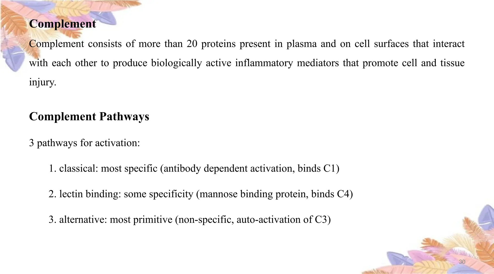
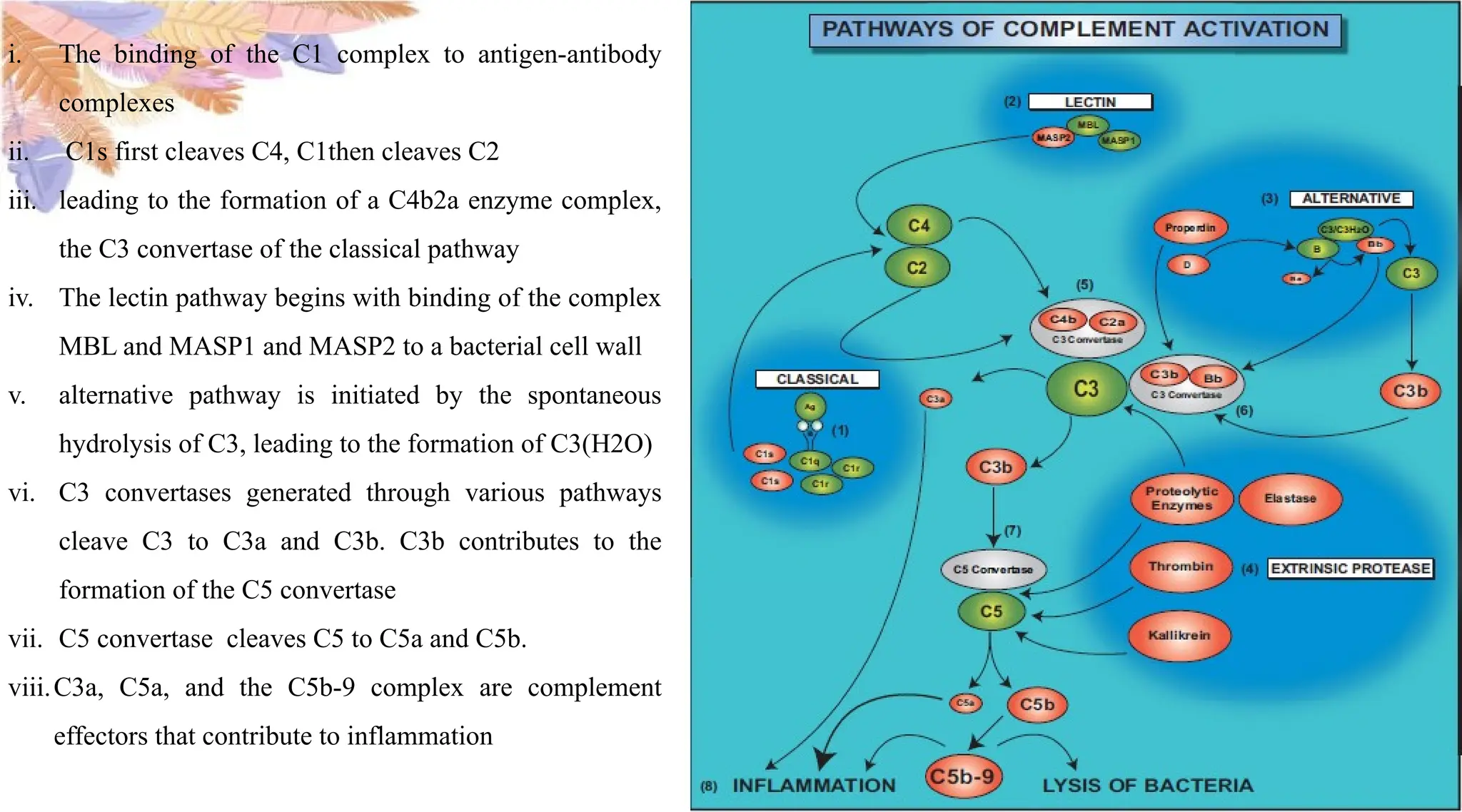
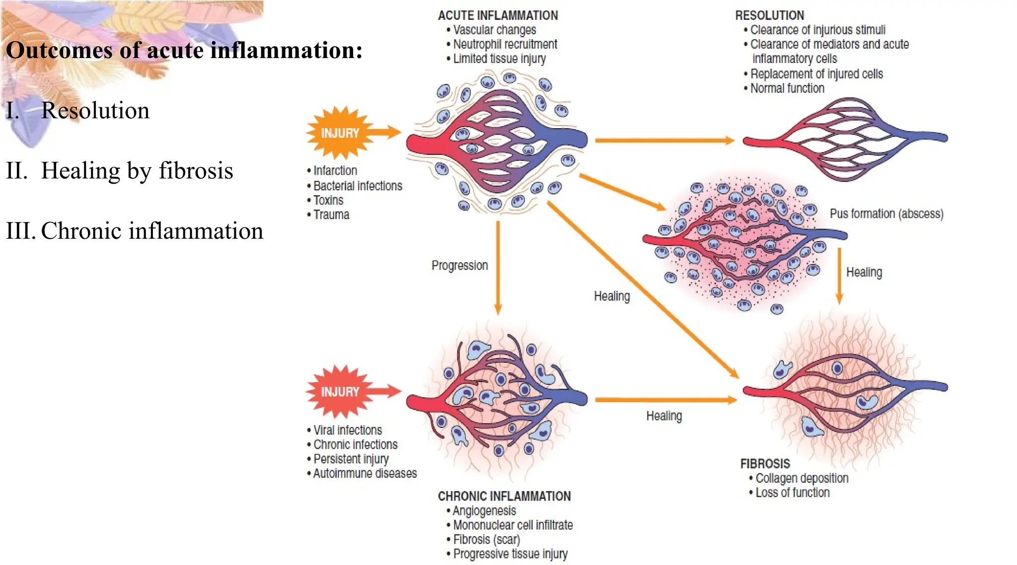
1
2
ROLE OF COMPLEMENT SYSTEM IN ACUTE INFLAMMATION
Presented by,
Dr. Nitha Willy
First year PG
Department Of Oral Medicine And Radiology
3
Contents
i. Overview of inflammation
ii. Acute inflammation
iii. Stimuli for acute inflammation
iv. Reactions of blood vessels in acute inflammation
Changes in vascular flow and caliber
Increased vascular permeability
Responses of lymphatic vessels
4
V. Reactions of leukocytes in inflammation
Recruitment of leukocytes to sites of infection and injury
Recognition of microbes and dead tissues
Removal of the offending agents
Release of leukocyte products and leukocyte mediated tissue injury
Termination of acute inflammatory response
5
Mediators of Inflammation
I. Cell-Derived Mediators
II. Plasma Protein–Derived Mediators
Complement System
Coagulation and Kinin Systems
Conclusion
References
6
Inflammation is fundamentally a protective response, designed to
rid the organism of both
the initial cause of cell injury (e.g., microbes, toxins) and
the consequences of such injury (e.g., necrotic cells and
tissues).
 Inflammation is a complex reaction in tissues that consists mainly
of responses of blood vessels and leukocytes.
INTRODUCTION
7
The body’s principal defenders against foreign invaders are:
plasma proteins
circulating leukocytes (white blood cells)
tissue phagocytes that are derived from circulating cells.
Microbes, necrotic cells (whatever the cause of cell death) and even hypoxia can trigger
the elaboration of inflammatory mediators, and thus elicit inflammation.
8
Inflammation may be
acute
chronic
depending on the nature of the stimulus and the effectiveness of the initial reaction in
eliminating the stimulus or the damaged tissues.
9
Acute inflammation
is rapid in onset (typically minutes)
short duration
lasting for hours or a few days
Its main characteristics
are the exudation of fluid and plasma proteins (edema)
the emigration of leukocytes, predominantly
neutrophils (also called polymorphonuclear leukocytes).
10
When acute inflammation is successful in eliminating the offenders the reaction subsides, but if
the response fails to clear the invaders it can progress to a chronic phase.
Chronic inflammation may follow acute inflammation
insidious in onset
it is of longer duration
associated with the presence of lymphocytes and macrophages, the proliferation of blood
vessels, fibrosis, and tissue destruction.
11
Rubor
Tumor
Calor
Dolor
Functio laesa
12
Stimuli for acute inflammation
Acute inflammatory reactions may be triggered by a variety of stimuli:
Infections (bacterial, viral, fungal, parasitic) and microbial toxins
Tissue necrosis from any cause, including ischemia, trauma, and physical and chemical injury
Foreign bodies (splinters, dirt, sutures)
Immune reactions (also called hypersensitivity reactions)
13
Acute inflammation has three major components:
(1) alterations in vascular caliber that lead to an
increase in blood flow
(2) structural changes in the microvasculature that
permit plasma proteins and leukocytes to leave
the circulation
(3)emigration of the leukocytes from the
microcirculation, their accumulation in the focus of
injury, and their activation to eliminate the offending
agent
14
Reactions of Blood Vessels in Acute Inflammation
Presence of exudates implies an increase in the normal
permeability of small blood vessels in an area of injury
and, therefore, an inflammatory reaction
Transudate is essentially an ultra filtrate of blood
plasma that results from osmotic or hydrostatic
imbalance across the vessel wall without an increase in
vascular permeability
15
Changes in Vascular Flow and Caliber
Vasodilation increased blood flow
Increased permeability of the microvasculature
Loss of fluid and increased vessel diameter lead to slower blood flow, concentration of red cells in
small vessels, and increased viscosity of the blood.
16
Increased Vascular Permeability(Vascular Leakage)
Mechanisms that are responsible for the increased vascular permeability
 Contraction of endothelial cells resulting in increased interendothelial spaces
Endothelial injury, resulting in endothelial cell necrosis and detachment
Increased transport of fluids and proteins, called transcytosis, through the endothelial cell.
17
18
Responses of Lymphatic Vessels
Lymph flow is increased
Drain edema fluid that accumulates due to increased vascular permeability
Lymphatic vessels, like blood vessels, proliferate during inflammatory reactions to handle
the increased load.
The lymphatics may become secondarily inflamed , as may the draining lymph nodes .
19
Reactions of Leukocytes in Inflammation
Critical function of inflammation is to deliver leukocytes to the site of injury and to activate the
leukocytes to eliminate the offending agents.
The most important leukocytes in typical inflammatory reactions are the ones capable of
phagocytosis, namely neutrophils and macrophages.
20
The journey of leukocytes from the vessel lumen to the interstitial tissue, called extravasation, can
be divided into the following steps:
i. Recruitment of Leukocytes to Sites of Infection and Injury
ii. Recognition of Microbes and Dead Tissues
iii. Removal of the Offending Agents
21
Recruitment of Leukocytes to Sites of Infection and Injury
1. In the lumen: margination, rolling, and adhesion to endothelium.
Vascular endothelium in its normal, inactivated state does not bind circulating cells or impede their
passage. In inflammation the endothelium is activated and can bind leukocytes, as a prelude to their
exit from the blood vessels.
2. Migration across the endothelium and vessel wall: transmigration or diapedesis.
3. Migration in the tissues toward a chemotactic stimulus : Chemotaxis of Leukocytes
22
23
Recognition of Microbes and Dead Tissues
The responses of leukocytes consist of two sequential sets of events:
(1) Recognition of the offending agents, which deliver signals that
(2) Activate the leukocytes to ingest and destroy the offending agents and amplify the inflammatory
reaction.
24
25
Removal of the Offending Agents
The functional responses that are most important for
destruction of microbes and other offenders are phagocytosis
and intracellular killing.
Phagocytosis involves three sequential steps:
(1) recognition and attachment of the particle to be ingested by
the leukocyte
(2) its engulfment, with subsequent formation of a phagocytic
vacuole; and
(3) killing or degradation of the ingested material
26
Release of leukocyte products and leukocyte mediated tissue injury
During activation and phagocytosis, neutrophils and macrophages release microbicidal and other
products not only within the phagolysosome but also into the extracellular space.
The most important of these substances are lysosomal enzymes, present in the granules, and
reactive oxygen and nitrogen species.
These released substances are capable of damaging normal cells and vascular endothelium, and
may thus amplify the effects of the initial injurious agent.
If unchecked or inappropriately directed against host tissues, the leukocyte infiltrate itself becomes
the offender.
27
As inflammation develops the process also triggers a variety of stop signals that serve to actively
terminate the reaction.
These active termination mechanisms include
a switch in the type of arachidonic acid metabolite produced, from proinflammatory
leukotrienes to anti-inflammatory lipoxins
 the liberation of anti-inflammatory cytokines, including transforming growth factor-β (tgf-β)
and il-10, from macrophages and other cells
 the production of anti-inflammatory lipid mediators, called resolvins and protectins, derived
from polyunsaturated fatty acids; and
neural impulses (cholinergic discharge) that inhibit the production of TNF in macrophages
28
Mediators of Inflammation
29
30
Complement
Complement consists of more than 20 proteins present in plasma and on cell surfaces that interact
with each other to produce biologically active inflammatory mediators that promote cell and tissue
injury.
Complement Pathways
3 pathways for activation:
1. classical: most specific (antibody dependent activation, binds C1)
2. lectin binding: some specificity (mannose binding protein, binds C4)
3. alternative: most primitive (non-specific, auto-activation of C3)
31
The complement system actively regulates various steps of an inflammatory response
The soluble components of complement are present not only in the circulation but also in body fluids
and tissues, ready to engage in defense reactions.
In addition to the specific activation induced by antigen-antibody complexes, complement is activated
through the pattern recognition receptors, present on the surface of pathogens.
The contribution of the pattern recognition system to complement activation assures the rapid initiation
of the complement cascade as a part of an early immune response and inflammation
Anaphylatoxins generated through complement activation interact with their receptors expressed on
various cells, thereby modulating their inflammatory properties.
32
i. The binding of the C1 complex to antigen-antibody
complexes
ii. C1s first cleaves C4, C1then cleaves C2
iii. leading to the formation of a C4b2a enzyme complex,
the C3 convertase of the classical pathway
iv. The lectin pathway begins with binding of the complex
MBL and MASP1 and MASP2 to a bacterial cell wall
v. alternative pathway is initiated by the spontaneous
hydrolysis of C3, leading to the formation of C3(H2O)
vi. C3 convertases generated through various pathways
cleave C3 to C3a and C3b. C3b contributes to the
formation of the C5 convertase
vii. C5 convertase cleaves C5 to C5a and C5b.
viii.C3a, C5a, and the C5b-9 complex are complement
effectors that contribute to inflammation
33
The biologic functions of the complement system fall into three general categories:
1) Inflammation
2) Phagocytosis: C3b and its cleavage product iC3b (inactive C3b), when fixed to a
microbial cell wall, act as opsonins and promote phagocytosis by neutrophils and
macrophages
3) Cell lysis: The deposition of the MAC on cells makes these cells permeable to
water and ions and results in death (lysis) of the cells
34
35
Contribution of complement anaphylatoxins to
vascular events of acute inflammation
A: Cytokine signaling contributes to an up-regulation of
anaphylatoxin receptors (C3aR, C5aR) by endothelial
cells in arterioles and on circulating leukocytes.
B: Binding of C3a and C5a to the reciprocal receptors
on these cells enhances the release of cytokines and
eicosanoids that contribute to an increase in vascular
permeability, vasodilation, and leukocyte extravasation.
36
Outcomes of acute inflammation:
I. Resolution
II. Healing by fibrosis
III. Chronic inflammation
37
CONCLUSION
Complement components that are activated in plasma and body fluids are engaged in the regulation
of virtually all phases of an acute inflammatory reaction, including changes in vascular flow and
caliber, the increase in vascular permeability, extravasation of leukocytes, and chemotaxis. Because
of its variety of activating mechanisms, complement can independently participate in the regulation
of inflammation, in either the presence or absence of an infection.
38
39
40
41
References
1. Robbins and Cotran Pathologic Basis of Disease
2. Maciej M. Markiewski and John D. Lambris,The Role of Complement in Inflammatory Diseases
From Behind the Scenes into the Spotlight, The American Journal of Pathology, Vol. 171, No. 3,
September 2007
3. Nathan C: Points of control in inflammation. Nature 2002, 420:846–852
42

ROLE OF COMPLEMENT SYSTEM IN ACUTE INFLAMMATION.pptx

  • 1.
  • 2.
    2 ROLE OF COMPLEMENTSYSTEM IN ACUTE INFLAMMATION Presented by, Dr. Nitha Willy First year PG Department Of Oral Medicine And Radiology
  • 3.
    3 Contents i. Overview ofinflammation ii. Acute inflammation iii. Stimuli for acute inflammation iv. Reactions of blood vessels in acute inflammation Changes in vascular flow and caliber Increased vascular permeability Responses of lymphatic vessels
  • 4.
    4 V. Reactions ofleukocytes in inflammation Recruitment of leukocytes to sites of infection and injury Recognition of microbes and dead tissues Removal of the offending agents Release of leukocyte products and leukocyte mediated tissue injury Termination of acute inflammatory response
  • 5.
    5 Mediators of Inflammation I.Cell-Derived Mediators II. Plasma Protein–Derived Mediators Complement System Coagulation and Kinin Systems Conclusion References
  • 6.
    6 Inflammation is fundamentallya protective response, designed to rid the organism of both the initial cause of cell injury (e.g., microbes, toxins) and the consequences of such injury (e.g., necrotic cells and tissues).  Inflammation is a complex reaction in tissues that consists mainly of responses of blood vessels and leukocytes. INTRODUCTION
  • 7.
    7 The body’s principaldefenders against foreign invaders are: plasma proteins circulating leukocytes (white blood cells) tissue phagocytes that are derived from circulating cells. Microbes, necrotic cells (whatever the cause of cell death) and even hypoxia can trigger the elaboration of inflammatory mediators, and thus elicit inflammation.
  • 8.
    8 Inflammation may be acute chronic dependingon the nature of the stimulus and the effectiveness of the initial reaction in eliminating the stimulus or the damaged tissues.
  • 9.
    9 Acute inflammation is rapidin onset (typically minutes) short duration lasting for hours or a few days Its main characteristics are the exudation of fluid and plasma proteins (edema) the emigration of leukocytes, predominantly neutrophils (also called polymorphonuclear leukocytes).
  • 10.
    10 When acute inflammationis successful in eliminating the offenders the reaction subsides, but if the response fails to clear the invaders it can progress to a chronic phase. Chronic inflammation may follow acute inflammation insidious in onset it is of longer duration associated with the presence of lymphocytes and macrophages, the proliferation of blood vessels, fibrosis, and tissue destruction.
  • 11.
  • 12.
    12 Stimuli for acuteinflammation Acute inflammatory reactions may be triggered by a variety of stimuli: Infections (bacterial, viral, fungal, parasitic) and microbial toxins Tissue necrosis from any cause, including ischemia, trauma, and physical and chemical injury Foreign bodies (splinters, dirt, sutures) Immune reactions (also called hypersensitivity reactions)
  • 13.
    13 Acute inflammation hasthree major components: (1) alterations in vascular caliber that lead to an increase in blood flow (2) structural changes in the microvasculature that permit plasma proteins and leukocytes to leave the circulation (3)emigration of the leukocytes from the microcirculation, their accumulation in the focus of injury, and their activation to eliminate the offending agent
  • 14.
    14 Reactions of BloodVessels in Acute Inflammation Presence of exudates implies an increase in the normal permeability of small blood vessels in an area of injury and, therefore, an inflammatory reaction Transudate is essentially an ultra filtrate of blood plasma that results from osmotic or hydrostatic imbalance across the vessel wall without an increase in vascular permeability
  • 15.
    15 Changes in VascularFlow and Caliber Vasodilation increased blood flow Increased permeability of the microvasculature Loss of fluid and increased vessel diameter lead to slower blood flow, concentration of red cells in small vessels, and increased viscosity of the blood.
  • 16.
    16 Increased Vascular Permeability(VascularLeakage) Mechanisms that are responsible for the increased vascular permeability  Contraction of endothelial cells resulting in increased interendothelial spaces Endothelial injury, resulting in endothelial cell necrosis and detachment Increased transport of fluids and proteins, called transcytosis, through the endothelial cell.
  • 17.
  • 18.
    18 Responses of LymphaticVessels Lymph flow is increased Drain edema fluid that accumulates due to increased vascular permeability Lymphatic vessels, like blood vessels, proliferate during inflammatory reactions to handle the increased load. The lymphatics may become secondarily inflamed , as may the draining lymph nodes .
  • 19.
    19 Reactions of Leukocytesin Inflammation Critical function of inflammation is to deliver leukocytes to the site of injury and to activate the leukocytes to eliminate the offending agents. The most important leukocytes in typical inflammatory reactions are the ones capable of phagocytosis, namely neutrophils and macrophages.
  • 20.
    20 The journey ofleukocytes from the vessel lumen to the interstitial tissue, called extravasation, can be divided into the following steps: i. Recruitment of Leukocytes to Sites of Infection and Injury ii. Recognition of Microbes and Dead Tissues iii. Removal of the Offending Agents
  • 21.
    21 Recruitment of Leukocytesto Sites of Infection and Injury 1. In the lumen: margination, rolling, and adhesion to endothelium. Vascular endothelium in its normal, inactivated state does not bind circulating cells or impede their passage. In inflammation the endothelium is activated and can bind leukocytes, as a prelude to their exit from the blood vessels. 2. Migration across the endothelium and vessel wall: transmigration or diapedesis. 3. Migration in the tissues toward a chemotactic stimulus : Chemotaxis of Leukocytes
  • 22.
  • 23.
    23 Recognition of Microbesand Dead Tissues The responses of leukocytes consist of two sequential sets of events: (1) Recognition of the offending agents, which deliver signals that (2) Activate the leukocytes to ingest and destroy the offending agents and amplify the inflammatory reaction.
  • 24.
  • 25.
    25 Removal of theOffending Agents The functional responses that are most important for destruction of microbes and other offenders are phagocytosis and intracellular killing. Phagocytosis involves three sequential steps: (1) recognition and attachment of the particle to be ingested by the leukocyte (2) its engulfment, with subsequent formation of a phagocytic vacuole; and (3) killing or degradation of the ingested material
  • 26.
    26 Release of leukocyteproducts and leukocyte mediated tissue injury During activation and phagocytosis, neutrophils and macrophages release microbicidal and other products not only within the phagolysosome but also into the extracellular space. The most important of these substances are lysosomal enzymes, present in the granules, and reactive oxygen and nitrogen species. These released substances are capable of damaging normal cells and vascular endothelium, and may thus amplify the effects of the initial injurious agent. If unchecked or inappropriately directed against host tissues, the leukocyte infiltrate itself becomes the offender.
  • 27.
    27 As inflammation developsthe process also triggers a variety of stop signals that serve to actively terminate the reaction. These active termination mechanisms include a switch in the type of arachidonic acid metabolite produced, from proinflammatory leukotrienes to anti-inflammatory lipoxins  the liberation of anti-inflammatory cytokines, including transforming growth factor-β (tgf-β) and il-10, from macrophages and other cells  the production of anti-inflammatory lipid mediators, called resolvins and protectins, derived from polyunsaturated fatty acids; and neural impulses (cholinergic discharge) that inhibit the production of TNF in macrophages
  • 28.
  • 29.
  • 30.
    30 Complement Complement consists ofmore than 20 proteins present in plasma and on cell surfaces that interact with each other to produce biologically active inflammatory mediators that promote cell and tissue injury. Complement Pathways 3 pathways for activation: 1. classical: most specific (antibody dependent activation, binds C1) 2. lectin binding: some specificity (mannose binding protein, binds C4) 3. alternative: most primitive (non-specific, auto-activation of C3)
  • 31.
    31 The complement systemactively regulates various steps of an inflammatory response The soluble components of complement are present not only in the circulation but also in body fluids and tissues, ready to engage in defense reactions. In addition to the specific activation induced by antigen-antibody complexes, complement is activated through the pattern recognition receptors, present on the surface of pathogens. The contribution of the pattern recognition system to complement activation assures the rapid initiation of the complement cascade as a part of an early immune response and inflammation Anaphylatoxins generated through complement activation interact with their receptors expressed on various cells, thereby modulating their inflammatory properties.
  • 32.
    32 i. The bindingof the C1 complex to antigen-antibody complexes ii. C1s first cleaves C4, C1then cleaves C2 iii. leading to the formation of a C4b2a enzyme complex, the C3 convertase of the classical pathway iv. The lectin pathway begins with binding of the complex MBL and MASP1 and MASP2 to a bacterial cell wall v. alternative pathway is initiated by the spontaneous hydrolysis of C3, leading to the formation of C3(H2O) vi. C3 convertases generated through various pathways cleave C3 to C3a and C3b. C3b contributes to the formation of the C5 convertase vii. C5 convertase cleaves C5 to C5a and C5b. viii.C3a, C5a, and the C5b-9 complex are complement effectors that contribute to inflammation
  • 33.
    33 The biologic functionsof the complement system fall into three general categories: 1) Inflammation 2) Phagocytosis: C3b and its cleavage product iC3b (inactive C3b), when fixed to a microbial cell wall, act as opsonins and promote phagocytosis by neutrophils and macrophages 3) Cell lysis: The deposition of the MAC on cells makes these cells permeable to water and ions and results in death (lysis) of the cells
  • 34.
  • 35.
    35 Contribution of complementanaphylatoxins to vascular events of acute inflammation A: Cytokine signaling contributes to an up-regulation of anaphylatoxin receptors (C3aR, C5aR) by endothelial cells in arterioles and on circulating leukocytes. B: Binding of C3a and C5a to the reciprocal receptors on these cells enhances the release of cytokines and eicosanoids that contribute to an increase in vascular permeability, vasodilation, and leukocyte extravasation.
  • 36.
    36 Outcomes of acuteinflammation: I. Resolution II. Healing by fibrosis III. Chronic inflammation
  • 37.
    37 CONCLUSION Complement components thatare activated in plasma and body fluids are engaged in the regulation of virtually all phases of an acute inflammatory reaction, including changes in vascular flow and caliber, the increase in vascular permeability, extravasation of leukocytes, and chemotaxis. Because of its variety of activating mechanisms, complement can independently participate in the regulation of inflammation, in either the presence or absence of an infection.
  • 38.
  • 39.
  • 40.
  • 41.
    41 References 1. Robbins andCotran Pathologic Basis of Disease 2. Maciej M. Markiewski and John D. Lambris,The Role of Complement in Inflammatory Diseases From Behind the Scenes into the Spotlight, The American Journal of Pathology, Vol. 171, No. 3, September 2007 3. Nathan C: Points of control in inflammation. Nature 2002, 420:846–852
  • 42.